Neonatal myocardial ischaemia is a rare event whose main aetiology is abnormality of coronary arteries. Septal hypertrophy is a well-known complication in neonates born in the context of a mother with diabetes. The reported septal myocardial hypertrophy incidence in infants from diabetic mothers varies from 10% to 50%.Reference Abu-Sulaiman and Subaih1–Reference Ullmo, Vial and Di Bernardo2 The association of a mother’s diabetes and neonatal infarction remains exceptional. We report the original case of a 35-week asymptomatic boy presenting both complications.
Case report
A 34-year-old pregnant woman with imbalanced diabetes was admitted at 35 weeks of pregnancy at the university hospital for hydramnios with a 11-millimetre septal myocardial hypertrophy associated with mitral valve regurgitation in the foetus. Despite an implanted insulin pump, diabetes was not under good control. A caesarian section was carried out at 35 weeks of pregnancy for major septal hypertrophy and anomaly of heart rate (tachycardia). A male infant was born with good extra-uterine adaptation – Apgar score was 10/10, cord blood pH = 7.32 – and normal clinical examination despite systolic murmur. The birth weight was 3.9 kilograms.
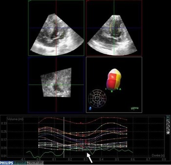
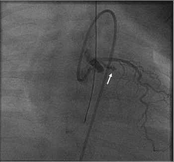
Two-dimensional echocardiography showed severe septal myocardial hypertrophy, 11 millimetres in diastole, associated with latero-basal myocardial akinesia. The mitral pillar was ischaemic with moderate mitral regurgitation. Despite septal hypertrophy, we did not observe a Doppler gradient through the left ventricular outflow. Three-dimensional echocardiography allowed global and regional left ventricular function analysis (Fig 1). Echocardiography showed a Q necrotic wave pattern in the latero-basal area. Troponin T was 0.253 nanogram per millilitre and brain natriuretic peptide was 48.1 nanograms per millilitre. Selective coronary angiography showed an occlusion of the medium part of the circumflex artery with parietal irregularities, downstream distension, and filiform ending (Fig 2). The implantation of abnormal coronary arteries or course was discarded. The biological standard check-up was normal. The thrombophilia check-up, which included platelet count, prothrombin, factor V, C and S proteins, antithrombin III, homocystein, plasminogen 1 and 2, was normal.
Global and regional left ventricular quantification by three-dimensional echocardiography (iE 33, paediatric probes 2–7 megahertz Philips) with automatic detection of outline (QLAB 4.2 system, Philips, Andover, MA, USA). The left ventricular ejection fraction was calculated at 50% (normal >60%). Note the poor contraction of latero-basal segment (white arrow).

Selective coronary angiography. The circumflex artery is occluded with parietal irregularities, downstream distension and filiform ending (white arrow).

Angiotensin-converting enzyme inhibitor, Lopril™ (Bristol-Myers Squibb, Rueil-Malmaison, Cedex, France), and aspirin therapy was initiated. Clinical and haemodynamic parameters remained normal all along his hospital stay. At discharge from hospital, echography showed a significant decrease in septum thickness measuring 8 millimetres with persistent dyskinesia and mitral insufficiency grade 1–2/4. At 3 months, the patient was asymptomatic and septum thickness was normal. At 15 months, L. weighed 8900 kilograms for 75 centimetres. He was asymptomatic. Electrocardiogram showed a persistent Q necrotic wave pattern in the latero-basal area and echography showed a limited akinesia of the latero-basal segment. Mitral insufficiency disappeared. Treatment still consisted in Lopril and aspirin.
Discussion
Classically, complications of the diabetic mother’s newborn are foetal asphyxia, macrosomia, and electrolytic disorders such as hypoglycaemia, hypocalcaemia and hypomagnesaemia, hypertrophic cardiomyopathy, respiratory distress syndrome, hyperbilirubinaemia, polyglobulia, and malformations (bone, genital, urinary, cutaneous, and brain malformations).Reference Schwartz and Teramo3 In all, 30–40% of perinatal deaths in insulin-dependent diabetic mothers’ pregnancies are caused by malformations, 20–30% by prematurity, and 20–30% by intrauterine asphyxia including late gestation foetal death. The incidence of abnormal foetal heart rate, cord blood acidosis, and low Agars score are increased in diabetic pregnancies.Reference Schwartz and Teramo3 The foetal heart is affected in maternal diabetes. The first incidence of cardiac malformations increases because cardiogenesis is impaired. Then, the incidence of hypertrophic cardiomyopathy in infants from diabetic mothers varies from 10% to 50%.Reference Abu-Sulaiman and Subaih1, Reference Ullmo, Vial and Di Bernardo2 It mainly affects the ventricular septum. The physiopathology of pathologic ventricular hypertrophy is not really known: foetal hyperinsulinism exasperates hyperplasia and hypertrophy of myocardial cells by protein and fat synthesis. Prognosis is usually good with spontaneous resolution of cardiac hypertrophy.Reference Ullmo, Vial and Di Bernardo2, Reference Sardesai, Gray, McGrath and Ford4, Reference Prefumo, Celentano, Presti, De Biasio and Venturini5 In rare cases, hydrops fetalis resulting from obstructive myocardial hypertrophy may be responsible of death.Reference Sardesai, Gray, McGrath and Ford4
Although septal myocardial hypertrophy is well known in the newborn of diabetic mothers,Reference Abu-Sulaiman and Subaih1 its association with cardiac infarction is uncommon. Myocardial infarction in the perinatal period is a rare occurrence, usually associated with congenital cardiac lesions. Other aetiologies are isolated coronary artery abnormalities as the abnormal origin of the left coronary artery from the pulmonary artery, or fibrous stenosis of a coronary ostium or the coronary artery wall.Reference Gault and Usher6, Reference Ferns, Khan, Firmin, Peek and Bu’Lock7 In the absence of morphological cardiac disease, the most common aetiologies of myocardial infarction are intrauterine asphyxia and thromboembolic coronary occlusion. A paradoxical embolus usually arises from a thrombus in the ductus venosus or umbilical vein and reaches the coronary circulation through normal foetal circulatory pathways.Reference Bernstein, Finkbeiner, Soifer and Teitel8 Neonates with high pulmonary vascular resistance are especially predisposed to this because of right–left shunt favourisation. Thromboembolic occlusion can be occurred by natural deficiency of anticoagulant factorsReference Lucas, Burchfield and Donnelly9 such as antithrombin III deficiency, protein S and protein C deficiency, complicating enteroviral myocarditis,Reference Murugan, Gnanapragasam and Vettukattil10 maternal toxic,Reference Bulbul, Rosenthal and Kleinman11 mediacalcosis of coronaries, coronary hypoperfusion in asphyxiated infants, and congenital abnormalities of the heart marked by hypertrophy of the left ventricular myocardium (endocardial fibroelastosis or non-obstructive hypertrophic cardiomyopathy).Reference Dimitriu, Scumpu and Brumariu12 In the last situation, perinatal asphyxia or coronary arteries compression by myocardium can lead to a low-output state and predispose to stasis and clot formation.Reference Kilbride, Way, Merenstein and Winfield13 Finally, neonatal myocardial infarction can remain as idiopatic infarction without any pathophyiological explanation.Reference Iannone, Duritz and McCarthy14
Our observation did not find any anatomical or biological causes of myocardial infarction. One hypothesis to link the septal hypertrophic and latero-basal ischaemic myocardium could be a steal blood. Because of the very thick septum, the anterior interventricular coronary artery was very developed and blood flow was preferentially directed to the septal wall rather than the lateral wall. As a consequence, the hypoperfusion of circumflex coronary artery could favour an ischaemia in the latero-basal segment. In the newborn of a diabetic mother with a 11-millimetre septal thickness and normal regional function, one should attempt a moderate-to-severe left ventricular obstruction assessed by the Doppler gradient. The absence of septal obstruction in our observation could be explained by the associated myocardial infarction of the opposite segment. Therefore, systolic septal motion was compensated by the absence of latero-basal motion and intraventricular reduction of the cavity was limited. Moreover, ischaemia of the mitral pillar could avoid anterior systolic motion of the mitral valve, which participates in the left ventricular obstruction.
Conflict of interest
None