INTRODUCTION
Trypanosoma cruzi is the aetiological agent of Chagas disease and presents atypical structures that could be targets for chemotherapeutic treatment (De Souza, Reference De Souza2009). One example is the kinetoplast, a specialized region of the mitochondrion that contains the mitochondrial or kinetoplast DNA (kDNA). The kDNA consists of thousands of interlocked DNA molecules, the minicircles and the maxicircles, which form a single network within the mitochondrial matrix. Kinetoplast Associated Proteins (KAPs) are small, basic proteins that pack the kDNA circles into a compact structure within the kinetoplast (Xu et al. 1993; Cavalcanti et al. Reference Cavalcanti, Shimada, Probst, Souto-Padrón, De Souza, Goldenberg, Fragoso and Motta2009). The maxicircles are analogous to the mitochondrial DNA of higher eukaryotes and encode rRNAs and subunits of respiratory complexes (e.g. cytochrome B, cytochrome oxidase, and NADH dehydrogenase) (Shapiro and Englund, Reference Shapiro and Englund1995; De Souza and Cavalcanti, Reference De Souza and Cavalcanti2007; Jensen and Englund, Reference Jensen and Englund2012). Most maxicircle transcripts are edited via insertions and/or deletions of uridylate residues to form functional open reading frames; the minicircles serve as guide RNAs in this RNA editing process (Stuart et al. Reference Stuart, Schnaufer, Ernst and Panigrahi2005; Aphasizhev and Aphasizheva, Reference Aphasizhev and Aphasizheva2011).
The replication of kDNA is complex and requires a repertoire of enzymes, including type II topoisomerase (topo II), DNA polymerase, primase, among others. At the beginning of this process, the minicircles are released from the network by topo II, and the minicircles initiate replication as a theta structure in the kinetoflagellar zone (KFZ) via the universal minicircle binding protein (UMSBP), Polymerase 1B and other proteins. Then, the daughter minicircles migrate to the antipodal sites, i.e. protein assemblies that flank the kDNA network and are located at opposite ends from one another. Primer removal, gap filling, and the sealing of nicks by DNA ligase occur at these sites. The replicated minicircles are attached to the network periphery adjacent to the antipodal sites by the action of topo II, and the relative motion of the kinetoplast distributes the newly replicated circles throughout the network (Guilbride and Englund, Reference Guilbride and Englund1998; Morris et al. Reference Morris, Drew, Klingbeil, Motyka, Saxowsky, Wang and Englund2001; Liu et al. Reference Liu, Liu, Motyka, Agbo and Englund2005; Liu and Englund, Reference Liu and Englund2007; Jensen and Englund, Reference Jensen and Englund2012). Little is known about the maxicircle replication process; however, unlike the minicircles, the maxicircles remain attached to the network during this process (Carpenter and Englund, Reference Carpenter and Englund1995; Liu et al. Reference Liu, Wang, Yaffe, Lindsay, Zhao, Zick, Shlomai and Englund2009). The unique characteristics of kDNA organization and replication suggest that kDNA could be an attractive target for selective chemotherapy (Das et al. Reference Das, Dasgupta, Sengupta and Majumder2004; Motta, Reference Motta2008).
Trypanosomatids can undergo the partial or even complete loss of kDNA, resulting in the formation of dyskinetoplastic (Dk) or akinetoplastic (Ak) cells, respectively. Dk organisms can be found in nature or induced in the laboratory via treatment with DNA-intercalating drugs (Schnaufer et al. Reference Schnaufer, Domingo and Stuart2002) or by RNA interference (RNAi) which may inactivate the kDNA replication process and the action of the editing proteins (Wang and Englund, Reference Wang and Englund2001; Wang et al. Reference Wang, Englund and Jensen2012). Using conventional staining techniques, kDNA is not detectable in Dk trypanosomatids; indeed, the kDNA in these cells is no longer organized into a network but appears dispersed in clumps throughout the mitochondrial matrix (Trager and Ruddzinska, Reference Trager and Rudzinska1964; Steinert and Assel, Reference Steinert and Van Assel1967; Simpson, Reference Simpson1968; Hill and Anderson, Reference Hill and Anderson1969; Stuart, Reference Stuart1971; Hajduk, Reference Hajduk1979).
Naturally occurring Dk cells were observed decades ago in Trypanosoma evansi and Trypanosoma equiperdum (Schnaufer et al. Reference Schnaufer, Domingo and Stuart2002). Nevertheless, the mechanism by which the Dk cells arise in nature is unknown, but they can only survive as bloodstream forms, not procyclic forms. This is probably related to the fact that a mutation in the nucleus-encoded γ-subunit of the F1-ATPase, which is a very rare event, is essential to permit kDNA loss (Schnaufer et al. Reference Schnaufer, Domingo and Stuart2002; Jensen and Englund, Reference Jensen and Englund2012).
As previously mentioned, dyskinetoplasty can be induced in the laboratory using DNA binding drugs. Such compounds are classified according to their mechanism of action, i.e. as non-intercalating drugs (e.g. berenil and pentamides) and intercalating drugs (e.g. acriflavine and ethidium bromide) (Motta, Reference Motta2008). Acriflavine, which is a mixture of two very closely related acridines, was discovered almost 100 years ago and has been traditionally used as an antimicrobial agent (Wainwright, Reference Wainwright2001). Early studies on the use of this intercalating agent in trypanosomatids showed the emergence of Dk cells in Crithidia fasciculata (Hajduk, Reference Hajduk1979; Hill and Anderson, Reference Hill and Anderson1969), Crithidia luciliae (Steinert and Assel, Reference Steinert and Van Assel1967), Trypanosoma brucei (Stuart, Reference Stuart1971), T. equiperdum (Hajduk, Reference Hajduk1979; Shapiro et al. Reference Shapiro, Klein and Englund1989), and Leishmania tarentolae (Trager and Rudzinska, Reference Trager and Rudzinska1964; Simpson, Reference Simpson1968). Electron microscopy studies have revealed that the kDNA loses its typical structure after treatment with intercalating compounds, but the double membrane of the mitochondrion and its cristae is maintained. Dk cells appear as a result of the selective inhibition of kDNA synthesis and the unequal distribution of the kDNA network during cell division (Simpson, Reference Simpson1968). The loss of kDNA gene sequences in Dk strains results in decreased O2 consumption and lower mitochondrial enzyme activity as demonstrated in C. fasciculata (Hill and Anderson, Reference Hill and Anderson1969; Manaia and Roitman, Reference Manaia and Roitman1977).
Although the effect of intercalating drugs on trypanosomatids has been demonstrated in early studies, the phenomenon of dyskinetoplasty has never been reported in T. cruzi and properly described using ultrastructural techniques. In this work, we show that the treatment of T. cruzi epimastigotes with acriflavine resulted in a decrease in cell proliferation and induced the appearance of Dk cells. Alterations in the kDNA topology were described via transmission electron microscopy (TEM), cytochemical approaches, and atomic force microscopy (AFM). The results suggest that the disruption of the kDNA network leads to the emergence of the Dk phenotype. In addition, we show the first images of the isolated kDNA network of T. cruzi as analysed by AFM.
MATERIALS AND METHODS
Cell culture
Epimastigotes of T. cruzi (Dm28c clone) were cultivated at 28 °C in liver infusion tryptose (LIT) medium that was supplemented with 10% fetal calf serum.
Drug treatment
A stock solution containing 1 mg mL−1 of acriflavine (Sigma Chemical Company) was prepared, sterilized by filtration, and protected from light before use. The drug was added to log-phase cultures of epimastigotes at concentrations of 1, 5, 10, 25 and 50 μg mL−1. Every 24 h, part of the control and treated cultures were removed for cell counting, and another portion was fixed for TEM studies. Dk cells were quantified using the panotic staining kit (Laborclin).
Transmission electron microscopy
Protozoa were fixed in 2·5% glutaraldehyde in 0·1 m cacodylate buffer, pH 7·2, and post-fixed in 1% OsO4 and 0·8% potassium ferricyanide in the same buffer. Cells were dehydrated in a graded series of acetone and embedded in epoxy resin. Ultrathin sections were stained with uranyl acetate and lead citrate and observed in a Zeiss900 transmission electron microscope operating at 80 KV.
Cytochemical assays
(a) The ethanolic phosphotungstic acid (E-PTA) method: Protozoa were fixed in 2·5% glutaraldehyde in 0·1 m cacodylate buffer, pH 7·2, for 2 h at room temperature, washed in the same buffer, and then dehydrated in a graded series of ethanol. Then, cells were incubated at room temperature in 2% phosphotungstic acid in 100% ethanol for 2 h, washed in absolute ethanol, and embedded in epoxy resin (Souto-Padrón and De Souza, Reference Souto-Padrón and De Souza1978).
(b) Ammoniacal silver (AS) method: This technique was performed as described by Souto-Padrón and De Souza (Reference Souto-Padrón and De Souza1978). Protozoa were fixed in 2·5% glutaraldehyde in 0·1 m cacodylate buffer, pH 7·2, for 2 h at room temperature, washed in distilled water, and incubated for 5 min at room temperature in the AS solution. The AS solution was prepared just before use by gradually adding a 10% silver nitrate solution to concentrated ammonium hydroxide until a slight turbidity appears and persists. Then, cells were washed in distilled water, kept for 5 min in a 3% formaldehyde solution, washed in distilled water again, and post-fixed in 1% OsO4 and 0·8% potassium ferricyanide in 0·1 m cacodylate buffer. Then, the cells were dehydrated in ethanol and embedded in epoxy resin.
kDNA isolation
The kDNA of the control cells and protozoa that were treated with 5 and 50 μg mL−1 of acriflavine for 48 h were isolated according to the protocol described by Pérez-Morga and Englund (Reference Pérez-Morga and Englund1993). Briefly, the protozoa were harvested via centrifugation and washed in NET 100 buffer (i.e. 10 mm Tris-HCl [pH 8·0], 100 mm NaCl and 100 mm EDTA). The cells were then lysed at 37 °C for 60 min in a solution containing 56·5 μL of 20% SDS and 11 μL of proteinase K (20 mg mL−1) in 1 mL of NET 100 buffer. The lysate was loaded onto a 20% sucrose cushion and centrifuged for 20 min at 18 000 g in an Eppendorf 5810R centrifuge. The kDNA pellet was resuspended in NET 100, loaded on top of a second sucrose cushion, and processed as previously described. The kDNA was dialysed against Tris-EDTA buffer (i.e. 10 mm Tris-HCl [pH 8·0] and 1 mm EDTA) overnight, centrifuged at 18 000 g for 30 min, and resuspended in 1 mm Tris–HCl, pH 8·0.
Atomic force microscopy
The kDNA networks from control and acriflavine treated protozoan cells were prepared for the AFM analyses as described by Cavalcanti et al. (Reference Cavalcanti, Gonçalves, Costa and De Souza2011). The solution containing the isolated kDNA networks was mixed with 25 mm magnesium chloride and then deposited onto a freshly cleaved mica surface. The sample was incubated for 5 min at room temperature, rinsed gently in Milli-Q water, rinsed gently in an ethanol/water mixture (1 : 1 vol), and then rinsed in ethanol. The sample was critical point dried in a Leica EM CPD030 apparatus. The network images were acquired with a Nanowizard AFM (JPK Instruments AG) in air using the intermittent mode. The AFM was mounted with a rectangular tip (AC240TS, Olympus, Tokyo) working in a nominal resonant frequency of about 75 kHz. The images were processed with JPK image processing software using line-wise levelling.
RESULTS
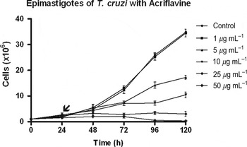
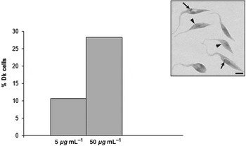
In this study, T. cruzi Dk epimastigotes were created via the addition of different concentrations of acriflavine to log-phase cultures. As shown in Fig. 1, this intercalating agent promoted a dose-dependent inhibitory effect on protozoal proliferation. The 1 μg mL−1 acriflavine treatment did not cause growth impairment, but higher concentrations, i.e. 5, 10, 25 and 50 μg mL−1, reduced cell proliferation by 5, 14, 40 and 55%, respectively, after 48 h of treatment. Higher drug concentrations (i.e. 25 and 50 μg mL−1) completely halted protozoal proliferation after 24 h of treatment (Fig. 1). Cultivation of protozoa in the presence of a low acriflavine concentration (i.e. 5 μg mL−1) promoted dyskinetoplasty in 10% of cells, and this increased to approximately 30% after treatment with 50 μg mL−1 of acriflavine (Fig. 2). Dk cells were generated even after a short period of treatment (i.e. 24 h) as observed with the Panotic staining (Fig. 2 inset).
Effect of acriflavine on the proliferation of Trypanosoma cruzi epimastigotes. The drug was added in concentrations of 1, 5, 10, 25 and 50 μg mL−1 to the log-phase culture 24 h after the initial inoculation (arrow). A dose-dependent inhibitory effect was observed. Standard deviations are indicated.

Quantification of Dk cells after treatment with 5 and 50 μg mL−1 of acriflavine for 24 h (n = 400 cells). Inset shows kinetoplast containing cells (arrows) and Dk cells (arrowheads) after the panotic staining of protozoa treated with 50 μg mL−1 of acriflavine for 24 h. Bar = 3 μm.

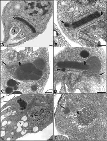
In order to evaluate the action of acriflavine on the ultrastructure of T. cruzi, TEM analysis was performed. The kinetoplast of non-treated protozoa is composed of DNA fibrils, which are aligned parallel to one another and densely packed within the matrix of a disc-shaped kinetoplast (Fig. 3A). In acriflavine treated cells, the kDNA topology is markedly affected (Figs 3B–F). When the lowest drug concentration (i.e. 5 μg mL−1) was used, we observed that the characteristic arrangement of kDNA was maintained; however, a round and electron-dense structure was seen next to one of the antipodal sites (Fig. 3B arrowhead). With higher concentrations of acriflavine, the kinetoplast lost its normal disc-shaped structure, and the fibrillar arrangement was transformed into a compact and amorphous mass within the mitochondrion (Figs 3C–F). Furthermore, fragmented parts of the network were often observed as clusters next to one of the antipodal sites (Figs 3C and D, arrows). Moreover, a complete disorganization of the kDNA network was observed after drug treatment with 50 μg mL−1 for 48 h (Fig. 3F).
Ultrastructural analysis of Trypanosoma cruzi epimastigotes treated with acriflavine. (A) control cells presenting a typical kDNA fibrillar arrangement; (B) cells treated with 5 μg mL−1 and (C–F) 50 μg mL−1 of acriflavine for 48 h. In treated cells, the kDNA distribution was altered, and formations of amorphous, compact and fragmented masses were observed within the mitochondrion matrix (C–F). In addition, an electron-dense and round structure was seen next to one of the antipodal sites (B and D – arrowheads), and clusters of electron-dense material were observed (C, D, F – arrows). Mitochondrial swelling (m) and cell vacuolization (v) were also observed (E). n = nucleus, f = flagellum, k = kinetoplast, m = mitochondrion. Bars = 0·25 μm.

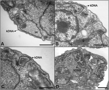
Drastic kDNA disorganization was also seen even when a low concentration of acriflavine was used for long periods (Figs 4A–D). In cells treated up to 168 h, the kDNA almost completely disappears (Figs 4A and B), culminating in the complete fragmentation of the network (Figs 4C and D). This indicates that the time of treatment, not just the drug concentration, contributes to promoting dyskinetoplasty in T. cruzi. The nucleus was not affected by the use of acriflavine (Fig. 4B); however, we observed mitochondrial swelling and intense vacuolization of cells treated with the high drug concentration (Fig. 3E).
The TEM analysis of epimastigotes treated with 5 μg mL−1 of acriflavine for long periods of time (A,B: 96 h; C,D: 168 h). The kDNA was almost completely lost, and an intense fragmentation of the network was seen. k = kinetoplast, n = nucleus, bb = basal body, f = flagellum, g = golgi, m = mitochondrion. Bars = 0·25 μm.

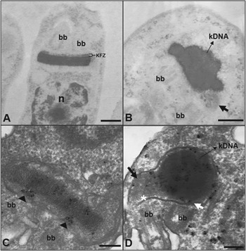
The acriflavine treatment also altered the distribution of basic proteins associated with the kDNA in the kinetoplast of T. cruzi. The employment of cytochemical techniques, such as the E-PTA (Figs 5A and B) and AS methods (Figs 4C and D), revealed that for the control cells (Figs 5A and C), staining was mainly observed over the kDNA disk and in the KFZ region, including the intramitochondrial filaments that link the inner face of the differentiated membrane zone to the kDNA and compose the tripartite attachment complex (TAC) (Fig. 5C – arrowheads). In acriflavine-treated cells, this staining pattern was modified; in fact, labelling was more intense in the electron-dense areas, which correspond to the condensed kDNA (Figs 5B and D, black arrows), whereas the fragmented network that was observed close to the antipodal sites was less visible (Figs 5B and D – white arrow). In treated protozoa, the staining in the TAC region was less evident in the electron-dense region (Fig. 5D, white asterisk) and absent in the area containing the fragmented network (Fig. 5D, white asterisk).
Detection of basic proteins in non-treated and acriflavine-treated Trypanosoma cruzi. (A-B) E-PTA and (C,D) AS technique. In control cells (A–C), we observed staining throughout the kinetoplast, but mainly in electron-dense areas and in the KFZ region. In protozoa treated with 50 μg mL−1 of acriflavine for 48 h (B,D), the basic proteins were concentrated in the electron-dense mass of the altered kDNA, but reduced staining was observed in less electron-dense regions (black arrows). Note the redistribution of filaments in the TAC region when comparing control cells (C, arrowheads) to acriflavine-treated protozoa (D, white arrow and asterisk). bb = basal body, KFZ = kinetoflagellar zone. Bars = 0·25 μm.

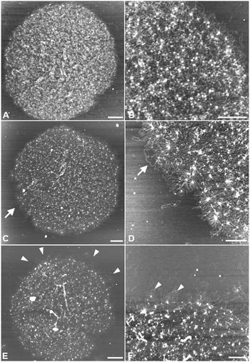
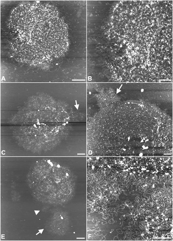
To better understand the effect of acriflavine on the kDNA topology, we isolated intact networks of treated and non-treated protozoa and analysed the samples using AFM, which has been extensively used to visualize morphological changes of DNA structure. In the present work, AFM helped us to understand the disorganization of the network that culminated in the Dk phenotype. The kDNA that was isolated from control cells appeared as an intact and massive network of interlocked DNA molecules (Figs 6A and B). When a low concentration of acriflavine was used (i.e. 5 μg mL−1) for 48 h, we observed that some DNA circles were released from the network periphery (Figs 6C–F). After using a high drug concentration (i.e. 50 μg mL−1), an intense release of circles from the network edge was seen (Figs 7A and B); we also observed frequent detaching of network bulges, which resulted in kDNA fragmentation (Figs 7C–F). The AFM data indicated that the dyskinetoplasty phenomenon begins with the release of minicircles from the edge of the network, culminating with network fragmentation and kDNA dispersion throughout the mitochondrial matrix of the protozoa.
AFM analysis of non-treated and acriflavine treated protozoa. The kDNA in control cells forms an intact and massive network (A, B). In protozoa treated with 5 μg mL−1 of acriflavine for 48 h, we observed an initial disorganization of the kDNA network edge (C and D – arrows) and the release of the circles from the network periphery (E and F, arrowheads). Bars = 1 μm (A, C, E) and 0·5 μm (B, D, F).

AFM analysis of epimastigotes treated with 50 μg mL−1 of acriflavine for 48 h. Using higher drug concentrations, an intense release of circles from the kDNA edge (A, B) and the detachment of pieces of the network (C, D and E – arrows) were observed. Fig. 7F is a magnification of the region indicated by the arrowhead in Fig. 7E and shows the loss of circles and the bulge detached from the kDNA network, which resulted in kDNA fragmentation. Bars = 1 μm (A, C, E) and 0·5 μm (B, D, F).

DISCUSSION
Early studies on the use of intercalating drugs, such as acriflavine and ethidium bromide, in trypanosomatids reported the emergence of cells in which the kinetoplast was no longer stained with Giemsa. This phenomenon was first named ‘akinetoplasty’ and was described in Bodo caudatus and T. evansi (Robertson, Reference Robertson1929; Inoki et al. Reference Inoki, Sakamoto, Ono and Kubo1961). Later on, this term was replaced by ‘dyskinetoplasty’ since electron microscopy analyses showed that the kinetoplast did not disappear as originally thought after observation via light microscopy. In fact, ultrastructural analyses revealed that the kDNA lost its fibrillar arrangement after drug treatment but still remained within the mitochondrial matrix (Trager and Rudzinska, Reference Trager and Rudzinska1964).
Crithidia fasciculata (Hill and Anderson, Reference Hill and Anderson1969; Hajduk, Reference Hajduk1979), C. luciliae (Steinert and Assel, Reference Steinert and Van Assel1967), T. brucei (Stuart, Reference Stuart1971), T. equiperdum (Hajduk and Cosgrove, Reference Hajduk and Cosgrove1979) and L. tarentolae (Trager and Rudzinska, Reference Trager and Rudzinska1964; Simpson, Reference Simpson1968) are markedly affected after treatment with acriflavine; in fact, a decrease in cell proliferation and the presence of Dk cells are observed after such treatment. In this work exposure to acriflavine also reduced T. cruzi cell growth and generated Dk epimastigotes that were able to survive and multiply for long periods, as also reported for T. brucei bloodstream forms (Stuart, Reference Stuart1971; Hajduk, Reference Hajduk1979).
Several studies reported the similarities in the phenotype of Dk cells from different species of trypanosomatids. In all species that have been analysed, the kDNA arrangement was modified due to alterations in its typical fibrillar arrangement, thereby giving rise to one or more compact and amorphous masses that spread throughout the kinetoplast matrix. Acriflavine also promotes the detachment of the packed kDNA fibres from the mitochondrial membrane. The inhibitory effect of acriflavine on cell proliferation appears to be highly selective since the nucleus of trypanosomatids is not affected by drug treatment. Tubbs et al. (Reference Tubbs, Ditmars and Van Winkle1964) demonstrated that the binding of acriflavine to DNA is particularly efficient in DNA with a high adenine-thymine base pair composition. Considering that the mitochondrial kDNA presents a higher A-T content than that of nuclear DNA (Riou and Paoletti, Reference Riou and Paoletti1967) partly explains why the kDNA topology is affected when the nuclear DNA compaction remains unaltered.
Dk T. cruzi showed the same pattern of kinetoplast alteration that has been reported for other trypanosomatids. In addition, by using cytochemical techniques that detect lysine, arginine, and histidine residues in basic proteins, we showed that acriflavine treatment altered the distribution of basic proteins throughout the kinetoplast of T. cruzi. Electron-dense areas of the altered kDNA mass presented a more intense staining than the areas of lower electron density, thereby confirming that the latter regions are composed of less condensed DNA fibres. In acriflavine treated cells, a redistribution of filaments in the TAC region was observed and may affect the segregation of minicircles, thereby resulting in a loss of kDNA and in dyskinetoplasty. This hypothesis is consistent with the assertion that TAC filaments link the basal body to the kinetoplast throughout the trypanosomatid cell cycle and play a structural role in mitochondrial positioning and during kDNA replication (Ogbadoyi et al. Reference Ogbadoyi, Robinson and Gull2003).
Interestingly, this present study revealed that the kinetoplast of treated T. cruzi contained regions with distinct electron density probably due to the loss of minicircles from the network. Indeed, when we analysed the isolated kDNA using AFM, we could see that the minicircles were detached from the network, thus complementing the data obtained by TEM. As the acriflavine concentration increased, an increase in the number of free circles was observed. This is the first report showing that the detachment of minicircles promotes the disassembly of the kDNA network, which generates Dk cells.
No evidence has indicated if acriflavine promotes the release of the circles from the network or if the circles released by topoisomerase II during the kDNA replication are unable to duplicate and reconnect to the network. Nevertheless, the latter hypothesis is seemingly the most plausible. In fact, when trypanosomatids are pulsed with [3H]-thymidine, silver grains are observed in the nucleus and in the kinetoplast of the protozoa, indicating sites of DNA replication (Carpenter and Englund, Reference Carpenter and Englund1995). However, an early report demonstrated that acriflavine-treated C. fasciculata did not incorporate this tracer into the kinetoplast but did incorporate it in the nucleus (Hill and Anderson, Reference Hill and Anderson1969). Acriflavine also inhibits the [3H]-thymidine incorporation into the kinetoplast of C. luciliae, thereby indicating that the replication process in the kinetoplast is directly affected; however, the nuclei of treated protozoa continue to synthesize DNA (Steinert and Assel, Reference Steinert and Van Assel1967).
A recent study clarified the mechanism by which an intercalating drug inhibits the kDNA replication in T. brucei (Roy Chowdhury et al. Reference Roy Chowdhury, Bakshi, Wang, Yildirir, Liu, Pappas-Brown, Tolun, Griffith, Shapiro, Jensen and Englund2010). In this study, the authors demonstrated that in the presence of ethidium bromide, the incorporation of BrdU (i.e. a thymidine analogue) was not observed in any minicircle. This result was not expected since BrdU should have been incorporated in gapped circles that had already replicated. In addition, they observed a dramatic accumulation of covalently closed free minicircles, i.e. the substrates for replication, in samples treated with ethidium bromide. Altogether, these data indicate that ethidium bromide inhibits the initiation of free minicircle replication. Once the released circles are not replicated, they are not reconnected to the network, which causes a loss of kDNA and its rearrangement. Acriflavine and other intercalating drugs may possibly act in the kinetoplast via a similar mechanism.
Our present observations strongly suggest that acriflavine inhibits T. cruzi multiplication via interference with kDNA replication or maintenance. However, we cannot rule out the possibility that acriflavine affects other essential cellular processes, which culminates in cell death. Interestingly, the kinetoplast is a structure that is only found in members of the Kinetoplastida order. Therefore, efforts should be focused on the synthesis of new and more specific compounds that interfere with the kinetoplast structure and function; indeed, the kinetoplast is a potentially important target for the development of new drugs. In addition, we reaffirm that TEM and AFM are powerful tools that can be used to provide information on the structural organization of the kinetoplast and the action of drugs that target kDNA.
ACKNOWLEDGEMENTS
The authors are grateful to Daniela Leão Gonçalves for technical assistance.
FINANCIAL SUPPORT
This investigation received financial support from Conselho Nacional de Desenvolvimento Científico e Tecnológico (CNPq) and Fundação Carlos Chagas Filho de Amparo à Pesquisa do Estado do Rio de Janeiro (FAPERJ).