Introduction
Since the early descriptions of mild cognitive impairment (MCI) (Petersen et al., Reference Petersen, Smith, Waring, Ivnik, Tangalos and Kokmen1999), there has been increasing interest in its clinical characterization and prognosis (Nelson and O'Connor, Reference Nelson and O'Connor2008; Werner and Korczyn, Reference Werner and Korczyn2008). In previous reports (Petersen et al., Reference Petersen, Smith, Waring, Ivnik, Tangalos and Kokmen1999), subjects with MCI exhibit poorer cognitive functioning than healthy subjects, but not as impaired as patients with dementia. Prognosis studies have stressed the use of this nosological entity as a risk or prodromic state for dementia, due to the high rate of conversion of MCI subjects to dementia (10%–15% of patients who meet the criteria of amnestic MCI develop Alzheimer's-type dementia per year, up to 80% at 5-year follow-up) (Amieva et al., Reference Amieva2004; Petersen, Reference Petersen2004; Gauthier et al., Reference Gauthier2006). Approximately 16% of the populations of older people who have not been diagnosed with dementia meet the current criteria for MCI (Petersen et al., Reference Petersen2010) and MCI prevalence increases with age.
Dementia inevitably involves a loss of functional independence, resulting in a progressive increase in disability (Sauvaget et al., Reference Sauvaget, Yamada, Fujiwara, Sasaki and Mimori2002). In parallel with the progressive loss of personal autonomy, increases in family/caregiver burden and in demand for health and social resource use have been reported (Potkin, Reference Potkin2002). However, disability, a milder form than dementia, is not included as an MCI diagnostic criterion. Furthermore, the diagnosis of MCI requires preservation of the person's ability to perform activities of daily living (ADL) (Petersen et al, Reference Petersen, Smith, Waring, Ivnik, Tangalos and Kokmen1999; Petersen and Negash, Reference Petersen and Negash2008), “or at least that impairment is minimal” in complex instrumental functions (Winblad et al., Reference Winblad2004). Recently, the lack of functional impairments of MCI in daily living as a diagnostic criterion has been questioned as the result of studies pointing to impairment in instrumental ADL rather than basic ADL (Tam et al., Reference Tam, Lam, Chiu and Lui2007; Pereira et al., Reference Pereira, Yassuda, Oliveira and Forlenza2008; Ahn et al., Reference Ahn2009; Burton et al., Reference Burton, Strauss, Bunce, Hunter and Hultsch2009; Schmitter-Edgecombe et al., Reference Schmitter-Edgecombe, Woo and Greeley2009; Aretouli and Brandt, Reference Aretouli and Brandt2010; Bangen et al., Reference Bangen2010; Reppermund et al., Reference Reppermund2010; Teng et al., Reference Teng, Becker, Woo, Cummings and Lu2010a; Reference Teng, Becker, Woo, Knopman, Cummings and Lu2010b). Although these studies were carried out in different countries and used different instruments to assess impairments in ADL, they all suggest that people who meet the other criteria described for MCI do show functional impairment of ADL.
Disability in dementia and other neurological and neuropsychiatric disorders has been largely associated, and partially attributed, to cognitive impairment (e.g. Millis et al., Reference Millis, Rosenthal and Lourie1994 in brain injury; Green, Reference Green1996 in schizophrenia). Those findings seem to support the a priori hypothesis that MCI would show some degree of functional impairment, which could be at least partially predicted by the cognitive impairment that characterizes MCI. However, to date there are very few studies that assess the potential associations between functional performance and cognitive impairment in MCI subjects or that attempt to determine which cognitive areas are more likely to predict functional abilities in MCI subjects.
In light of the above, we aimed to (1) determine whether MCI is associated with functional impairment; (2) identify the role of sociodemographic, emotional, and cognitive variables as independent predictors of functional impairment in elderly healthy subjects, single-domain amnestic MCI subjects and multi-domain amnestic MCI subjects; and (3) discuss the extent to which the two former issues are instrument-dependent. We hypothesized that (1) MCI patients will show impaired independent functioning; (2) among subjects with MCI, such impairment will be associated with the degree of cognitive impairment, and to a lower degree with sociodemographic and anxiety-depression symptoms, whereas in healthy controls, impaired functioning will be only associated with sociodemographic and anxiety-depression symptoms; and (3) in MCI subjects the degree of impairment and the nature and effect size of associations will partially depend on the type of independent functioning, in such a way that quantitative measures based on performance (Craig Handicap Assessment and Reporting Technique, CHART) will be more reliable than qualitative measures (World Health Organization Disability Assessment Schedule, second version, WHO-DAS-II) based on a subjective judgment of disability.
Methods
Participants
Recruitment of participants was conducted during 2010 and the beginning of 2011 in three clinical or university settings in three regions of Spain: the University of Oviedo (northern Spain), Consorci Sanitari of Terrassa (Barcelona, northeastern Spain), and the University of Granada (southern Spain). Recruitment in the Consorci Sanitari of Terrassa was done by review of clinical history and application of inclusion and exclusion criteria to outpatients attending its memory clinic. The two universities recruited participants by inviting subjects attending community services for healthy elderly people to participate in a free memory-training program. The memory training program was conducted as part of a randomized clinical trial, aimed to compare the efficacy of two neuropsychological rehabilitation strategies (restitution versus compensation and substitution) in improving functional independence and cognition in MCI subjects.
A total of 328 elderly people living in the community were recruited. Inclusion criteria included: age over 55 years, meeting the diagnostic criteria for MCI as defined in Petersen et al. (Reference Petersen, Smith, Waring, Ivnik, Tangalos and Kokmen1999), living in the community, and signing informed consent. Exclusion criteria were: living in an assisted living residence, cognitive functioning suggesting a possible diagnosis of dementia (see below), previous diagnosis of dementia, previous psychiatric disorder according to DSM-IV-TR at the time of recruitment, presenting a moderate or higher degree of disability due to other conditions than MCI, and severe language impairments that would compromise their active participation in the neuropsychological rehabilitation clinical trial.
Neuropsychological assessment
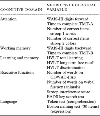
Cognitive assessment was performed by means of a neuropsychological battery designed to comprehensively evaluate attention, working memory, memory, executive functioning, and language (see Table 1). In order to obtain a summary score for each cognitive domain (mean of variables that compounded a cognitive domain as presented in Table 1) and a global score for cognition (average of the four cognitive domains), raw test scores were converted to T-scores (mean = 50, SD = 10) based on normative data published in the Spanish handbooks of the tests (WAIS-III digits, Stroop) and normative data obtained in an ongoing normative study with an independent healthy sample (N = 173). In this ongoing normative study, normative data were obtained for the following age groups: 56–65 years (n = 56), 66–75 (n = 71), 76 or older (n = 46). T-scores were truncated at 10 (–4 standard deviations), to avoid outlying values (as suggested in Rousseeuw and Leroy, Reference Rousseeuw and Leroy1987). Results are reported for cognitive areas summary scores, in order to avoid an excess of data (results of single test variables available upon request). Language summary score was not included in statistical analyses because none of the recruited subjects showed language impairment and distribution of this variable was very asymmetric due to ceiling effect of the language measures employed. Neuropsychological assessment was performed by seven experienced neuropsychologists. Reliability in administering and scoring the neuropsychological tests were evaluated prior to baseline estimation in an independent sample of ten subjects (inter-rater reliability exceeded 0.80 for all instruments).
Neuropsychological tests and variables grouped by cognitive domain

WAIS-III = Wechsler Adult Intelligence Scale, 3rd Edition; TMT-A = Trail Making Test, part A; TMT-B = Trail Making Test, part B; HVLT = Hopkins Verbal Learning Test; COWAT = Control Oral Word Association Test. Total phonetic cueing (F+A+S); BADS = Behavioral Assessment of the Dysexecutive Syndrome.
Memory complaints were registered by means of the Spanish version (García Martínez and Sánchez Cánovas, Reference García Martínez and Sánchez Cánovas1994) of the Memory Failures Everyday (MFE) test (Sunderland et al., Reference Sunderland, Harris and Gleave1984), a 28-item questionnaire about experiences in daily living associated with memory and other cognitive area failures. It is scored according to a 3-point likert-scale about the frequency of such failure (0 = never or almost never; 1 = sometimes or rarely; 2 = many times).
Sample subdivision
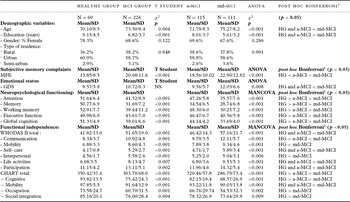
Based on the results of neuropsychological assessment, the sample was divided in six groups: healthy subjects (control group); subjects with single domain amnestic MCI (a-MCI); subjects with multi-domain amnestic MCI (md-MCI); single non-amnestic MCI; multi-domain non-amnestic MCI; and subjects with a dementia-like cognitive functioning. Sample sociodemographic and clinical characteristics of the groups are described in Table 2. The classification of subjects to different groups was made according to performance in four cognitive domains (attention, memory, working memory, and executive function):
• Healthy Group (HG; n = 69): subjects who, after neuropsychological assessment, presented a performance within normal limits. A cutoff point of T-score equal or higher than 40 (z ≥ −1 SD) in all the four cognitive domains was established. All healthy subjects were recruited at the above cited community services for healthy elderly.
• Subjects with MCI (n = 238): subjects with at least one cognitive domain summary T-score equal to or below 35 (z ≤ −1.5), but whose possible diagnosis of dementia was not supported. This group was subdivided into four subgroups, according to the four types of MCI proposed by Petersen et al. (Reference Petersen2004):
- Amnestic Single-domain MCI Group (a-MCI; n = 115): subjects whose T-score in the cognitive domain of memory or in the long-term free-recall of the Hopkins Verbal Learning Test (HVLT) was equal to or below 35 (z ≤ −1.5), and the other cognitive domain summary T-scores above 35 (z > −1.5).
- Amnestic multi-domain MCI Group (md-MCI; n = 111): subjects with T-scores below 35 (z ≤ −1.5) in at least two cognitive domain summary T-scores, including memory summary score or long-term free recall of HVLT.
- Non-amnestic single-domain MCI Group (n = 10): subjects with a unique cognitive domain other than memory or HVLT long-term free-recall T-score equal to or below 35 (z ≤ −1.5), and the other cognitive domain summary T-scores above 35 (z > −1.5).
- Non-amnestic multi-domain MCI Group (n = 2): subjects with T-scores below 35 (z ≤ −1.5) in at least two cognitive domains, other than memory summary score or long-term free recall of HVLT.
Given the low sample size of the non-amnestic single-domain (n = 10) and the non-amnestic multi-domain (n = 2) MCI groups, these two groups were not included in further analyses.
• Subjects with a dementia-like cognitive functioning (n = 21): subjects with at least two cognitive domain summary T-score below 25 (z < −2.5). The diagnosis of dementia was not proven due to the lack of neuroimaging data, but this conservative strategy was adopted in order to minimize a potential magnifying effect of differences on functional ability between groups exclusively due to outliers.
Comparison of demographic, emotional, neuropsychological, and functional variables between healthy and MCI subjects

Note: GDS = Geriatric Depression Scale; MFE = memory failures everyday; WHODAS II = the World Health Organization Disability Assessment Schedule; SD = Standard Deviation; CHART = Craig Handicap Assessment and Reporting Technique. HG = Healthy Group; a-MCI = amnestic-MCI group; md-MCI = multidomain-MCI group.
1Bonferroni post hoc test for the ANOVA. 2Bonferroni post hoc test for the MANCOVA after controlling for age, years of education, and GDS score.
Functional independence assessment
Assessment of functional independence was performed by means of two different scales, one based on subjective judgment of disability – the World Health Organization Disability Assessment Schedule, second version (WHO-DAS-II) – and another based on frequency rates of time spent performing ADL – Craig Handicap Assessment and Reporting Technique (CHART). The WHO-DAS II (Federici et al., Reference Federici, Meloni and Lo Presti2009) is a 36-item inventory based on the World Health Organization's (WHO) International Classification of Functioning, Disability and Health Framework (WHO-ICF) that evaluates six domains of functioning in daily life: understanding and communicating, mobility, self-care, interpersonal, life activities, and participation in society. The participants interviewed were asked to rate the experienced level of “difficulty” (none, mild, moderate, severe, extreme), by taking into account the way in which they normally perform a given activity, and including the use of whatever support or/and help by a person (aids). For every item receiving a positive answer, the subsequent question asks the number of days (“in the last 30 days”) in which the interviewee has found such a difficulty; however, for the present study only the 36-item answers were analyzed. A total WHODAS-II functional impairment score was obtained by adding up all functional areas subscores, except for the work subscale (possible total score range: 36–180), higher values indicating higher degree of functional impairment. The WHO-DAS II is available at http://www.who.int/icidh/whodas/whodasversions/36sa.pdf, and examples of items are “How much difficulty did you have in: Moving around inside your home? Washing your whole body? Getting along with people who are close to you? Taking care of your household responsibilities?”
The CHART was designed to provide a simple, objective measure of the degree of impairments and disabilities (Whiteneck et al., Reference Whiteneck, Charlifue, Gerhart, Overholser and Richardson1992). Each CHART dimension is characterized by directly observable qualities which lend themselves to easy quantification. CHART consists of six subscales assessing (1) physical independence; (2) cognitive independence; (3) mobility/accessibility to activities and resources inside and outside the home; (4) occupation, including household activities, caring activities, volunteer activities, entertainment, etc.; (5) quantity and quality of interpersonal relationships, and (6) financial independence (this subscale was not administered). For each subscale a percentage score (0%–100%) is calculated by raw scores conversions and mathematical operations, indicating a 100% absolute independence for that domain. A total CHART functional impairment score was obtained by adding up all functional areas subscores, except for the Physical Independence and Economic Self-Sufficiency subscales (possible total score range: 0–400), with higher values indicating lower degree of functional impairment. The Physical Independence subscore was not included in the summary score because presence of a moderate or higher degree of disability due to other conditions than MCI was an exclusion criterion, so all subjects scored 100. The economic self-sufficiency was excluded in order to preserve participants' privacy given the fact that the fulfillment of this subscale requires providing data of annual income. The CHART is available at http://www.craighospital.org/Research/CHART%20Manual.pdf, and examples of items are “How many hours per week do you spend in active homemaking including parenting, housekeeping, and food preparation?”; “In a typical week, how many days do you get out of your house and go somewhere?”; How many friends. . . do you visit, phone, or write to at least once a month?”
Both functional independence scales, which gather information about both basic and instrumental ADL, were administered by trained neuropsychologists within the context of an interview in such a way that information was provided by subjects and a caregiver together when available, or by subjects alone (some of these subjects lived alone, or with a spouse with higher degrees of disability).
Depression symptoms
The presence and severity of depression symptoms were assessed by the Geriatric Depression Scale (GDS) (Yesavage, Reference Yesavage1988). The GDS is a screening depression scale for geriatric populations comprising 30 items to which the subject provides a “yes” or “no” answer. Each item is scored as 1 (presence of a depression/anxiety symptom) or 0 (absence of a depression/anxiety symptom). According to previous reports, scores ranging 0–10 indicate the absence of depression; 11–20 would suggest a mild possible depression; and scores higher than 20 would be suggestive of severe depression. The GDS was administered by the neuropsychologists who performed the cognitive and functional evaluation.
Statistical analyses
Mean and standard deviation are provided for continuous variables. Discrete variables are expressed as frequencies and/or percentages. Distribution of variables was tested by the Kolmogorov–Smirnov test, and when dividing the sample into the three subgroups (Healthy, a-MCI, md-MCI), only the variables of years of education and WHO-DAS II total score showed non-normal distributions. On the basis of the normal distribution of most variables, the sample sizes, the robustness of the parametric tests based on analysis of variance, and the need to use post hoc analyses, these techniques were chosen for statistical analyses. For sociodemographic data Student's t-test for two independent samples (Healthy Group versus MCI subjects) were used to compare means for continuous variables. When more than two groups were considered (for sociodemographic and clinical data), one-way analysis of variance (ANOVA) was conducted. When significant differences were detected the Bonferroni post hoc test was used to identify which specific groups differed. The χ2 statistics were used for comparison of categorical measures. As healthy control and MCI groups differed for age and years of education, the healthy group differed from the md-MCI group in age and years of education, analyses on neuropsychological and functioning variables were performed controlling for these variables when required. For doing so, full factorial multivariate analysis of covariance (MANCOVA) models were performed, using group (MCI/control and a-MCI/md-MCI/controls) as fixed factors, cognitive summary T-scores and WHO-DAS-II and CHART scores as dependent variables, and age, years of education, and GDS as covariates. Again, the Bonferroni post hoc test was performed to identify specific differences/effects when the MANCOVA was significant.
Receiver operating characteristic (ROC) curve analyses were conducted to test diagnostic accuracy (Healthy vs. all MCI as a group) of the WHO-DAS II, CHART, MFE, and total cognition variables. The estimate of the area under the ROC curve was computed using a binegative exponential model.
Multiple regression analyses were performed separately with the total sample together and for the three groups independently (a-MCI, md-MCI, and controls), with total WHO-DAS II and CHART scores as dependent variables, and age, years of education, GDS, and cognitive domain summary scores as independent variables or predictors. The step-wise method was used in the regression analyses due to the fact that it provides a final model including only significant predictors, which are presented decreasingly according to the amount of variance explained.
All statistical analyses were performed with SPSS v13 (Statistical Package for the Social Sciences, Chicago, Illinois), and a 2-tailed p-value lower than 0.05 was considered statistically significant.
Results
Differences in demographic and clinical variables
Comparisons of demographic, cognitive, depression symptoms, and functional independence variables are presented in Table 2. Both healthy subjects and a-MCI were younger and had more years of education and less depression symptoms than md-MCI, with no differences in these variables between the two former groups. Similarly, the md-MCI group showed higher rates of depression symptoms than both healthy and a-MCI subjects. Given these results, for the following comparisons between the three groups (MANCOVA) age, years of education, and GDS were introduced as covariates. MANCOVA analyses results suggested that healthy subjects had a better cognitive functioning than both a-MCI and md-MCI in all cognitive areas assessed, and that cognitive performance of the a-MCI group was superior to that of md-MCI in all cognitive areas. Frequency and severity of subjective memory complaints followed an identical pattern: more prominent in md-MCI than in a-MCI, and in both MCI groups than in healthy subjects.
As to functional independence, the healthy group showed better functional adjustment than the MCI group according to both WHO-DAS II and CHART total scores. When the MCI group was subdivided into a-MCI and md-MCI, CHART total score and cognitive domain, and WHO-DAS II communication subscale showed a similar profile as cognitive functioning, after controlling for age, education, and GDS score: md-MCI was more functionally impaired than a-MCI, showing that both MCI groups had higher rates of disability than the healthy group. The md-MCI group showed a higher degree of functional impairment than both healthy subjects and a-MCI in the WHO-DAS II total score and self-care, life activities, and participation subscales, and in the CHART mobility domain. The remaining functional ability measures showed a consistent functional impairment only of md-MCI in comparison with healthy subjects (for more details, see Table 2).
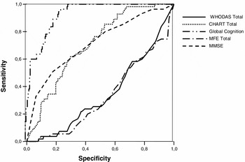
In comparing the ROC curves, the CHART appears with a larger area under the curve (AUC = 0.707) than the WHO-DAS II (AUC = 0.347) and the MFE (AUC = 0.331), but less than the global cognition score (AUC = 0.923), and very close to MMSE (AUC = 0.714) (Figure 1).
ROC curve to distinguish normal from the MCI using total CHART, total WHO-DAS II, total MFE, and global cognitive score.

Predictors of functional status (regression analyses)
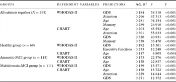
The results of the regression analyses are shown in Table 3. When the whole sample was analyzed together, attention and memory summary scores explained a total variance of 8.2% and 0.8% of the WHO-DAS II, respectively, and 9.8% and 1.8% of the CHART, respectively. When depression and age were entered in both former models, the WHO-DAS II score was predicted by depression symptoms as measured by the GDS (19.2%) and by executive functions (8.1%) only in the healthy group. By contrast, CHART total score was only predicted by age among healthy subjects. For both MCI groups, WHO-DAS II was predicted by depression symptoms only, with a total variance explained of 17.3% for a-MCI and 13.9% for md-MCI. CHART score was predicted only by age on a-MCI subjects, and by age, attention, and GDS score on md-MCI subjects. Attention explained 9.1% variance of CHART total score.
Regression analyses with total WHO-DAS II and CHART as dependent variables

Method: Step-wise. Independent variables: age, years of education, GDS, attention, memory, working memory and executive functions.
GDS = Geriatric Depression Scale; WHODAS-II = the World Health Organization Disability Assessment Schedule, second edition; CHART = Craig Handicap Assessment and Reporting Technique; Adj. R2 = adjusted R2.
Discussion
Our results show that functional impairment is a defining feature of MCI, and that the impairment showed by MCI subjects is partially dependent on the degree of their cognitive impairment. Furthermore, measures of functional ability show adequate psychometric properties (i.e. discriminant power) to contribute to the MCI diagnosis. Functional measures based on quantitative rates of number and quality of ADL performed seem to be more sensitive to identifying functional impairment in MCI than those based on a subjective judgment of disability.
The idea of a functional compromise associated with MCI is not new, and previous studies have reported a higher degree of functional impairment in MCI subjects when compared with matched healthy subjects (Tam et al., Reference Tam, Lam, Chiu and Lui2007; Pereira et al., Reference Pereira, Yassuda, Oliveira and Forlenza2008; Ahn et al., Reference Ahn2009; Burton et al., Reference Burton, Strauss, Bunce, Hunter and Hultsch2009; Schmitter-Edgecombe et al., Reference Schmitter-Edgecombe, Woo and Greeley2009; Aretouli and Brandt, Reference Aretouli and Brandt2010; Bangen et al., Reference Bangen2010; Reppermund et al., Reference Reppermund2010; Teng et al., Reference Teng, Becker, Woo, Cummings and Lu2010a; Reference Teng, Becker, Woo, Knopman, Cummings and Lu2010b). Independent of the instrument used to evaluate functional ability, most of these studies agree that higher order ADL are more likely to be impaired than those ADL with a lower degree of cognitive involvement/demand (Burton et al., Reference Burton, Strauss, Bunce, Hunter and Hultsch2009; Aretouli and Brandt, Reference Aretouli and Brandt2010; Reppermund et al., Reference Reppermund2010). In our results, the highest effect sizes between healthy controls and MCI subjects were functioning areas related to cognitive difficulties (CHART cognitive subscale; WHO-DAS II communication subscale), and participation in productive and social activities (CHART occupation and mobility subscales; WHO-DAS II life activities and participation subscales). All these data support the a priori intuitive notion that highly cognitive-dependent living skills are more likely to be affected as a consequence of cognitive impairment, and that MCI subjects show significant impairment in these functional domains. On the other hand, it is noteworthy that differences between healthy and MCI subjects were not restricted to higher order ADL, but also to functional areas such as moving around and self-care, usually referred to as basic ADL.
The early descriptions of MCI excluded the presence of disability or functional impairment as a characteristic, perhaps to differentiate from dementia. However, as a consequence of the enrichment and improvement of the conceptual definitions of functional independence and participation, and the increasing importance of these concepts in the sphere of health and well-being (see WHO International Classification of Functioning, Disability and Health: WHO-ICF), instruments to assess independent living abilities have improved their ability to identify impairments included in WHO-ICF activity and participation levels. In WHO-ICF, the concept of disability is no longer restricted to the notion of “inability to perform basic ADL,” but includes quantitative (inability, reduction in the number of times the ADL is performed) or qualitative (increased time or difficulty in performing ADL) difficulties with both basic and instrumental ADL (see Wade, Reference Wade, Halligan and Wade2005 for an interesting approach to the application of the WHO-ICF model to patients with cognitive deficits). As a result of this paradigm shift, and in the light of previous and the present results, it would be very helpful for clinicians, caregivers, and health-system managers if MCI definitions included the presence of impairment of functional abilities as a clinical feature inherent to MCI.
Moreover, given the moderately good psychometric properties demonstrated in our study of the CHART in discriminating healthy from MCI subjects, assessing functional ability would improve the identification of MCI subjects, and the use of qualitative and/or quantitative impairment of functional abilities as an MCI diagnostic criterion should be further explored. Goldberg et al. (Reference Goldberg2010) found that a sensitive performance-based measure they developed (the University of California, San Diego Performance-Based Skills Assessment; UPSA) had a remarkably good discriminant power to distinguish healthy from MCI (area under the curve 0.84), and to distinguish MCI subjects from Alzheimer's disease patients (area under the curve 0.88). Hence, the inclusion of functional competence measures seems convenient for the screening and early identification of neurodegenerative processes characterized by cognitive impairment.
As to the type of functional assessment instrument, our results suggest that performance-rate measures of functioning are superior to those based on a subjective judgment of disability. The latter was consistently more biased by depressive symptoms, and the CHART showed better discriminant power between healthy and MCI groups than the WHO-DAS II, suggesting that the more objective the approach to measuring independent functioning, the more sensitive it was to to functioning loss secondary to cognitive impairment. In this regard, the study by Pereira et al. (Reference Pereira, Yassuda, Oliveira and Forlenza2008) also reported high effect sizes between healthy subjects, MCI subjects, and Alzheimer's disease patients with an objective measure of functioning; the Direct Assessment of Functional Status Scale (DAFS-R) (Loewenstein et al., Reference Loewenstein1989), which evaluates functional competence by simulating ADL (time orientation, communication skills, ability to deal with finances, shopping, grooming, eating, and transportation). Additionally, the associations found between the DAFS-R and cognitive measures of executive functions were quite robust (r = −0.872, p < 0.001). The previously noted study of Goldberg et al. (Reference Goldberg2010) showed that their performance-based measure of functional ability was more discriminant of healthy-MCI- Alzheimer's disease groups than a questionnaire based on a caregiver response. Hence, independent functioning assessment instruments should be designed in a way that places emphasis on the subject's ability to perform complex ADL and the frequency with which such activities are performed, rather than on subjective judgments of difficulties.
Our results also noted that subjective judgments of disability (i.e. WHO-DAS II) are more influenced by depressive symptoms, suggesting depression may play a confounding role in this measure. That the most robust predictor of the WHO-DAS II score was the severity of depressive symptoms, and that the amount of variance explained was close to 20% support this hypothesis. By contrast, the more objective measure of functional ability was less influenced by depression, although it is noteworthy that in md-MCI subjects, depression symptoms influenced functional impairment as measured by both WHO-DAS II and CHART, and that this group showed higher rates of depressive symptoms than the other two (healthy and a-MCI groups). Hence, it may be the case that when depressive symptoms reach syndromic levels, they play a more important role in independent functioning. The alternative hypothesis would be that depressive symptoms are partially secondary to functional impairment. The relationship between depression and functional impairment in MCI subjects requires further examination, because our study design does not allow us to disentangle the relationship between depression and functional impairment. On the other hand, the influence of depression on functioning measures may have been partially increased in those cases in which these scales were administered to subjects without an independent informant to contrast information.
We found several results supporting the association of the degrees of functional impairment with cognitive impairment. The evidence supporting this association is the finding that the higher the number of impaired cognitive domains the higher the degree of functional impairment, so that healthy controls showed more functional independence than a-MCI, and these more than md-MCI. A similar gradual profile of global cognitive functioning can be found between the three groups. The ability of cognitive functioning to predict performance rates of functional activities (CHART) was restricted to MCI subjects also supports this notion, suggesting that the impact of cognition on functional ability gets higher as cognitive impairment increases. In other words, variability on cognitive functioning is irrelevant when cognitive functioning is within normal limits, but when cognitive resources are impaired they show a higher weight on functional independence. The higher contribution of cognitive measures on the regression model of all subjects together (around 11.6%) is putatively due to the heterogeneity of the entire sample, whereas no cognitive measure seems to predict functional ability in a-MCI subjects because these subjects have a very similar cognitive profile (i.e. all subjects present memory impairment alone). In other words, the stratification based on their degree of cognitive impairment is likely to be the best predictor of functional impairment.
Limitations of the study include the potential measure error derived from the fact that in many cases the MCI subjects were the informants of their own functional independence levels. Although functional assessment was conducted in an interview context with the collaboration of a caregiver when available, for approximately half the sample the subject was the key informant and the accuracy of the subjects' reports might have been mediated by their cognitive impairment and/or depression symptoms. However, given that recruitment was mostly conducted in community locations, a close informant was not always available with enough knowledge of the subject's daily routine. Unfortunately, the exact number of measures gathered from subjects only versus those gathered from subjects and caregivers together was not recorded. A second methodological issue is that the sample recruitment may have been biased as invitation to participate in the study was addressed to people willing to participate in a memory training program, and hence it could be argued that people with subjective memory complaints were more likely to enroll in the study. As a consequence, our healthy subjects might have presented more subjective memory complaints than the general population. However, given the large sample size, the effect size differences between healthy and MCI groups on the subjective memory complaints scale (MFE), and that most participants (including all healthy subjects) were recruited in community settings, we consider the sample to be representative of the elderly living in the community. Finally, cognitive and functional assessments were in most cases performed by the same person, and although group assignment (healthy vs. a-MCI vs. md-MCI) was not undertaken until the conclusion of all data gathering, this may have led to possible bias in functional ability assessment.
In summary, these and previous results emphasize the presence of qualitative and quantitative functional impairments of both basic and instrumental ADL in MCI as a logical consequence of cognitive impairment. Although dementia is characterized by a more severe degree of disability than MCI, the WHO-ICF conceptualization of disability would include MCI as a disabling condition, although to a lower degree. The need for a better definition of disability as a diagnostic criterion (putatively, by shifting from a categorical notion of able/disable to a more spectrum/gradual approach) to discriminate MCI from dementia must not conceal the fact that MCI subjects have their own health/functional assistance needs. Researchers and clinicians have the responsibility and opportunity of designing, testing, and implementing effective therapeutic strategies targeted to improve, or at least preserve, both functional and cognitive functioning in MCI.
Conflict of interest
None.
Description of authors' roles
I. Bombin designed the study, supervised the data collection, and wrote the paper. S. Santiago-Ramajo collected the data and assisted with writing the paper. M. Garolera, E. M. Vega-González, N. Cerulla, A. Caracuel, A. Cifuentes, and M. T. Bascarán collected the data and assisted with preparation of data for analyses. J. Bobes assisted with design of the study. All authors reviewed the paper and contributed to its final version.
Acknowledgments
This study was financially supported by the Spanish Ministry of Science and Innovation, Instituto de Salud Carlos III, and European Regional Development Fund (ERDF).