Implications
Physiological function of the muscle, postmortem muscle to meat conversion and meat quality are influenced by myofiber type composition. Myosin heavy chain (MyHC) is a polymorphic key motor protein of the muscle showing different expression and transition between wild or domesticated (white) pigs from the early postnatal life up to adulthood. The differences in the pattern of MyHC expression and transition in porcine skeletal muscle elucidates the effects of genetic selection pressure in terms of growth and lean meat content and could help designing breeding programmes targeting improved skeletal muscle characteristics.
Introduction
Skeletal muscles are composed of functionally specialized myofibers, and their diversity primarily depends on the composition of their MyHCs. The locomotor skeletal muscles of adult mammals express one slow (MyHC-I) and three fast MyHC isoforms (MyHC-IIa, -IIx and -IIb). The MyHCs are characterized by different shortening velocities that increase in the following order: I<IIa<IIx<IIb (Bottinelli et al., Reference Bottinelli, Betto, Schiaffino and Reggiani1994). Transitions between MyHC expressions follow a reversible obligatory pathway from MyHC-I to IIa, IIa to IIx and IIx to IIb. The metabolic activity of myofibers generally matches the energetic demands of each MyHC. Greater oxidative capacity is characteristic for type I myofibers, which have to support sustained contraction and contain lipids as the main energy source. The IIb myofibers are predominantly glycolytic and use glycogen for quick, transitory contractions, whereas IIa and IIx myofibers represent the metabolically intermediate type between I and IIb myofibers (Pette and Staron, Reference Pette and Staron2000).
The expression of fast MyHC isoforms differs between species and muscles. In the skeletal muscles of rodents and lagomorphs, all three fast MyHC isoforms, namely IIa, IIb and IIx, are expressed (Schiaffino et al., Reference Schiaffino, Gorza, Sartore, Saggin, Ausoni, Vianello, Gundersen and Lomo1989), whereas only two fast isoforms (IIa and IIx) were demonstrated in the locomotor skeletal muscles of large mammals including humans (Smerdu et al., Reference Smerdu, Karsch-Mizrachi, Campione, Leinwand and Schiaffino1994) and some large animals, such as cats (Talmadge et al., Reference Talmadge, Grossman and Roy1996), dogs (Strbenc et al., Reference Strbenc, Smerdu, Zupanc, Tozon and Fazarinc2004), cattle (Tanabe et al., Reference Tanabe, Muroya and Chikuni1998), horses (Eizema et al., Reference Eizema, van den Burg, Kiri, Dingboom, van Oudheusden, Goldspink and Weijs2003) and bears (Smerdu et al., Reference Smerdu, Cehovin, Strbenc and Fazarinc2009). As an exception to this rule, all three fast MyHC isoforms (IIa, IIx and IIb) were identified in pigs (Lefaucheur et al., Reference Lefaucheur, Hoffman, Gerrard, Okamura, Rubinstein and Kelly1998) and llamas (Graziotti et al., Reference Graziotti, Menendez, Rios, Cossu, Bosco, Affricano, Ceschel, Moisa and Basso2001). In the pig, spatial distribution of myofibers resembles rosette clusters, each consisting in a central islet of type I myofibers surrounded by three concentric rings of IIa, IIx and IIb myofibers from the centre to the periphery of each rosette (Lefaucheur et al., Reference Lefaucheur, Hoffman, Gerrard, Okamura, Rubinstein and Kelly1998). This spatial distribution typically uncounted in pig skeletal muscle is consistent with the MyHC transition rule I↔IIa↔IIx↔IIb and proceeds from the centre to the periphery of each rosette (Lefaucheur et al., Reference Lefaucheur, Ecolan, Lossec, Gabillard, Butler-Browne and Herpin1995; Herpin et al., Reference Herpin, Lossec, Schmidt, Cohen-Adad, Duchamp, Lefaucheur, Goglia and Lanni2002). The MyHC-IIb-positive myofibers were especially numerous in the so-called white muscles of the domestic pig (DP), in which they were located in the peripheral region of the rosettes (Lefaucheur et al., Reference Lefaucheur, Hoffman, Gerrard, Okamura, Rubinstein and Kelly1998). Initially, it was postulated that the MyHC-IIb isoform expression in the DP was related to genetic improvement and breeding conditions; however, recent studies dismissed this assumption as the MyHC-IIb isoform expression was also demonstrated in the muscles of wild pigs (WPs), although at lower levels than in domestic ones (Fazarinc et al., Reference Fazarinc, Ursic, Kantura, Vukicevic, Skrlep and Candek-Potokar2013).
In pigs, myogenesis is described as a biphasic phenomenon and comprises the formation of primary and secondary myofibers. The primary generation of myofibers develops between 35 and 55 days of gestation, followed up to 90 days of gestation by the development of secondary myofibers that are positioned around the primary myofibers, thus using them as a scaffold (Lefaucheur et al., Reference Lefaucheur, Ecolan, Lossec, Gabillard, Butler-Browne and Herpin1995). Primary myofibers express only slow MyHC, whereas developmental MyHCs (embryonic/foetal) are present in the secondary myofibers. Expression of developmental isoforms of MyHCs decreases dramatically by the end of gestation and then disappears during the first postnatal weeks. In the early postnatal period, de novo expression of slow MyHC was detected in a subpopulation of secondary myofibers that were in contact with the primary myofibers, whereas the remaining secondary myofibers matured into IIa, IIx and IIb myofiber types (Chang and Fernandes, Reference Chang and Fernandes1997). Furthermore, evidence was also provided for the third generation of myofibers that contribute to the ultimate total myofiber number in different species (Mascarello et al., Reference Mascarello, Stecchini, Rowlerson and Ballocchi1992; Wilson et al., Reference Wilson, McEwan, Sheard and Harris1992; Lefaucheur et al., Reference Lefaucheur, Ecolan, Lossec, Gabillard, Butler-Browne and Herpin1995; Gagniere et al., Reference Gagniere, Picard and Geay1999). The third-generation myofibers observed in the DP after birth had a very small diameter, were randomly scattered among the normal-sized myofibers and displayed a positive immunohistochemical reaction with antibodies raised against foetal and embryonic MyHC. These data suggested that the total myofiber number in pig is determined after birth, mainly between birth and 3 weeks of age (Berard et al., Reference Berard, Kalbe, Losel, Tuchscherer and Rehfeldt2011).
Genetic selection of modern pig breeds resulted in high growth rates and muscle tissue deposition. Morphological and physiological properties of myofibers, a major constituent of skeletal muscle, may underlay the antagonism between growth performance and meat quality traits (Rehfeldt et al., Reference Rehfeldt, Henning and Fiedler2008). Therefore, the aim of this study was to further elucidate the influence of domestication on the differentiation of myofiber types. Particular emphasis was placed on the MyHC expression patterns during postnatal growth in the DP breed raised in conventional conditions and its comparison with the WP. We selected the longissimus dorsi muscle, which is most frequently used as an indicator of muscularity in pigs. Comparative knowledge of the MyHC temporal expression/transition pattern in porcine skeletal muscle could help design a breeding programme that would allow a balance between growth performance, muscularity and meat quality.
Material and methods
Animals and muscle samples
Muscle samples were collected from DPs (Large White, n=20) and European WPs (Sus scrofa scrofa, n=20). Muscle samples were taken from pigs at different stages of life, namely 24 to 48 h postpartum (1-day piglets, five DP with body mass from 1.12 to 1.51 kg and five WP with body mass from 0.96 to 1.28 kg), at 21 to 23 days of age (3-week pigs, five DP with body mass from 3.72 to 4.11 and five WP with body mass from 1.92 to 2.43 kg), at ~7 months of age (five DP with body mass 85 to 112 kg and five WP with body mass between 19 and 24 kg) and at ~2 years of age (five DP with body mass between 185 and 200 kg and five WP with body mass between 46 and 65 kg). Only female pigs were included in the study. The DP were raised and slaughtered under approved standards, and the WP were reared in a large hunting enclosure (>1000 ha) that simulates their natural habitat. The confined WP population varied in number between 100 and 140, that is 10 boars, 20 to 25 sows, around 35 yearlings and around 70 piglets. WP were seeking for their food but were additionally fed on ground daily with corn. WP piglets were collected during the spring over a period of 2 years. The professional hunter monitored pregnant sows and status of the farrowing nest on a daily basis. WP piglets used in the study were found dead (drained nest), injured due to overlaying or were abandoned by the sow, which is frequently observed in young wild sows. Out of 20 collected piglets five 1-day and five 3-week-old piglets were selected for the analysis (based on age, body mass and gender). The WP aged ~7 months and 2 years were shot on the basis of the regular annual bag during the hunting season. The professional hunter estimated their age.
DPs were obtained from the pig farrowing farm. They were overlaid and were selected according to their age, gender and body mass and did not show any clinical locomotor dysfunction according to the animal keeper, neither were found any pathological changes during autopsy, which could affect locomotion ability of the animals. All animal procedures were in accordance with the guidelines set by the Slovenian Law of Animal protection. The study was not subject to ethical protocols according to Directive 2010/63/EU (2010).
Approximately 1 cc muscle sample was taken from the central part of the longissimus dorsi muscle, at the level of the last rib within 24 h postmortem. The samples were frozen in liquid nitrogen and stored at –80°C. Transverse serial cryosections (10 μm) of muscle tissue were cut with a cryostat Leica CM 1800 (Leica Microsystems, Nussloch, Germany) at –17°C, mounted on APES (3-aminopropyl-triethoxysilane)-coated slides and air-dried.
Immuno/enzyme histochemistry
Three different monoclonal antibodies (MAb) specific for adult MyHC isoforms were used: NCL-MHCs, specific for slow-twitch MyHC-I; A4.74, specific for rat MyHC-IIa; and BF-F3, specific for MyHC-IIb. The suitability of the listed MAb for pig myofiber classification was confirmed previously (Lefaucheur et al., Reference Lefaucheur, Ecolan, Plantard and Gueguen2002; Graziotti et al., Reference Graziotti, Rios and Rivero2011). In addition, MAb F158.4C10, originally raised against foetal bovine myosin, was used. Mab BF-F3 originated from the Developmental Studies Hybridoma Bank, University of Iowa. The MAb NCL-MHCs were purchased from Novocastra Laboratories (Newcastle upon Tyne, UK) and the F158.4C10 and A4.74 from Alexis Corporation (Lausen, Switzerland). Serial cryosections were incubated with the primary antibody diluted according to the manufacturer’s recommendations in a humidified chamber at 4°C overnight. The immunohistochemical reaction was accomplished with the peroxidase-conjugated secondary antibody kit using the Dako REALTMDAB chromogen (Dako, Copenhagen, Denmark). The metabolic profile of the myofibers was estimated on the basis of the activity of mitochondrial succinate dehydrogenase (SDH), which was histochemically demonstrated according to Nachlas et al. (Reference Nachlas, Tsou, de Souza, Cheng and Seligman1957). The sections were counterstained with haematoxylin, dehydrated and mounted with Shandon Synthetic Mountant (Thermo Fischer Scientific Inc., MA, USA).
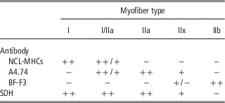
On the basis of antibody reactivity and in combination with SDH activity, the presence of four adult MyHC isoforms was demonstrated in the pig longissimus dorsi muscle, and the myofibers were subsequently classified into I, I/IIa, IIa, IIx and IIb (Table 1). Slow myofibers (type I) displayed a positive reaction only with the MAb NCL-MHCs. The NCL-MHC-negative myofibers were fast type II myofibers, which were further classified into subtypes according to the other antibodies and SDH-staining reactivities. The MAb A4.74 antibody recognized both the MyHC-IIa and MyHC-IIx in the pig; however, its affinity for the MyHC-IIa isoform was higher (Graziotti et al., Reference Graziotti, Rios and Rivero2011). Consequently, myofibers that reacted strongly with the A4.74 were classified as type IIa, and those that reacted moderately were classified as IIx. Myofibers that reacted variably with both MAb, that is, NCL-MHCs and A4.74 were designated as hybrid I/IIa. BF-F3-positive myofibers were divided into two subgroups: pure IIb myofibers that reacted strongly with the BF-F3 and were SDH negative and myofibers that reacted moderately with both BF-F3 and A4.74 and displayed weak or moderate SDH activity. The latter were designated as IIx myofibers, although they probably co-express MyHC-IIx and IIb. The SDH activity was very high in type I and IIa myofibers and moderate in IIx, whereas IIb myofibers remained unstained (see Table 1).
Characterization of myofiber types in pig longissimus dorsi muscle according to the specificity of the antibody set used and the succinate dehydrogenase (SDH) activity

+and ++ Denote a moderate and strong positive reactions, respectively. −Denotes a negative reaction.
Analysis of serial cryosections
Approximately 400 to 500 myofibers in randomly selected complete muscle fascicles were analysed per muscle to determine the pattern of MyHC expression. The selected fascicles were the same for all antibodies on serial sections. The average proportion of myofiber types was determined according to the MyHC isoform expression, and the cross-sectional area (CSA) of myofiber types was measured. The stereological analysis of the serial sections was carried out on a Nikon Microphot FXA microscope equipped with a DS-Fi1 camera and the Imaging Software NIS Elements D.32 (Nikon Instruments Europe B.V., Badhoevedorp, The Netherlands). Data were subjected to two-way ANOVA performed by SigmaPlot 11.0 statistical software (Systat Software Inc., San Jose, CA, USA). The model included the fixed effects of age (1 day, 3 weeks, 7 months, 2 years), breed (WP v. DP) and their interaction. Data shown in tables are least squares means. Individual differences between groups were tested by Student’s t-test. A P-value of <0.05 was considered statistically significant.
Results
Classification of myofiber types by immuno/enzyme histochemistry
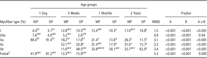
The results of immuno/enzyme histochemistry of the longissimus muscle in the WP and the DP at different ages are shown in Figures 1 to 4, and the morphometric data are summarized in Tables 2 and 3. In 1-day-old wild and domestic piglets, all type II myofibers reacted positively with the A4.74 antibody and were classified as IIa myofibers (Figure 1, WP b and DP b). In the WP, individual smaller, less intensely stained myofibers were observed in the periphery of the rosettes (Figure 1, WP b), which could indicate co-expression with the MyHC-IIx. However, due to a small difference in the staining intensity, they were not classified as an additional myofiber subtype. MyHC-IIb isoform could not be detected at this age because all myofibers reacted negatively with the BF-F3 antibody (data not shown). MyHC-I was expressed in myofibers that originated from the primary myofibers located in the centre of the muscle fasciculus and in the subpopulation of secondary myofibers in the direct vicinity of the primary myofibers (I/IIa myofibers). Some type I myofibers of domestic piglets still showed small hollow centres that probably indicate the end of the myotube phase in myofiber formation (Figure 1, DP a). All myofibers showed comparable SDH activity (not shown). Morphometrical analysis showed that 1-day-old WP piglets had a significantly higher proportion of intermediate I/IIa and a substantially lower proportion of foetal myofibers than the DP, whereas the proportion of type I myofibers was not statistically significant between breeds.
Serial sections of the longissimus dorsi muscle of a 1-day-old wild pig (WP) and a domestic pig (DP) stained for monoclonal antibodies NCL-MHCs (WP a, DP a) and A4.74 (WP b, DP b). 1, 2 and 3 correspond to myofiber types I, I/IIa and IIa, respectively. The asterisk marks the myofibers with less intense A4.74 reactivity in the periphery of muscle bundles. The arrow indicates the hollow centre in type I myofibers. Scale bar=100 µm.

Serial sections of the longissimus dorsi muscle of a 3-week-old wild pig (WP) and a domestic pig (DP) stained for monoclonal antibodies NCL-MHCs (WP a, DP a), A4.74 (WP b, DP b), BF-F3 (WP c, DP c) and succinate dehydrogenase (WP d, DP d). 1, 2, 3, 4 and 5 correspond to myofiber types I, I/IIa, IIa, IIx and IIb, respectively. Scale bar=100 µm.

Serial sections of the longissimus dorsi muscle of a 7-month-old wild pig (WP) and a domestic pig (DP) stained for monoclonal antibodies NCL-MHCs (WP a, DP a), A4.74 (WP b, DP b), BF-F3 (WP c, DP c) and succinate dehydrogenase (WP d, DP d). 1, 3, 4 and 5 correspond to myofiber type I, IIa, IIx and IIb, respectively. Scale bar=200 µm.

Sections of the longissimus dorsi muscle of a 1-day (a), 3-week (b) and 7-month-old (c) wild pig (WP) and domestic pig (DP) stained with monoclonal antibodies F158.4C10 against the foetal myosine heavy chain (MyHC). The number of foetal MyHC-positive myofibers declined with age, and the decline was faster in the WP than in the DP. Scale bar=100 µm (WP a, DP a, WP b, DP b); scale bar=200 µm (DP c).

The proportion (%) of myofiber types in the longissimus muscle of the wild pig (WP) and the domestic pig (DP) in different age groups

RMSE=root mean square error; A=age; B=breed; A×B=age×breed interaction.
Values are given as least squares means; n=5 per group; in total n=40.
aSignificantly different between breeds within age group.
bSignificantly different between age groups within breed: b1significantly different between 1-day and 3-week-old pigs; b2significantly different between 3-week and 7-month-old pigs; b3significantly different between 7-month and 2-year-old pigs.
1 Foetal myofibers that displayed distinctive immunohistochemical reaction were included in morphometrical analysis.
Differences between groups were considered significant at P<0.05 (Student’s t-test).
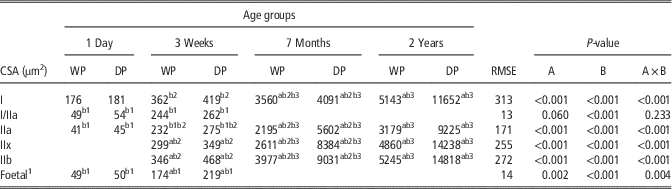
The cross-sectional area (CSA, µm2) of myofiber types in the longissimus muscle of the wild pig (WP) and the domestic pig (DP) in different age groups

RMSE=root mean square error; A=age; B=breed; A×B=age×breed interaction.
Values are given as least squares means; n=5 per group; in total n=40.
aSignificantly different between breeds within age group.
bSignificantly different between age groups within breed: b1significantly different between 1-day and 3-week-old pigs; b2significantly different between 3-week and 7-month-old pigs; b3significantly different between 7-month and 2-year-old pigs.
1 Foetal myofibers that displayed distinctive immunohistochemical reaction were included in morphometrical analysis.
Differences between groups were considered significant at P<0.05 (Student’s t-test).
In the older pig groups, all four adult MyHC isoforms could be immunohistochemically recognized, although the expression patterns were age- and breed-dependent. At 3 weeks of age, obvious differences in the muscle myofiber type composition between the WP and the DP were observed (Figure 2). In the WP, the proportion of IIb myofibers was significantly lower than in the DP, whereas the proportion of type I, I/IIa and IIx myofibers was significantly higher in the WP than in the DP (Table 2). It is evident that myofibers differentiated into subtype IIb in the periphery of the rosettes in the DP during the first 3 postnatal weeks (Figure 2, DP c), whereas in the WP, the shift mostly occurred to type IIx myofibers exhibiting a higher SDH staining than IIb myofibers. The higher proportion of type I and hybrid I/IIa myofibers in the WP suggests that the transformation from type IIa to type I myofibers was more important in the wild than in the domestic piglets during the first 3 postnatal weeks. The SDH activity was detected in myofiber types I, IIa and IIx; however, the number of positive myofibers was higher in WP than DP (Figure 2, WP d and DP d).
In 7-month and 2-year-old pigs, the coexistence of different MyHCs in the same myofiber was less obvious, which enabled clear immunohistochemical distinction of myofiber types on the basis of MAb reactivity (Figure 3). In 7-month-old pigs, the difference in the muscle myofiber type composition was even more pronounced between the breeds. The proportion of oxidative type I, IIa and IIx myofibers was significantly higher in the longissimus muscle of the WP than in the DP. On the contrary, the proportion of glycolytic IIb myofibers was almost twice higher in the DP. The percentage of hybrid I/IIa myofibers was <0.5% and was therefore excluded from the quantitative analysis in these two age groups. From 7 months to 2 years, the proportion of type I myofibers significantly increased from 13.4% to 17% and the proportion of type IIb myofibers decreased from 33.8% to 25.7% in the WP. On the contrary, there were no statistically significant differences in the proportion of myofiber types between the age groups (7 months v. 2 years) in the DP. This indicates that the MyHC transformation in DP is mostly finished by the age of 7 months (Table 2).
Myofiber cross-sectional area
The measurement of the myofiber CSA in different age groups revealed that myofiber hyperthropy was significantly faster in the DP, particularly after 3 weeks of age (Table 3). In 1-day-old piglets, the CSA of type I myofibers was substantially larger than that of type I/IIa and IIa myofibers. The CSA of all myofiber types was slightly larger in the DP, although the differences were not statistically significant. During the first 3 weeks, the CSA of all myofibers increased, with the most pronounced CSA enlargement observed in the myofibers that expressed MyHC-IIx and, especially, IIb. The CSA of type IIx and type IIb myofibers was significantly larger in the 3-week-old DP than in the WP of the same age. From 3 weeks to 7 months, intense hypertrophy of all myofiber types was observed. The CSA increased by around 10-fold in type I, IIa, IIx and IIb myofibers in the WP, while the CSA of IIx and IIb myofibers increased by up to 20-fold in the DP. From 7 months to 2 years, the hypertrophy of all myofiber types slowed in both pig breeds, with the process being most evident in myofibers that expressed MyHC-IIb. Interestingly, during this period, an ~2.5 and 1.5-fold increase in the CSA of type I myofibers was detected in the DP and WP, respectively. Finally, at the age of 2 years, the CSA of all myofiber types was 1.5- to 2.5-fold smaller in the WP than in the DP.
Expression pattern of foetal myosine heavy chain
Regarding the immunohistochemical detection of foetal MyHC using MAb F158.4C10, we identified two myofiber subtypes in 1-day-old piglets. Strongly stained myofibers were localized in the direct vicinity of type I myofibers (which were negative) and moderately stained smaller myofibers in the periphery of the rosettes. The expression of foetal MyHC in 1-day-old pigs was higher in domestic than in wild piglets (Figure 4). During the first 3 postnatal weeks, the expression of foetal MyHC declined in both the WP and the DP, although the percentage of positive myofibers was still higher in the DP than in the WP (Table 2). In addition, some smaller, irregularly scattered myofibers that strongly reacted with MAb F158.4C10 were observed in this age group and appear to be more numerous in the DP (Figure 4b). In the 7-month-old DP, only isolated, smaller myofibers reacted with anti-foetal MAb. MyHC immunoreactivity was observed in a very small percentage (<0.5%) and was not present in all specimens (Figure 4c). No foetal MyHC-positive myofibers were observed in the 7-month-old WP, 2-year-old WP and 2-year-old DP (data not shown). It should be noted that all foetal MyHC-positive myofibers also expressed type IIa isoform, but not vice versa.
Discussion
Several studies have compared the muscle characteristics of different pig breeds. In general, these studies revealed that domestication and breeding selection are associated with myofiber hypertrophy and changes in fibre type composition with a prevalent age-dependent increase in the proportion of fast glycolytic myofibers (Szentkuti and Schlegel, Reference Szentkuti and Schlegel1985; Weiler et al., Reference Weiler, Appell, Kremser, Hofacker and Claus1995; Muller et al., Reference Muller, Rutten, Moser, Reiner, Bartenschlager and Geldermann2002; Ruusunen and Puolanne, Reference Ruusunen and Puolanne2004). Most of these studies were based on the myosin ATPase reaction and metabolic enzyme histochemistry with a simplified myofiber type classification scheme (slow-twitch oxidative, fast-twitch oxidative, fast-twitch glycolitic). We used a more reliable immunohistochemical approach for myofiber classification and performed a detailed comparative study of the age-dependent MyHC expression in the longissimus dorsi of the WP and the DP from birth until adulthood. We observed differences in (i) the expression/transition pattern of the MyHCs, (ii) the proportion of individual myofiber types and (iii) the hypertrophic potential of myofibers between the WP and the DP.
The rosettes of myofibers observed in adult pig skeletal muscle is primarily determined by the prenatal successive development of primary and secondary myofibers. The primary myofibers act as a framework on which myoblasts align and fuse to form the secondary myofibers. This biphasic process results in the unique rosette distribution of myofibers consisting of islets of type I myofibers surrounded by the fast type II myofibers (Lefaucheur et al., Reference Lefaucheur, Ecolan, Lossec, Gabillard, Butler-Browne and Herpin1995; Bee, Reference Bee2004). During the first postnatal days and weeks, secondary myofibers that do not express MyHC-I, mature into type II myofibers in which MyHC-IIa, IIx and IIb are present. Adult fast MyHC-IIa and IIx are present in the secondary myofibers at birth, whereas IIb appears after birth (Chang and Fernandes, Reference Chang and Fernandes1997). Hence, the early transformation of some type II myofibers into type I, the decline in the expression of foetal MyHC and its subsequent replacement with the adult fast MyHC isoforms in remaining type II myofibers are strong criteria to characterize postnatal maturation of myofibers. Our immunohistochemical results were largely consistent with the proposed MyHC transition scheme. In 1-day-old WP, the proportion of transitional I/IIa myofibers was significantly higher than in the DP. There were no differences in the proportion of type I myofibers between the two breeds. The absence of MyHC-IIb fibres was confirmed by real-time PCR; in addition, MyHC-IIx was highly expressed at the mRNA level at 1 day (data not shown), suggesting that A4.74-positive fibres are not only the MyHC-IIa fibres as presented in Table 2 but likely co-express the MyHC-IIx. This could indicate that at birth, the expression of slow MyHC in the longissimus dorsi muscle is comparable between the WP and the DP; however, the transformation from fast MyHC to slow MyHC during the first hours postpartum was accelerated in the WP. During the first 3 postnatal weeks, all three fast MyHCs (IIa, IIx, IIb) were sequentially expressed in the longissimus dorsi muscle of the DP and the WP. Co-expression of two MyHCs in the same myofiber was frequent, indicating that myofiber functional specialization was intense at this stage. In the WP, the distinct shift towards myofibers expressing MyHC-I, IIa and IIx was detected, whereas the number of MyHC-IIb-positive myofibers started to prevail in the DP. The MyHC differentiation was accompanied by the CSA enlargement of all myofiber types, in particular of MyHC-IIx and IIb-positive myofibers in the DP. Changes in the proportion of muscle fibre types were less intense in older animals (age groups 7 months and 2 years), which was also confirmed by a lower percentage of intermediate I/IIa myofibers. Our results also showed that the myofiber hypertrophy was especially intense from 3 weeks to 7 months of age. In this period, the myofiber CSA increased up to 10- and 20-fold in the WP and the DP, respectively. In the DP, the hypertrophy of all myofiber types was more intense than in the WP, especially that of MyHC-IIx and IIb myofibers. The intensity of myofiber hypertrophy markedly declined after 7 months of age.
As shown in this study, the MyHC-IIb was the predominant isoform in the longissimus dorsi muscle, especially in the DP and was the main factor that contributed to the increased muscle mass. The co-expression of the MyHC-IIb and the MyHC-IIx was frequently regarded as the fine regulation of IIx and IIb gene expression in white muscles and is primarily influenced by the breed (Wimmers et al., Reference Wimmers, Ngu, Jennen, Tesfaye, Murani, Schellander and Ponsuksili2008). On the other hand, the published data about the proportion of type I myofibers in DP and WP are contradictory. Similarly as Bogucka et al. (Reference Bogucka, Kapelanski, Elminowska-Wenda, Walasik and Lewandowska2008), we observed a significantly higher proportion of type I myofibers in 3-week, 7-month and 2-year-old WP than in DP. This is in contradiction with the findings of other studies, in which no differences in the proportion of type I myofibers were detected (Losel et al., Reference Losel, Franke and Kalbe2013) or type I myofibers were even more numerous in the longissimus dorsi muscle of DP than of WP (Muller et al., Reference Muller, Rutten, Moser, Reiner, Bartenschlager and Geldermann2002; Rehfeldt et al., Reference Rehfeldt, Henning and Fiedler2008). The detection of type I myofibers is reliable, regardless of whether immunohistochemistry or myosin ATPase-based methods are used. Therefore, the reported differences cannot be attributed to the techniques used to identify type I myofibers. According to Szentkuti and Schlegel (Reference Szentkuti and Schlegel1985), the proportion of type I myofibers in the longissimus dorsi muscle is genetically determined. However, recent studies have revealed that the muscle MyHC composition can be influenced by additional factors, such as animal nutrition, physical activity and environmental temperature (Herpin et al., Reference Herpin, Lossec, Schmidt, Cohen-Adad, Duchamp, Lefaucheur, Goglia and Lanni2002). The pigs used in our study were raised in different conditions. While the DP were kept in standard (indoor) breeding conditions and fed with a conventional commercial diet ad libitum, the WP were raised in a large hunting enclosure and were highly physically active on a daily basis while they were seeking food. This is probably the main reason for a higher percentage of slow-twitch type I myofibers in the WP than that found in some other studies in which WP were kept in group housing with diminished locomotor activity and were fed ad libitum (Muller et al., Reference Muller, Rutten, Moser, Reiner, Bartenschlager and Geldermann2002) or were obtained from a zoological garden (Losel et al., Reference Losel, Franke and Kalbe2013). In addition to physical activity, cold exposure is also known to influence muscle MyHC composition. After birth, thermogenesis in piglets almost exclusively depends on muscle shivering. Piglets raised in the natural habitat experience an increased exposure to cold compared with those raised in farm conditions with lamp heating. Cold exposure during the early postnatal period and the consequent muscle activity (shivering) dramatically increase the expression of MyHC-I and decrease foetal MyHC (Lefaucheur et al., Reference Lefaucheur, Le Dividich, Mourot, Monin, Ecolan and Krauss2001). This phenomenon is the most likely explanation for the accelerated myofiber transformation towards type I observed in the WP during the first hours postpartum. Muscle thermogenic adaptation in terms of MyHC composition was also described in older pigs. An increased percentage of type I myofibers in pig muscles, as revealed by the myosin ATPase staining, has been observed after longer periods of cold exposure ranging, from 3 to 7 weeks (Harrison et al., Reference Harrison, Rowlerson and Dauncey1996) and 1 to 6 months (Lefaucheur et al., Reference Lefaucheur, Edom, Ecolan and Butler-Browne1991). Therefore, thermoregulation in the early postnatal period in combination with higher physical activity are likely the main environmental factors triggering the myofiber transformation towards oxidative myofibers in which MyHC-I, IIa and IIx were predominantly expressed in the WP.
Faster myofiber maturation in the WP was also confirmed with the anti-foetal MyHC antibody (F158.4C10). In our case, the antibody reacted strongly with the myofibers that also reacted with the A4.74 antibody, specific against MyHC-IIa. The reactivity of anti-foetal antibody disappeared in older age groups, and the reactivity of the A4.74 was sustained; we expect these results to be due to the co-expression of foetal and IIa MyHCs in the myofibers and not due to cross reactivity of antibodies with the structurally similar epitopes. In 1-day-old piglets, the MAb F158.4C10 antibody labelled all fast myofibers but with different intensities. In the WP, the myofibers in the periphery of the muscle fasciculus were less intensely stained than in the DP, thus providing additional evidence that the process of MyHC transformation from foetal to adult MyHCs was accelerated in the WP. Strong immunohistochemical reaction was mostly restricted to myofibers that were in the vicinity of type I myofibers, suggesting that foetal MyHC expression decreases from the periphery towards the centre of the rosettes in the early postnatal period. A decrease in the foetal MyHC was first observed in the peripheral myofibers in which expression of MyHC-IIx and IIb was subsequently observed. A similar pattern of developmental MyHC disappearance during the first 2 to 4 postnatal weeks was also described in rat (Schiaffino et al., Reference Schiaffino, Gorza, Pitton, Saggin, Ausoni, Sartore and Lomo1988). Our results are generally in agreement with the findings that established the substitution of foetal MyHC with the IIa and IIx during the first days postpartum (Chang and Fernandes, Reference Chang and Fernandes1997). In 3-week-old pigs, the myofibers co-expressing MyHC foetal and IIa were more numerous in the DP than in the WP. Small, irregularly scattered foetal MyHC-positive myofibers observed in 3-week-old pigs could belong to the third generation of the myofibers that were demonstrated in pig muscles in older studies (Mascarello et al., Reference Mascarello, Stecchini, Rowlerson and Ballocchi1992; Lefaucheur et al., Reference Lefaucheur, Ecolan, Lossec, Gabillard, Butler-Browne and Herpin1995) and in a more recent study (Berard et al., Reference Berard, Kalbe, Losel, Tuchscherer and Rehfeldt2011). This myofiber population seems to underlie an extraordinary muscle growth potential in DP breeds and probably contributes to the postnatal increase in the total myofiber number. Isolated small foetal MyHC-positive myofibers observed in 7-month-old DP could represent activated satellite cells, as suggested by Mascarello et al. (Reference Mascarello, Stecchini, Rowlerson and Ballocchi1992); however, their number was too low to substantially contribute to the postnatal myofiber hyperplasia. The total myofiber number in the longissimus dorsi muscle on the basis of the muscle CSA was not determined because myofibers are not parallel to the longitudinal axis of the muscle. Therefore, we could not estimate the postnatal effect of foetal MyHC-positive myofibers on the hyperplastic muscle growth potential in the WP or the DP, although it is obvious that the elimination of foetal MyHC is faster in the WP than in the DP denoting an accelerated early postnatal muscle maturation in WP.
To summarize, the comparison of growing DP with wild ancestors of the same age showed that domestication and breeding conditions lead to substantial changes in the direction and intensity of postnatal MyHC transformation in the pig longissimus. In the DP, the transformation of MyHC was shifted towards myofibers that expressed MyHC-IIb, which resulted in accelerated myofiber hypertrophy and protein accumulation with clear glycolytic metabolic properties of the muscle as a whole. In the WP, the maturation of the longissimus muscle was accelerated and characterized by a faster elimination of foetal MyHC and differentiation towards oxidative myofibers in which type I, IIa and IIx MyHCs prevailed.
Acknowledgements
The authors acknowledge funding from the Slovenian Research Agency programme (P4-0053, P4-0133) and thank American Journal Experts for proofreading the English usage in the manuscript. The authors also acknowledge the reviewers for their valuable comments which helped to improve this article.