INTRODUCTION
Antipatharians, commonly known as black corals, represent a small taxon of hexacorals counting up to 230 species subdivided into 42 genera, all characterized by six tentacle polyps and a spiny endoskeleton. Despite their abundance in shallow tropical environments, their ecology is largely unknown (Tazioli et al., Reference Tazioli, Bo, Boyer, Rotinsulu and Bavestrello2007).
The majority of the ecological observations about the group have been made in the black coral communities of New Zealand (Grange, Reference Grange1985, Reference Grange1988; Grange & Singleton, Reference Grange and Singleton1988), Hawaii (Grigg, Reference Grigg1965, Reference Grigg1974) and the Caribbean (Warner, Reference Warner1981; Sánchez et al., Reference Sánchez, Zea and Díaz1998; Sánchez, Reference Sánchez1999). These studies have dealt with bathymetric distribution, population structure, associated fauna and response to different environmental constraints.
Recently we have conducted an extended series of ecological observations on a community of Indonesian black corals, showing both the high species diversity and the ecological role of black corals as a centre of symbiotic relationships for the tropical reefs (Tazioli et al., Reference Tazioli, Bo, Boyer, Rotinsulu and Bavestrello2007).
Black corals are generally considered slow growing organisms, with a long life span and an older age of maturity (Montgomery, Reference Montgomery2002). Radial growth rates of the skeletons have been recently recorded in a radiometric study using 14C (Roark et al., Reference Roark, Guilderson, Dunbar and Ingram2006). This research analysed a deep specimen of Leiopathes glaberrima (Esper, 1792), which resulted extremely longevous (2377 years old with a radial growth rate of less than 10 µm y−1) and several shallow-water specimens of Antipathes dichotoma, which resulted much younger and fast-growing (12–32 years old with a radial growth rate of 130–1140 µm y−1).
Regarding growth in height, observations made on Antipathella aperta (Totton, 1923) in the New Zealand shallow-water fjords indicated values comprised between 2.9 and 6.4 cm y−1 (Grigg, Reference Grigg1976; Grange, Reference Grange1985; Grange & Singleton, Reference Grange and Singleton1988). In the same sites Antipathella fiordensis (Grange, Reference Grange1990) grows at a rate of 1.6 cm y−1 (Grange & Goldberg, Reference Grange, Goldberg and Battershilh1993). Some experiments conducted in Hawaii (Grigg, Reference Grigg1976) indicated, for Antipathes dichotoma Pallas, 1766 a linear growth of 6.4 cm y−1 and for Antipathes grandis Verrill, 1928 6.1 cm y−1. The same author recorded 9.3 cm y−1 for a colony of Antipathella aperta in Palau (Grigg, Reference Grigg1975). In Jamaica it has been estimated a growth rate of 0.51 cm y−1 for a 35 year-old colony of Plumapathes pennacea (Pallas, 1766) settled on a wreck (Oakley, Reference Oakley1988), while radiometric analysis indicated a growth rate of 1.5 cm y−1 for a deep centenarian colony of Antipathes dendrochristos Opresko, 2005 (Love et al., Reference Love, Yoklavich, Black and Andrews2007). The only data concerning a whip black coral is that of Warner (Reference Warner2005), recording a very fast growth rate (76 cm y−1) for a specimen 395 days old of Stichopathes lütkeni Brook, 1889, and living in the shallow waters of Jamaica.
This work deals with the data set obtained by medium-term experiments on the growth of two species of whip black corals, Cirrhipathes cfr. anguina and Stichopathes cfr maldivensis, very abundant in mixed assemblages in the Marine Park of Bunaken. The study has been organized in order to get information on the specific growth rates both of natural and transplanted colonies and therefore to assess the feasibility of transplantation of these species.
MATERIALS AND METHODS
Data were obtained through different field experiments conducted in the Marine Park of Bunaken (Celebes Sea) and monitored for over one year.
A first set of observations was made in the site of Tanjung Pisok, a site characterized by a large community of whip black corals starting from 20 m depth. In order to have information on the growth of the undisturbed colonies in natural conditions 20 specimens (10 Cirrhipathes cfr anguina and 10 Stichopathes cfr maldivensis) were tagged a few centimetres above their anchorage and periodically measured in their natural environment. Only adult specimens of both Cirrhipathes (height comprised between 50 cm and 160 cm, basal diameter >0.5 cm) and Stichopathes (height comprised between 30 cm and 130 cm, basal diameter >0.5 cm) were chosen. Unfortunately the tag loss prevented the monitoring of seven colonies of Cirrhipathes so that at the end of the observation period (14 months, from November 2006 to January 2008) we were able to monitor all the colonies of Stichopathes (S1–S10) and only three of Cirrhipathes (C1, C2, C3).
Growth data have been checked with the F-test and analysed with Student's t-test.
A second set of measurements was made to determine the feasibility of transplanting juvenile and adult colonies and to assess the growth rates of Cirrhipathes cfr anguina. Ten specimens of this species were used: seven entire juvenile colonies with anchorage, less than 15 cm high, and a basal diameter of <0.5 cm, and three portions of adult colonies with a length between 11 and 18 cm, basal diameter >0.5 cm, cut immediately under the apical tip. These pieces were transplanted on to a support at 11 m depth at the site of Siladen Onong from June 2006 until January 2008.
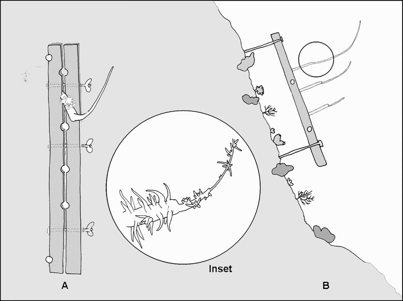
A third experiment was conducted at the site of Siladen Jeti, where six adult entire colonies of Cirrhipathes cfr anguina and six adult entire colonies of Stichopathes cfr maldivensis were collected in the surrounding area. From each specimen three pieces, about 20 cm long, were cut from the base, the centre and the apex of the colony and mounted on several supports fixed to the vertical wall at 25 m depth (Figure 1A). The transplant survival and growth were monitored over a period of 14 months from November 2006 to January 2008.
Scheme of transplants in Siladen Jeti. (A) Frontal view of a support; (B) lateral view of a mounted support with a Cirrhipathes colony. The differential growth between apical, central and basal pieces is shown. Inset: apical growth of a Cirrhipathes colony.

Data, square root transformed, were analysed by analysis of variance (ANOVA) and Tukey test.
In all the experiments the linear growth was measured both directly in the field with a caliper and through the digital analysis of the underwater photographs using the software ImageJ (by Wayne Rasband, National Institutes of Health).
RESULTS
The employed species
The employed species belong to the genera Cirrhipathes (Blainville, 1834) and Stichopathes Brook, 1889, both characterized by a simple stem corallum, without branches and pinnules. The two genera can be distinguished on the base of the polyps' arrangement, in one series in Stichopathes and all around the stem in Cirrhipathes.
The species determination of whip corals is very uncertain: in accordance with the existing literature we have assigned our species to Cirrhipathes cfr anguina (Dana, 1846) and Stichopathes cfr maldivensis (Forster-Cooper, 1903).
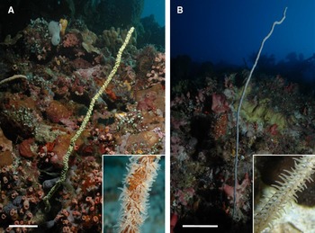
Cirrhipathes cfr anguina (Dana, 1846) is a straight or curved unbranched unpinnulated species showing different colours (varying from brown, to white, yellow, red or orange). Depending on the habitat, in Bunaken it may show variable adult sizes (50–200 cm high) (Figure 2A). The stem of the colony shows nodes, possibly corresponding to the major growing moments of the skeleton. The spines, equal on both sides of the stem, are triangular, with a slightly papillose tip. They are arranged in numerous longitudinal rows, are 0.15–0.2 mm high and show a mutual distance of 0.4–0.5 mm. The polyps are up to 1.5 mm in transverse diameter and are situated on every side of the stem except, occasionally, for a narrow streak on one side of the axis.
Employed species. Underwater photographs in their natural environment of: (A) Cirrhipathes cfr anguina with detail of the irregular arrangement of polyps; and (B) Stichopathes cfr maldivensis, with detail of the uniserial arrangement of polyps. Scale bars: A, 5 cm; B, 20 cm.

Stichopathes cfr maldivensis (Forster-Cooper, 1903) presents a single straight corallum, characterized by only one line of brown or white polyps along its stem (Figure 2B). In Bunaken the colonies are usually around 50–100 cm high, but in peculiar conditions of strong current, they result higher (2–3 m). Spines are typically distinguished in stout, blunt, conical, rough primary spines and small, triangular, smooth secondary spines scattered between the first ones. Primary spines, 0.2–0.5 mm high, distanced 0.6–0.7 mm apart, are arranged in 8–9 longitudinal rows (visible from one aspect). Polyps have a transverse diameter of 1.5–3 mm and their average mutual distance is 1.4 mm.
Growth of intact colonies
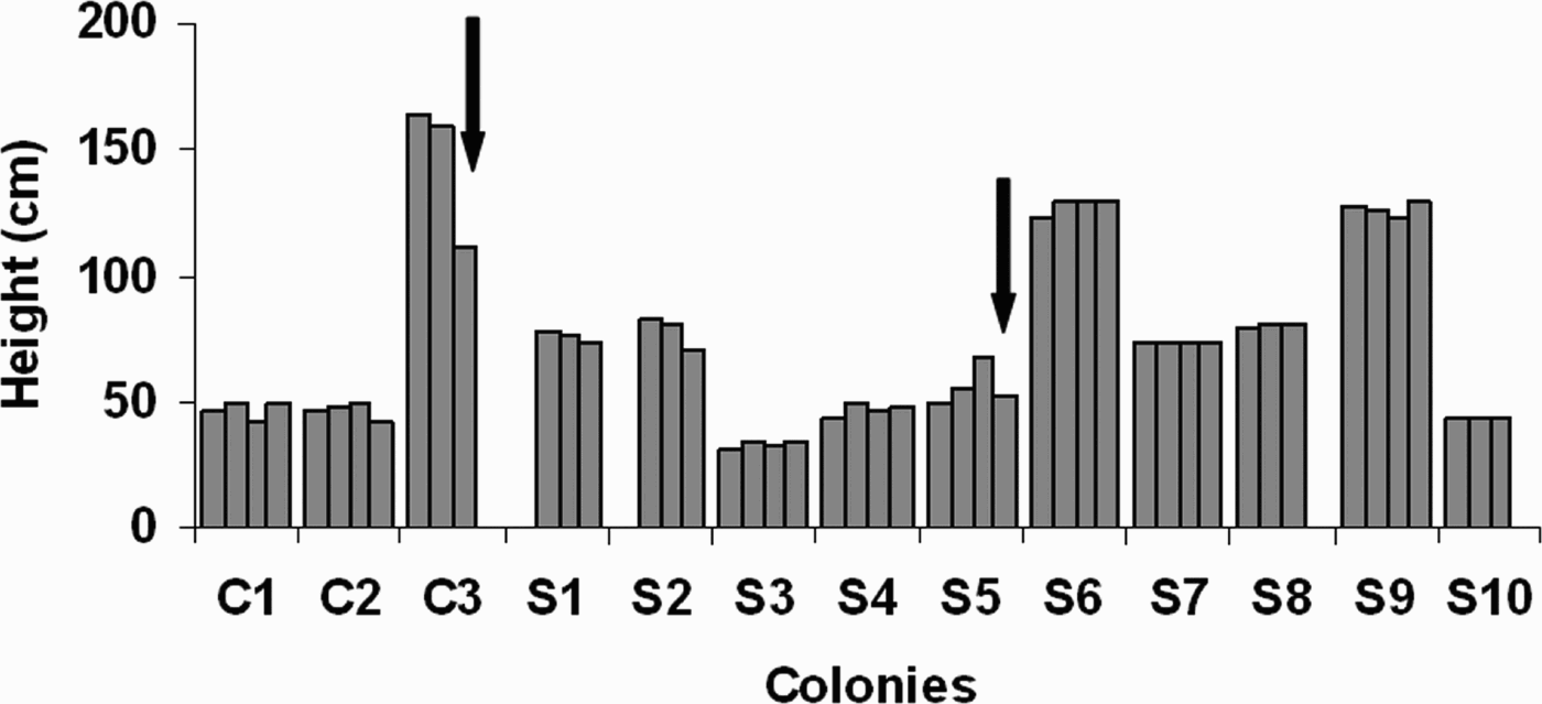
The analysis of the data recorded over 14 months of observation of adult, undisturbed colonies of both Stichopathes cfr maldivensis and Cirrhipathes cfr anguina did not show significant differences between the two species (P > 0.05, t-test).
Nevertheless in Cirrhipathes colony C3 the size strongly decreased in March 2007 due to a breakage. Also the Stichopathes colony S5, showed a gradual increase from November 2006 to March 2007, broke down in January 2008 (Figure 3).
Growth of Cirrhipathes and Stichopathes colonies in their natural environment (Tanjung Pisok). C1–C3 are colonies of Cirrhipathes cfr anguina, while S1–S10 are Stichopathes cfr maldivensis colonies. Black arrows indicate the rupture of the apical portion of the colonies. The monitored months are: November 2006, February 2007, March 2007 and January 2008; each column corresponds to a different month.

The microscopical observation of the skeleton tip of several undisturbed specimens of Cirrhipathes cfr anguina showed signs of fracture, with evidence of re-growing, suggesting that the breakage due to water movement is a frequent episode during the whip coral growth (Figure 4A–D).
Apical growth. Scanning electron microscopy images of the apical portion of the skeleton of different Cirrhipathes cfr anguina specimens showing signs of fracture and skeleton regeneration: (A) apical portion of the skeleton lacking spines; (B) regenerating skeleton with roughs of spines; (C) evident skeletal fracture; (D) detail of the growing area of C showing the abrupt change in spine deposition; (E) underwater photographs of a transplanted portion of C. cfr anguina showing the positive phototropism of the growing apex. Scale bars: A–D, 1 mm; E, 5 cm.

Growth and shape in transplanted colonies
At the site of Siladen Onong only the pieces of adult colonies of Cirrhipathes cfr anguina survived while the entire juvenile colonies died one month after the transplantation.
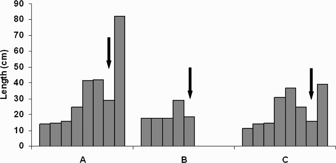
The growth rate of the three surviving specimens (A–C) showed very high maximal values, respectively 13.25, 2.75 and 5.75 cm month−1.
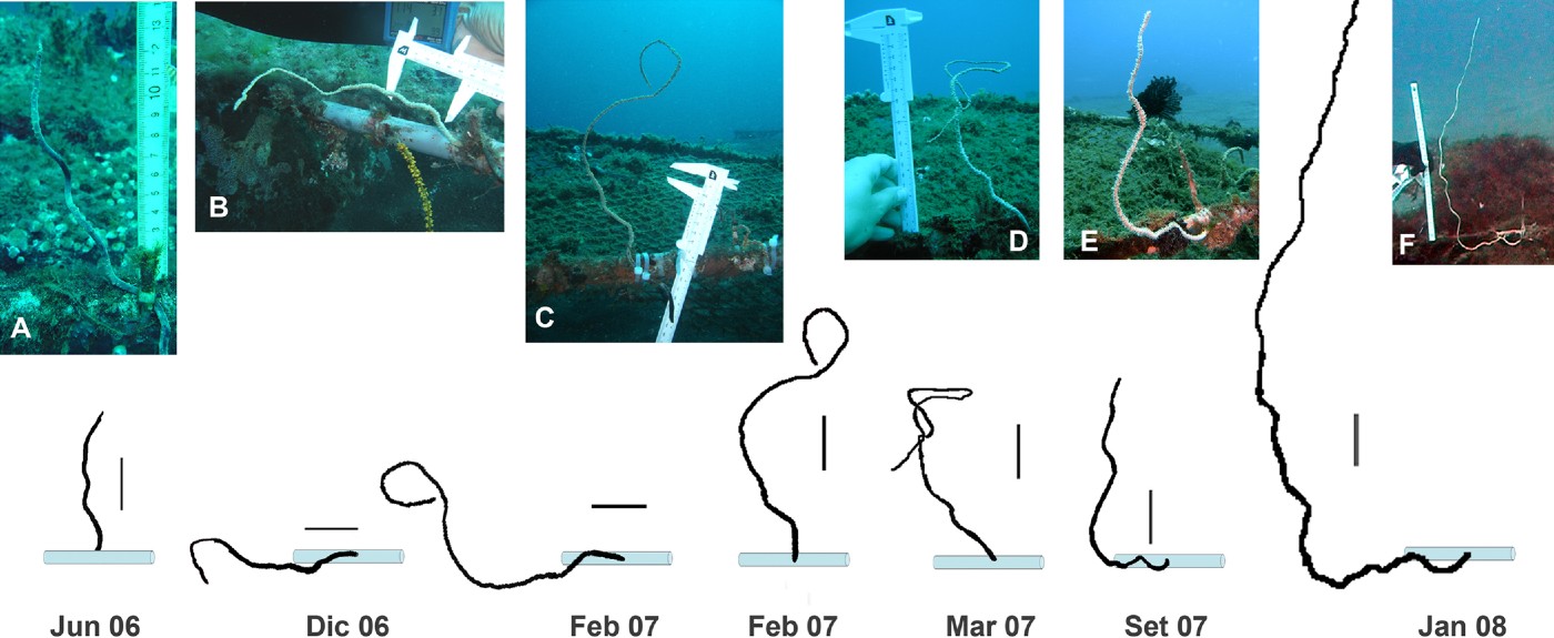
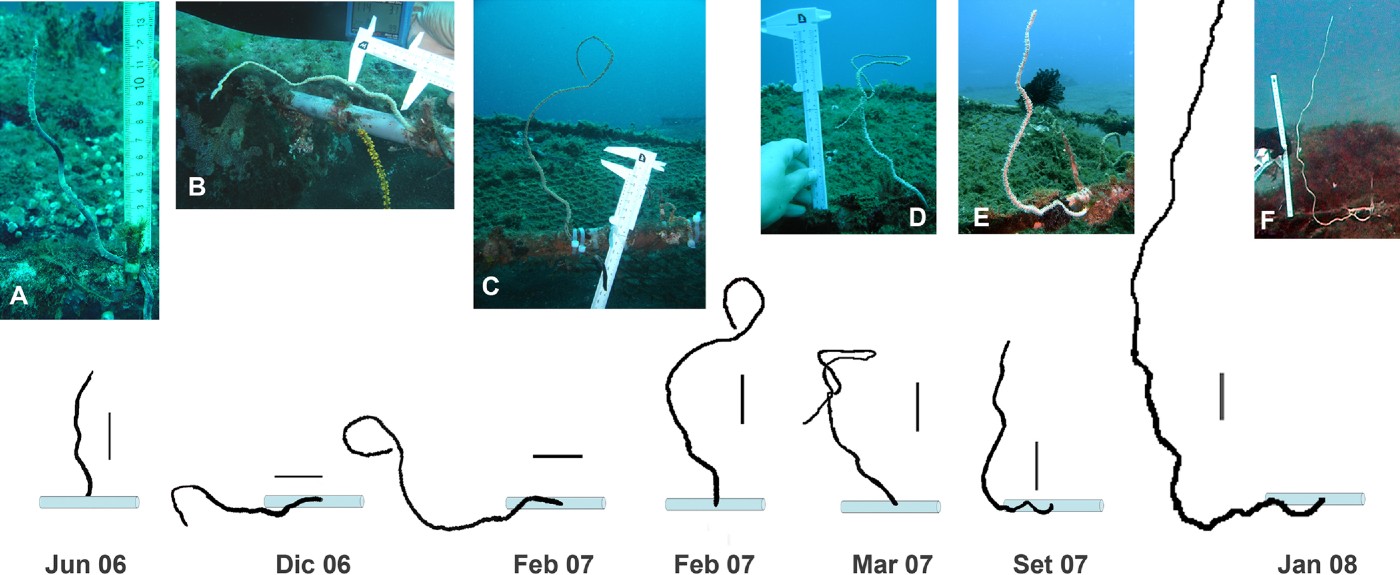
The growth rate was variable, gradually increasing in the months following the transplantation. In the colonies A and C, the apical portions were broken by the action of current and after the rupture both colonies showed the highest growth rate (Figure 5). In two cases (A and B) the transplanted pieces had been knocked over by the action of current in December 2006. This caused their apex to be turned downwards so that the two colonies underwent an evident contortion of the corallum in order to be always growing upwards. Both specimens were re-positioned upwards, but colony B died in February 2007, while colony A, after the breaking of the contorted loop in September 2007, continued to grow straight (Figure 6A–F).
Growth of Cirrhipathes cfr anguina in Siladen Onong. A–C are the three Cirrhipathes cfr anguina specimens. The graphic shows the gradual increase of the growth rate during the monitored months, which are: June 2006, July 2006, August 2006, December 2006, February 2007, March 2007, September 2007, and January 2008. Black arrows indicate the rupture of the apical portion of the colonies.

Growth of Cirrhipathes cfr anguina in Siladen Onong. Photographic monitoring of colony A. The photograph C, putting in evidence the contortion of the corallum, was taken after repositioning upwards the transplant, that was displaced from its original position by the action of current (see text for details). The drawings are schematic representations of the corallum growth during the considered months. Scale bar drawings: 5 cm.
In Siladen Jeti we have compared the growth rates of pieces of Cirrhipathes cfr anguina and Stichopathes cfr maldivensis coming from different portions of the colonies (base, centre and apex). At the end of the experiment the average percentages of survival were 80% and 68% respectively for Stichopathes and Cirrhipathes. All the pieces were fixed on the supports horizontally, but their growing apex, after turning 90°, continued to grow vertically (Figure 4E).
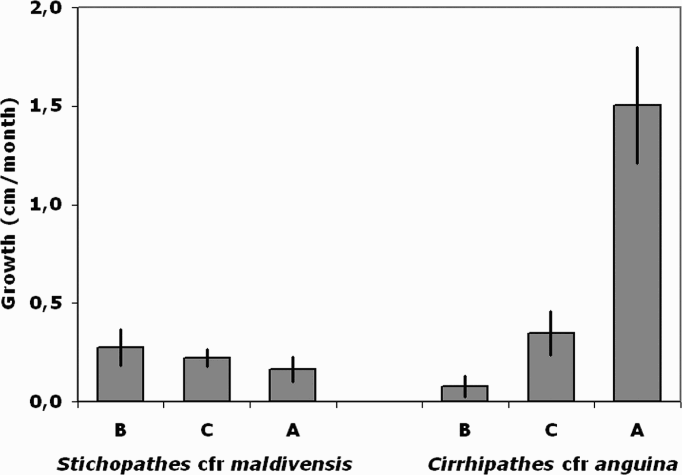
The analysis of the recorded data indicates that the two species show significantly different growth rates in the three analysed portions (ANOVA, P < 0.001). Inside each species, Stichopathes does not show any significant differences among the three portions (0.3 ± 0.09, 0.2 ± 0.04, 0.2 ± 0.06 cm month−1 respectively for base, centre and apex), but in Cirrhipathes significant differences arise (ANOVA, P < 0.001), in particular the growth rate of the apical portion is significantly greater than those recorded for the basal and central portions (0.1 ± 0.05, 0.3 ± 0.11, 1.5 ± 0.29 cm month−1 respectively for base, centre and apex) (Tukey test, P < 0.05) (Figure 7).
Growth of transplanted pieces in Siladen Jeti. Graphic showing the differential growth (means±SE) of the transplanted pieces of Stichopathes cfr maldivensis and Cirrhipathes cfr anguina during 14 months. B, base; C, centre; A, apex.

DISCUSSION
The over-exploitation for commercial aims of the shallow stocks of black corals in the last decades, lead to the inclusion of all the species of the order Antipatharia in the IUCN list and the Appendix 2 of the CITES list. These raise the need to assess the feasibility of black coral transplantation, in order to start replacement projects of damaged reefs (Montgomery, Reference Montgomery2002). Our data suggest that un-branched black corals can be easily handled and transplanted with a high per cent of survival of the transplanted colonies. The recorded survival rates indicate that the transplant stress is bigger for juvenile colonies probably because their thin tissues can be severely damaged by the manipulation. In a short span of time after transplantation the healthy conditions of the colonies are recognizable for the expanded polyps and the possible presence of typical epibionts like the shrimp Pontonides unciger (Tazioli et al., 2006). The present study therefore supports the use of antipatharians in the coral restoration of damaged reefs and suggests their inclusion, with hard and soft corals, in reef recovery projects.
The growth data obtained from the undisturbed adult colonies in their natural habitat show negligible variations in length both inside each species and between the two species in contrast with the high growth recorded in transplanted colonies. This evidence suggests that the final length of whip corals in their habitat is probably reached in some years and after the further growth is constrained by the environmental conditions of the specimens. Studying the length variation of the single colonies and their apex morphology it is clear that the breakages of the growing skeleton are a common event in the life history of black corals. These evidences allow us to hypothesize that in their habitat the length of adult colonies of whip black corals is the result of continuous apical fractures, inflicted by the local water movement, and consequent episodes of re-growth.
Although the two tested species live in mixed assemblages in the same habitats, their growth rates, recorded on transplanted portions of the colonies, are very different. In particular in Cirrhipathes cfr anguina we have observed one of the highest growth rates ever recorded both for antipatharians and sea fans, comparable with Warner's (2005) observations for Stichopathes lütkeni and very different with that observed in Stichopathes cfr maldivensis and in ramified antipatharians (Montgomery, Reference Montgomery2002).
Our data put also in evidence important differences in the regenerative ability between the two species and inside each species: in S. cfr maldivensis no significant differences were recorded between different portions of the colony while in C. cfr anguina growth decreases from the apical to the basal portion. This differential regenerative ability of the different portions of the colony, inversely related to the age of the tissues, probably indicates a different concentration of stem cells in younger and older parts of the colony.
From this study other details related to black corals' growth strategies arise. Both the studied species clearly show a strong phototropic growth that orients upward the colonies settled on substrata of different inclinations. These data confirm the important role played by light in black coral development as already suggested by Grigg (Reference Grigg1965).
In the monopodial sea fans of the genus Juncella the asexual reproduction of colonies via broken portions is a common strategy (Walker & Bull, 1983). In our work we have never recorded the regeneration of anchorages in the transplanted cut portions. This fact suggests that reproduction in black corals is more likely due to settling of larvae rather than through asexual strategies.
ACKNOWLEDGEMENTS
We would like to thank Stefano Vignando for his help in the placement of the supports, Igor Bartolucci for help with the references, Clara Serra, Federico Betti, the dive guides Jemmy and Tony, and all the staff of Celebes Divers for their support during underwater works.