Introduction
Understanding three-dimensional spatial anatomy is paramount in interventional and surgical planning for CHD. Relying on two-dimensional imaging data to understand complex anatomy can be suboptimal. Two-dimensional imaging can be complimented with various forms of three-dimensional imaging and imaging reconstruction if necessary.Reference Mori, Spicer and Anderson 1 – Reference Mori, Tretter, Spicer, Bolender and Anderson 3 In this case report of a child with an obstructed pulmonary venous baffle following a double switch procedure for congenitally corrected transposition, we highlight two time-efficient and cost-effective forms of volume rendering three-dimensional reconstruction techniques using cardiac computed tomographic data. These techniques can be applied to CHD to maximise imaging data for improved procedural planning.
Case presentation
The patient was diagnosed prenatally and confirmed postnatally by transthoracic echocardiography to have congenitally corrected transposition with usual atrial arrangement and left-hand ventricular topology, a large perimembranous ventricular septal defect, and pulmonary atresia. In the first few days of life, she underwent placement of a right-sided modified Blalock–Taussig–Thomas shunt. At 8 months old, she had takedown of the shunt with a Senning atrial switch and Rastelli procedure using a 14-mm Contegra conduit. Her post-operative course was complicated by complete heart block requiring placement of a dual-chamber epicardial permanent pacemaker. Due to continued biventricular dysfunction and an increasing conduit gradient, a diagnostic cardiac catheterization was performed 9 months post-operatively. This demonstrated elevated right and left ventricular end-diastolic pressures with moderate biventricular systolic dysfunction and dyssynchronous left ventricular contraction. She was transitioned to a dual-chamber multi-site epicardial permanent pacemaker for resynchronization therapy and started on Enalapril and Digoxin with some improvement in left ventricular systolic function.
At 16 months old, she developed progressively worsening work of breathing and lethargy. A transthoracic echocardiogram demonstrated stable moderately depressed left and mildly depressed right ventricular systolic function. However, the systemic and pulmonary venous baffles were not well delineated. Subsequent diagnostic cardiac catheterization demonstrated a wedge pressure to left ventricular end-diastolic pressure gradient of 8–12 mmHg. It was unclear if the gradient was due entirely to pulmonary venous baffle stenosis or if there was additional mitral valve stenosis.
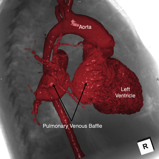
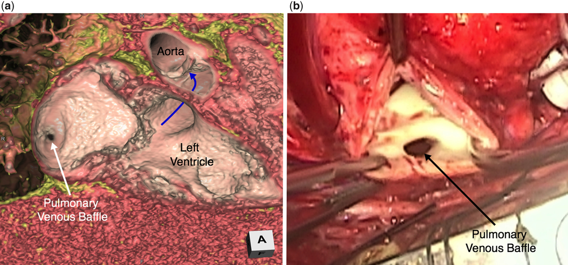
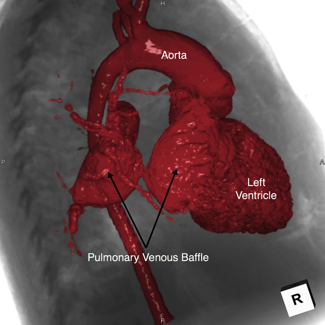
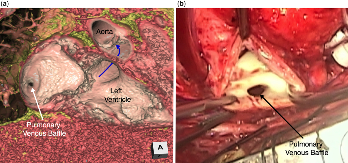
She was referred for cardiac computed tomographic angiography. The study was performed using a 320-detector row system (Genesis Dynamic Volume CT; Toshiba Medical Systems, Tochigi-ken, Japan). A bolus of 18 ml of Ioversol 68% (Optiray 320 mg/mL; Mallinckrodt Medical, St Louis, Missouri, United States of America) was administered through a right antecubital vein at 3 mL/s. This was followed by 14 ml of a 50:50 solution of Ioversol 68% and saline at 3 mL/s and then by a 10-ml saline chaser. Monitoring began 5 seconds after the start of the contrast administration, and scanning was triggered when contrast was seen in the descending thoracic aorta. Volume scanning technique was used with a section collimation of 0.5 mm. A tube potential of 80 kVp and an effective tube current of 320 mA were used for the study. The study showed marked focal narrowing of the pulmonary venous baffle measuring 8 × 4 mm in diameter. Three-dimensional reconstructions were created to guide management planning (Figs 1 and 2a; Video 1 and Video 2 in Supplementary material) using a commercially available workstation (Ziostation2 V.2.4.2.3, Ziosoft, Belmont, California, United States of America). The patient was taken to the operating theatre where the surgeon confirmed that a membrane had formed in the patch portion of the pulmonary venous baffle leaving a restrictive opening in the baffle proximal to the mitral valve (Fig 2b). The surgeon resected the fibrotic membrane and incised the baffle vertically towards the pulmonary veins. The pulmonary venous baffle was patched with a large bovine pericardial patch. She had an uneventful post-operative course with a resolution of presenting symptoms. At 1-year follow-up, she has remained asymptomatic with improved biventricular systolic function on Enalapril and Digoxin, now with mildly depressed left and low normal right ventricular systolic function.
This endocast reconstruction overlaid on a fluoroscopic-appearing image both reconstructed from the cardiac computed tomographic dataset visualises the severe pulmonary venous baffle narrowing in a right anterior oblique projection. The pulmonary veins are visualised to drain into a dilated pulmonary venous baffle proximal the severe discrete narrowing.
(a) This virtual dissection reconstruction from the cardiac computed tomographic dataset viewed with the anterior free walls of the pulmonary venous baffle and morphological left ventricle removed visualises the severe narrowing opening into the distal aspect of the pulmonary venous baffle, with the surrounding membrane formed as a depression adjacent to the interatrial septal wall. The baffle from the left ventricle to aorta (blue curved arrow) is unobstructed. (b) A similar view of the severe narrowing in the pulmonary venous baffle with surrounding membrane was obtained intra-operatively, albeit with a much smaller field of view.
Discussion
The advantages of both virtual and printed three-dimensional reconstructions are well established for improved understanding of unrepaired and repaired CHD. This specific case provides no exception. While printed models can provide added benefit in specific scenariosReference Yoo, Thabit and Kim 2 , Reference Bhatla, Tretter and Ludomirsky 4 , Reference Bhatla, Tretter, Chikkabyrappa, Chakravarti and Mosca 5 more often, the virtual reconstruction is sufficient to provide the appropriate three-dimensional information necessary for the surgeon or interventionalist at a fraction of the cost, resources, and time needed to complete the reconstruction process.Reference Mori, Spicer and Anderson 1 , Reference Mori, Tretter, Spicer, Bolender and Anderson 3
In this case, while the cardiac computed tomographic images were not obtained for the purpose of three-dimensional reconstruction, we demonstrated how these time and cost-efficient methods maximised the utility of the data already obtained. We also emphasised the benefits of the more commonly used endocast imaging reconstruction technique with the additional value of overlaying on a fluoroscopic-like image (Fig 1; Video 1 in Supplementary material). Additionally, we highlighted a relatively novel volume-rendering virtual dissection technique (Fig 2a; Video 2 in Supplementary material).
Endocast, or diecast imaging, visualises the contrast-enhanced vessels and chambers and shows their relationship to each other. We also created a separate volume from the cardiac computed tomographic data into a fluoroscopic-like appearing image. This technique of fluoroscopic-like imaging with endocast overlay is extremely useful for interventionalists. It creates views similar to those seen by fluoroscopy in the catheterization laboratory with the ability to plan the optimal angles to profile specific structures.Reference Mori, Spicer and Anderson 1 , Reference Mori, Tretter, Spicer, Bolender and Anderson 3 Once the technique is learned, the fluoroscopic images can be created in a few minutes, and the endocast images can be generated in 10–30 minutes depending on the complexity of the lesion.
In the virtual dissection technique, the contrast-enhanced blood pool is removed from the image. This allows visualization of intracardiac structures including valves, walls, defects, and trabeculations. These images are like those seen by the pathologist in the autopsy suite or the surgeon in the operating theatre. Additionally, this reconstruction provides the ability to dissect into any plane repeatedly while viewing the structures in their in vivo hemodynamic state, and with a much wider field of view than that seen by the surgeon (compare Fig 2a and b). The images can be efficiently created in 10–20 minutes depending on the complexity of the lesion. These added benefits argue for this technique as the gold standard for viewing intracardiac anatomy, surpassing both autopsy and intraoperative assessment.Reference Mori, Spicer and Anderson 1 , Reference Mori, Tretter, Spicer, Bolender and Anderson 3
High-quality three-dimensional reconstructions are at the mercy of the two-dimensional source images; as the saying goes, “garbage in, garbage out.” Additionally, an accurate reconstruction is dependent on the understanding and interpretation of the reconstructor.Reference Anderson, Bolender, Mori and Tretter 6 Both ECG-gated magnetic resonance whole heart and cardiac computed tomographic angiography images can serve as the source data for excellent three-dimensional reconstructions. Cardiac computed tomographic data sets, however, are preferred in many situations that call for more detailed intracardiac anatomy.Reference Yoo, Thabit and Kim 2 , Reference Bhatla, Tretter and Ludomirsky 4 This is related to the ability for a higher spatial resolution, often between approximately 0.3–0.7 mm in computed tomography, compared to approximately 1 mm in magnetic resonance imaging.Reference Yoo, Thabit and Kim 2 Endocast imaging commonly serves as the basis for rapid prototyping and often can be adequately reconstructed with magnetic resonance imaging. The ability to create a fluoroscopic-appearing image, however, may be limited in magnetic resonance due to decreased ability to image bone and lung parenchyma in sequences intended for cardiac imaging.
While the endocast reconstruction in this particular case helped understand the pulmonary venous baffle narrowing, the virtual dissection reconstruction better prepared the surgeon for what he would encounter when opening the anterior aspect of the pulmonary venous baffle distal to the stenosis (compare Fig 2a and 2b). The surgeon commented that the virtual dissection reconstruction appeared identical to the actual surgical inspection. Additionally, the understanding given by both three-dimensional reconstructions allowed for preparation of a detailed plan prior to entering the operating theatre. This technique of virtual dissection relies on the high spatial resolution afforded by cardiac computed tomographic datasets.Reference Mori, Spicer and Anderson 1 , Reference Mori, Tretter, Spicer, Bolender and Anderson 3 Although the clear disadvantage of radiation exposure afforded by cardiac computed tomography must be considered, with modern technology high-quality images can be obtained at less than 1 millisievert.Reference Han, Rigsby and Leipsic 7
Conclusion
The use of three-dimensional reconstruction techniques improves the understanding of CHD in procedural planning. This case demonstrates the utility of maximizing previously obtained two-dimensional cardiac computed tomographic dataset for procedural planning to address pulmonary venous baffle obstruction, of utilizing time and cost-efficient techniques of endocast reconstruction overlaid on a fluoroscopic-appearing image, and of the more novel virtual dissection reconstruction.
Author ORCIDs
Justin T. Tretter 0000-0001-8926-3752
Supplementary material
To view supplementary material for this article, please visit https://doi.org/10.1017/S1047951119001501
Acknowledgements
We are indebted to Dr. Shumpei Mori from Kobe University Graduate Medical School of Medicine in Kobe, Japan, for his excellent mentorship in teaching these reconstruction techniques.
Financial Support
This research received no specific grant from any funding agency, commercial or not-for-profit sectors.
Conflicts of interest
None.