Introduction
Oocyte quality has been defined as its abilities to be fertilized, mature, and give rise to normal offspring (Duranthon & Renard, Reference Duranthon and Renard2001). Good quality of oocytes is essential for fertilization and subsequent embryonic development. However, if fertilization does not occur for a prolonged period of time after ovulation, oocytes progressively undergo a time-dependent deterioration in quality, referred to as post-ovulatory ageing (Miao et al., Reference Miao, Kikuchi, Sun and Schatten2009). Post-ovulatory ageing displays a range of morphological, cellular and molecular changes, including spontaneous activation, partial cortical granule exocytosis, zona pellucida hardening, mitochondrial dysfunction, spindle abnormalities and loss of chromosome integrity (Dodson et al., Reference Dodson, Minhas, Curtis, Palmer and Robertson1989; Ducibella et al., Reference Ducibella, Duffy, Reindollar and Su1990; Xu et al., Reference Xu, Abbott, Kopf, Schultz and Ducibella1997; Longo, Reference Longo1981; Wakayama et al., Reference Wakayama, Thuan, Kishigami, Ohta, Mizutani, Hikichi, Miyake and Wakayama2004; Takahashi et al., Reference Takahashi, Takahashi, Igarashi, Tezuka and Kurachi2003; Tatone et al., Reference Tatone, Di Emidio, Barbaro, Vento, Ciriminna and Artini2011; Zhang et al., Reference Zhang, Wakai and Fissore2011; Premkumar & Chaube, Reference Premkumar and Chaube2013). These abnormalities may affect the developmental competence and fertilization rates, increasing abnormal and/or retarded development of embryo/fetuses (Szollosi, Reference Szollosi1971; Winston et al., Reference Winston, Braude and Johnson1993; Tarin et al., Reference Tarin, Perez-Albala, Aguilar, Minarro, Hermenegildo and Cano1999; Fissore et al., Reference Fissore, Kurokawa, Knott, Zhang and Smyth2002; Takahashi et al., Reference Takahashi, Takahashi, Igarashi, Tezuka and Kurachi2003). Moreover, severe deterioration in oocyte quality ultimately leads to apoptosis (Fujino et al., Reference Fujino, Ozaki, Yamamasu, Ito, Matsuoka, Hayashi, Nakamura, Ogita, Sato and Inoue1996). Although various factors influence the post-ovulatory ageing of oocytes, oxidative stress is considered to be one of the key factors involved in deterioration of oocyte quality (Lord & Aitken, Reference Lord and Aitken2013). Indeed, ROS levels have been found to increase with oocyte age and oxidative stress-induced apoptosis during in vitro culture of oocytes (Takahashi et al., Reference Takahashi, Takahashi, Igarashi, Tezuka and Kurachi2003; Chaube et al., Reference Chaube, Prasad, Thakur and Shrivastav2005). Consistent with this, it has been shown that the addition of antioxidants, such as glutathione, vitamin E, and vitamin C, to the culture medium improves the rate of in vitro maturation and subsequent embryonic development (Eppig et al., Reference Eppig, Hosoe, O'Brien, Pendola, Requena and Watanabe2000; de Matos & Furnus, Reference de Matos and Furnus2000; Tao et al., Reference Tao, Zhou, Xia, Wang, Wu and Fu2004; Luberda, Reference Luberda2005; Choi et al., Reference Choi, Banerjee, Falcone, Bena, Agarwal and Sharma2007). Therefore, it is of interest to develop methods that protect oocytes from oxidative stress to preserve oocyte quality.
Artemisia asiatica Nakai ex Pamp. has been widely used in East Asia to treat inflammation, cancer, and microbial infections (Huh et al., Reference Huh, Kwon, Shin, Kim, Ahn, Oh and Kim2003; Song et al., Reference Song, Shin, Oh and Sohn2008). Recent studies have shown that A. asiatica exhibits antioxidative and anti-inflammatory effects, which contribute to its protective effects against various pathophysiological conditions such as gastric damage, liver damage, pancreatitis, and tumour promotion (Hahm et al., Reference Hahm, Kim, You, Kim, Cho, Yim, Ahn and Kim1998; Oh et al., Reference Oh, Ahn, Choi, Ahn and Kim2005; Ryu et al., Reference Ryu, Ahn, Oh, Kim, Kim and Lee1998; Seo et al., Reference Seo, Park, Han, Chung, Son, Kim and Surh2002). Currently, a formulated ethanol extract of A. asiatica, DA-9601 (StillenTM) is clinically used to treat gastric mucosal ulcers and inflammation (Seol et al., Reference Seol, Kim, Ryu, Choi, Shin and Ahn2004). Moreover, eupatilin, an active compound isolated from A. asiatica, has been reported to have antioxidant and anti-inflammatory effects against gastric mucosal injury (Oh et al., Reference Oh, Lee, Ahn, Cho, Kim, Kim, Surh, Cho, Lee and Hahm2001).
Given that ROS is a primary factor involved in deterioration of oocyte quality and that A. asiatica has antioxidant activity, it is possible to hypothesize that A. asiatica may relieve post-ovulatory ageing of oocytes by reducing oxidative stress during in vitro culture of oocytes. Therefore, in the present study we investigated the effects of A. asiatica on post-ovulatory ageing of oocytes. We found that A. asiatica could effectively reduce the ROS level in oocytes during in vitro culture, delaying deterioration in quality and the onset of apoptosis. Therefore, our results provide the first evidence that A. asiatica may have beneficial effects on oocyte quality.
Materials and methods
Materials
A 95% ethanol extract (Code No: CA02-070) of A. asiatica Nakai ex Pamp. was purchased from the Plant Extract Bank of the Plant Diversity Research Center (Daejeon, Korea). A stock solution (350 mg/ml) of A. asiatica ethanol extract was prepared in 100% dimethyl sulfoxide (DMSO) and diluted to 0–200 μg/ml with M16 medium for in vitro culture. All other reagents were purchased from Sigma-Aldrich (St. Louis, MO, USA), unless otherwise stated.
Animals
Female CD-1 mice (3- to 4-week-old) were purchased from Koatech (Pyeongtaek, Korea). All procedures for mouse care and use were conducted in accordance with the guidelines and approved by the Institutional Animal Care and Use Committees of Sungkyunkwan University (approval ID:SKKU 12–37).
Oocyte collection and culture
Mice were superovulated by injection of pregnant mare's serum of gonadotrophin (PMSG, 5 IU) followed by human chorionic gonadotrophin (hCG, 5 IU) 46–48 h later. Oocytes were collected in M2 medium at 13–14 h after hCG injection, and cumulus cells were removed using hyaluronidase. After washing, cumulus-free oocytes were cultured in M16 medium in a 5% CO2 in air atmosphere at 37°C with or without ethanol extract of A. asiatica.
Immunoblotting analysis
Oocytes were lysed in SDS sample buffer and subjected to SDS-PAGE. After transfer, the membranes were blocked in TBST (0.1% Tween-20, 3% BSA) at room temperature for 1 h, and then incubated with primary antibodies overnight at 4°C. After washing three times in TBST, membranes were incubated with secondary antibodies for 1 h. The blots were developed with the ECL Plus Western Blotting Detection kit (GE Healthcare Life Science, Marlborough, MA, USA). The primary antibodies were anti-Bax (Cell Signaling, Beverly, MA, USA, 1:1000), anti-Bcl-2 (Cell Signaling, 1:1000) and anti-β actin (Cell Signaling, 1:500). The HRP-labelled mouse and rabbit antibodies (Jackson ImmunoResearch, Westgrove, PA, USA) were used as the secondary antibodies for immunoblotting.
Measurement of intracellular ROS level
Intracellular ROS levels were detected using dihydrorhodamine 123 (DHR-123). Briefly, oocytes were incubated with 20 μM DHR-123 for 30 min before visualization under fluorescence microscopy. The fluorescence signals were acquired using a Nikon Eclipse Ti inverted microscope with a CCD-cooled camera (DS-Qi1Mc, Nikon, Tokyo, Japan) and quantified using ImageJ software (National Institutes of Health, MD, USA).
Annexin V staining
Oocytes were incubated with FITC annexin V (BD Pharmingen, San Diego, CA, USA) for 15 min and washed three times in M2 medium. Fluorescent signals were measured using a Nikon Eclipse Ti inverted microscope with a CCD-cooled-camera (DS-Qi1Mc, Nikon) and quantified using ImageJ software (National Institutes of Health).
Immunostaining and confocal microscopy
Oocytes were fixed in 4% paraformaldehyde in phosphate-buffered saline (PBS) for 20 min, permeabilized in PBS with 0.1% Triton X-100 for 15 min, and blocked in PBS containing 3% BSA for 1 h. Immunostaining was performed using primary antibodies against acetylated α-tubulin (1:500), followed by Alex Fluor-conjugated 488 secondary antibodies (Jackson ImmunoResearch). DNA was counterstained with DAPI. For mitochondrial staining, oocytes were stained with CytoPainter MitoRed (Abcam, Cambridge, UK) for 30 min. After washing, the oocytes were mounted on glass slides and examined with a confocal laser-scanning microscopy (LSM 700; Zeiss, Berlin, Germany) equipped with a C-Apochromat ×63/1.2 water immersion objective. Data analysis was performed using the ZEN 2010 LSM software (Zeiss) and ImageJ software (National Institutes of Health).
Statistical analysis
Statistical analysis was performed with GraphPad Prism (GraphPad Software, San Diego, CA, USA). Data are representative of at least three independent experiments unless otherwise specified and each experimental group included at least 20 oocytes. The significance of differences between groups was analysed by Student's t-test; P-values < 0.05 were considered to be statistically significant.
Results
Artemisia asiatica reduces morphological changes during post-ovulatory oocyte ageing
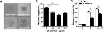
To investigate the effect of A. asiatica on ageing-induced morphological changes, we treated oocytes with ethanol extracts of A. asiatica during in vitro culture. After 12 h of culture, oocytes showed various morphological defects, including granulation, fragmentation, parthenogenetic activation and degeneration (Fig. 1 A). While untreated oocytes exhibited various abnormal morphologies, oocytes treated with A. asiatica showed a significant decrease in abnormal morphologies in a dose-dependent manner (Fig. 1 B). However, the preventive effects of A. asiatica on ageing-induced morphological changes were not further increased at 200 μg/ml of A. asiatica, suggesting that 100 μg/ml of A. asiatica is the optimal concentration to reduce morphological changes during post-ovulatory oocyte ageing.
Effects of Artemisia asiatica on oocyte morphology during post-ovulatory ageing. (A, B) Oocytes were cultured for 12 h with 0, 50, 100 or 200 μg/ml of Artemisia asiatica ethanol extract. (A) Representative abnormal morphologies of oocytes after in vitro culture are shown. (B) Oocytes with abnormal morphologies were quantified. (C) Oocytes were cultured for up to 24 h with 100 μg/ml A. asiatica ethanol extract. Oocytes with abnormal morphologies were quantified. *P < 0.05.

We next determined the time-dependent effect of A. asiatica on ageing-induced abnormal morphologies. Oocytes were cultured for up to 24 h with or without 100 μg/ml of A. asiatica and morphological changes in oocytes were assessed. After 8 h of culture with A. asiatica, oocytes with abnormal morphologies were significantly reduced. Similarly, we also observed reduced morphological defects after 24 h of culture (Fig. 1 C). These results suggest that A. asiatica reduces morphological changes during post-ovulatory ageing of oocytes.
Artemisia asiatica decreases ROS accumulation in ageing oocytes
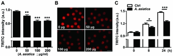
To further examine the effects of A. asiatica on oocyte quality, we investigated intracellular ROS generation during post-ovulatory ageing of oocytes. After 12 h of in vitro culture, we observed that A. asiatica treatment resulted in a significant decrease in ROS accumulation in a dose-dependent manner (Fig. 2 A, B). We next assessed the time-dependent effects of A. asiatica on ROS accumulation in ageing oocytes. Similarly, the elevation of ROS level was retarded in oocytes treated with A. asiatica (Fig. 2 C).
Effects of Artemisia asiatica on ROS generation in ageing oocytes. (A, B) Oocytes were cultured for 12 h with 0, 50, 100 or 200 μg/ml of Artemisia asiatica ethanol extract. (A) The fluorescence intensity was measured following incubation with DHR-123 for 30 min. (B) Representative images are shown. Bar, 100 μm. (C) Oocytes were cultured for up to 24 h with 100 μg/ml of A. asiatica ethanol extract. The fluorescence intensity was measured following incubation with DHR-123 for 30 min. *P < 0.05, **P < 0.001, ***P < 0.0001.

Artemisia asiatica delays the onset of apoptosis during post-ovulatory ageing of oocytes
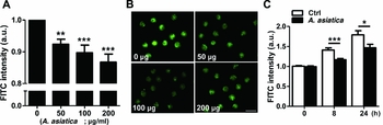
Because oxidative stress has been shown to accelerate the onset of apoptosis in oocytes, we examined whether A. asiatica reduced the onset of apoptosis. Similar to levels of ROS, the levels of annexin V, a hallmark of the early phase of apoptosis (Martin et al., Reference Martin, Reutelingsperger, McGahon, Rader, van Schie, LaFace and Green1995), were decreased in a dose-dependent manner following A. asiatica treatment for 12 h (Fig. 3 A, B). We also observed that A. asiatica significantly delayed the increase in annexin V intensity during in vitro culture of oocytes (Fig. 3 C). In conjunction with reduced ROS levels in oocyte culture with A. asiatica, this result demonstrates that A. asiatica delays the onset of apoptosis during post-ovulatory oocyte ageing.
Effects of Artemisia asiatica on oocyte apoptosis during post-ovulatory ageing. (A, B) Oocytes were cultured for 12 h with 0, 50, 100 or 200 μg/ml of Artemisia asiatica ethanol extract. (A) The fluorescence intensity was measured following incubation with annexin V for 15 min. (B) Representative images are shown. Bar, 100 μm. (C) Oocytes were cultured for up to 24 h with 100 μg/ml of A. asiatica ethanol extract. The fluorescence intensity was measured following incubation with annexin V for 15 min. *P < 0.05, **P < 0.001, ***P < 0.0001.

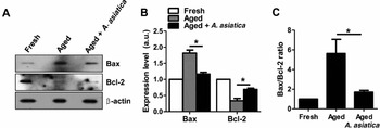
We next determined the levels of Bax and Bcl-2 following A. asiatica treatment. Immunoblot analysis revealed that Bax expression increased, whereas Bcl-2 expression decreased after in vitro culture of oocytes. Thus, the ratio of Bax/Bcl-2 was significantly increased during post-ovulatory ageing of oocytes. However, A. asiatica reduced upregulation of Bax and downregulation of Bcl-2 during in vitro culture of oocytes, thereby reducing the elevation of the Bax/Bcl-2 ratio (Fig. 4 A–C). These results suggest that A. asiatica delays the onset of apoptosis during post-ovulatory ageing of oocytes.
Effects of Artemisia asiatica on the expression of anti- and pro-apoptotic proteins during post-ovulatory ageing. Oocytes were cultured for 12 h with or without 100 μg/ml of Artemisia asiatica ethanol extract. (A) The expression levels of Bax and Bcl-2 were determined by immunoblot analyses. β-Actin was used as a loading control. (B) The normalized levels of Bax and Bcl-2 are shown. (C) The relative Bax/Bcl-2 expression ratio is shown. *P < 0.05.

Artemisia asiatica attenuates ageing-induced spindle and chromosome abnormalities
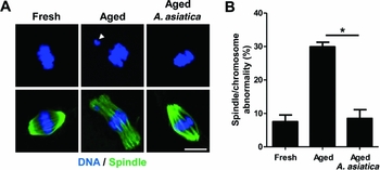
We next determined the effect of A. asiatica on the chromosome alignment and spindle organization, which are the most critical events involved in ensuring developmental competency of the oocytes. Whereas fresh oocytes displayed a typical barrel-shaped spindle, spindles became elongated and less integrated in aged oocytes. Furthermore, aberrant chromosome alignment was increased in aged oocytes. However, these abnormalities in spindle organization and chromosome alignment were significantly reduced after A. asiatica treatment (Fig. 5 A, B).
Effects of Artemisia asiatica on spindle and chromosome misalignment defects during post-ovulatory ageing. Oocytes were cultured for 12 h with or without 100 μg/ml of Artemisia asiatica ethanol extract. (A) Spindle and chromosome abnormalities were determined by immunostaining with anti-tubulin antibody and DAPI. The representative images are shown from three independent experiments. Arrowhead indicates the misaligned chromosomes. Bar, 10 μm. (B) The incidence of spindle and chromosome abnormality is shown. *P < 0.05.

Artemisia asiatica reduces mitochondrial aggregation during post-ovulatory ageing
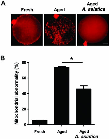
Because mitochondria distribution is correlated with oocyte quality and developmental competency in aged oocytes, we examined whether mitochondrial distribution was affected by A. asiatica treatment during post-ovulatory ageing of oocytes (Nagai et al., Reference Nagai, Mabuchi, Hirata, Shoda, Kasai, Yokota, Shitara, Yonekawa and Hoshi2006). Confocal analysis with mitochondria probe revealed that mitochondria were evenly distributed in the cytoplasm in fresh oocytes, but extensively aggregated after 12 h of in vitro culture. However, these aggregations were significantly reduced in oocytes treated with A. asiatica (Fig. 6 A, B). Taken together, our data demonstrate that A. asiatica prevents the deterioration of oocyte quality during post-ovulatory ageing of oocytes.
Effects of Artemisia asiatica on mitochondrial distribution during post-ovulatory ageing. Oocytes were cultured for 12 h with or without 100 μg/ml of Artemisia asiatica ethanol extract. (A) Mitochondrial distribution was determined by immunostaining with CytoPainter MitoRed. Bar, 10 μm. (B) The incidence of mitochondrial aggregation is shown. *P < 0.05.

Discussion
The effect of A. asiatica on oocyte quality, especially during post-ovulatory ageing, has not been investigated. In this study, we demonstrated that A. asiatica has the potential to attenuate the deterioration in oocyte quality during post-ovulatory ageing.
Post-ovulatory ageing has been shown to induce many morphological changes, such as increased cytoplasmic granules, fragmentation and spontaneous activation. Moreover, the increase in incidence of oocytes exhibiting spindle defects, chromosome misalignment and mitochondrial aggregation is a hallmark of ageing-related decline in oocyte quality (Lord and Aitken, Reference Lord and Aitken2013). Artemisia asiatica has been shown to have various beneficial effects on health by exhibiting anti-inflammatory and antioxidant activities as well as anti-cancer effects (Hahm et al., Reference Hahm, Kim, You, Kim, Cho, Yim, Ahn and Kim1998; Oh et al., Reference Oh, Ahn, Choi, Ahn and Kim2005; Ryu et al., Reference Ryu, Ahn, Oh, Kim, Kim and Lee1998; Seo et al., Reference Seo, Park, Han, Chung, Son, Kim and Surh2002). However, the effects of A. asiatica on reproductive health have not been reported. Our results showed that morphological and cellular abnormalities were significantly decreased in oocytes treated with A. asiatica. These results suggest that A. asiatica also has beneficial effects on reproductive health.
To further examine the effects of A. asiatica on oocyte quality during post-ovulatory ageing, we measured the levels of ROS in ageing oocytes. It was reported that ROS accumulate during in vitro culture of oocytes and are considered to be a key factor that influences the oocyte quality (Takahashi et al., Reference Takahashi, Takahashi, Igarashi, Tezuka and Kurachi2003; Lord & Aitken, Reference Lord and Aitken2013). Consistent with this, we observed that the extended culture of oocytes in vitro results in increased ROS levels. However, the accumulation of ROS was significantly reduced when oocytes were treated with A. asiatica, implying that A. asiatica has antioxidant effects. Indeed, it has been well documented that A. asiatica has antioxidant effects (Hahm et al., Reference Hahm, Kim, You, Kim, Cho, Yim, Ahn and Kim1998; Ryu et al., Reference Ryu, Ahn, Oh, Kim, Kim and Lee1998; Seo et al., Reference Seo, Park, Han, Chung, Son, Kim and Surh2002; Oh et al., Reference Oh, Ahn, Choi, Ahn and Kim2005).
Overproduction of ROS may cause oxidative stress and ultimately result in apoptosis in oocytes (Agarwal et al., Reference Agarwal, Gupta, Sekhon and Shah2008). To investigate the effects of A. asiatica on apoptosis associated with oocyte ageing, we used annexin V, a phospholipid-binding protein that detects the onset of apoptosis (Martin et al., Reference Martin, Reutelingsperger, McGahon, Rader, van Schie, LaFace and Green1995). Our results suggest that A. asiatica delays the onset of apoptosis, showing reduced levels of annexin V. To further investigate the effects of A. asiatica on ageing-induced apoptosis, we examined the level of apoptosis-related proteins, Bax and Bcl-2. It has been reported that Bcl-2 expression decreases, whereas Bax expression increases in the oocytes aged in vitro (Perez et al., Reference Perez, Jurisicova, Matikainen, Moriyama, Kim, Takai, Pru, Kolesnick and Tilly2005; Steuerwald et al., Reference Steuerwald, Steuerwald and Mailhes2005). Consistent with this, we observed a decrease in Bcl-2 and increase in Bax expression, thereby elevating the Bax/Bcl-2 ratio during in vitro culture of oocytes. However, after A. asiatica treatment, the expression ratio of Bax/Bcl-2 was not elevated during post-ovulatory ageing of oocytes. Therefore, our data suggest that A. asiatica delays the onset of apoptosis during in vitro culture of oocytes by preventing elevation of the Bax/Bcl-2 ratio.
Assisted reproductive technology (ART) refers to all procedures that involve handling oocytes or embryos in vitro to achieve successful pregnancy. In humans, the post-ovulatory ageing of oocytes is one of the causes of failure in ART. Therefore, the establishment of methods to prevent oocyte ageing might enhance progress in ART procedures. In this study, we have found that supplementation with A. asiatica in the culture medium relieves multiple facets of oocyte ageing and apoptosis in vitro, thereby preventing the age-associated decline in oocyte quality. Therefore, our data provide the first evidence that A. asiatica may be a promising treatment for oocyte ageing during prolonged culture times, which is particularly important for ART procedures.
Acknowledgements
This paper was supported by Sungkyun Research Fund, Sungkyunkwan University, 2016, Korea.
Conflict of interest
The authors declare no competing financial interests.
Authors’ contributions
J.O. conceived and designed the experiments. H.J., S.Y. and D.K. performed the experiments. J.O. analysed the data. H.J. contributed reagents and materials. J.O. wrote the paper. All authors reviewed the manuscript.