Introduction
Clinical depression is a common disorder that affects approximately 15% of the population at some time in their lives and shows a tendency to become recurrent or persistent (Thornicroft & Sartorius, Reference Thornicroft and Sartorius1993). The disorder is predicted to become the leading cause of worldwide disability (Murray & Lopez, Reference Murray and Lopez1997) and has significant costs for affected individuals, their families and society at large. Despite the considerable burden, the neurobiology of depression is poorly understood although genetic factors and stressful life events both play a part in its etiology (Levinson, Reference Levinson2006).
Brain imaging has provided important insights into the biological basis of depression, with evidence of modest gray-matter reductions in frontotemporal regions using both region of interest and voxel-based methods (Drevets et al. Reference Drevets, Price, Simpson, Todd, Reich, Vannier and Raichle1997; Videbech, Reference Videbech1997; Kumar et al. Reference Kumar, Jin, Bilker, Udupa and Gottlieb1998; Videbech & Ravnkilde, Reference Videbech and Ravnkilde2004). Depression has not, however, typically been associated with a single cortical or subcortical region (Arnone et al. Reference Arnone, McIntosh, Ebmeier, Munafo and Anderson2012) and this could suggest that the disorder is caused by abnormal interactions between different brain regions due to disrupted connectivity. In support of this idea, some studies have found evidence of reduced white-matter integrity in the prefrontal and temporal lobes, and in several other regions (Taylor et al. Reference Taylor, MacFall, Gerig and Krishnan2007; Cullen et al. Reference Cullen, Klimes-Dougan, Muetzel, Mueller, Camchong, Houri, Kurma and Lim2010; Dalby et al. Reference Dalby, Frandsen, Chakravarty, Ahdidan, Sorensen, Rosenberg, Videbech and Ostergaard2010). Many of these white-matter tracts, for example the uncinate fasciculus and cingulum, connect prefrontal areas with the medial temporal lobe. Disrupted connectivity of these emotion-related areas may therefore lead to emotional dysregulation and possibly an increased risk of depression. The extent to which these findings are secondary to the effects of clinical psychiatric illness or its treatment is unclear.
To overcome the issue of confounding by treatment-related factors, an alternative approach to studying clinical depression directly is to examine the relationship of white mater tract integrity to depressive symptoms and known risk factors in a non-clinical population cohort. Trait liability to depression has long been associated with neuroticism (Kendler et al. Reference Kendler, Neale, Kessler, Heath and Eaves1993a, Reference Kendler, Neale, Kessler, Heath and Eaves1994, Reference Kendler, Gardner and Prescott2002; Matthews et al. Reference Matthews, Deary and Whiteman2009), a heritable and relatively stable personality trait. Family and twin studies indicate that there is a high genetic correlation between neuroticism, clinical depression (Kendler et al. Reference Kendler, Gatz, Gardner and Pedersen2006) and depressive symptoms in non-clinical population samples (Ivkovic et al. Reference Ivkovic, Vitart, Rudan, Janicijevic, Smolej-Narancic, Skaric-Juric, Barbalic, Polasek, Kolcic, Biloglav, Visscher, Hayward, Hastie, Anderson, Campbell, Wright, Rudan and Deary2007), with an overlap of more than 50% (Kendler et al. Reference Kendler, Neale, Kessler, Heath and Eaves1993b; Levinson, Reference Levinson2006) in their underlying architecture. Lower extraversion has also been proposed as a risk factor for depression, although the literature (Barnett et al. Reference Barnett, Huang, Perlis, Young, Rosenbaum, Nierenberg, Sachs, Nimgaonkar, Miklowitz and Smoller2011) implicating this dimension of personality in the etiology of depression is sparse.
In the current study we first examined the relationship of white-matter tract fractional anisotropy (FA), derived from diffusion tensor imaging (DTI) as a proxy for tract integrity, with depressive symptoms in individuals from a population-based sample, the Lothian Birth Cohort 1936 (LBC1936). Second, we examined whether trait liability to depression was also associated with reductions in white-matter tract FA in the same tracts using the personality traits of (higher) neuroticism and (lower) extraversion. Third, because we hypothesized that reduced frontotemporal white-matter tract FA confers greater risk of depressive symptoms through effects on trait liability, we conducted a mediation analysis specifically to test this directional hypothesis.
Method
Subjects
The LBC1936 comprises 1091 community-dwelling relatively healthy individuals without dementia, residing around the city of Edinburgh, Scotland, most of whom had participated in the Scottish Mental Survey 1947 (SMS1947). The LBC1936 undertook personality, medical and cognitive testing at a mean age of about 70 years (for full details on participant recruitment and cognitive testing, see Deary et al. Reference Deary, Gow, Taylor, Corley, Brett, Wilson, Campbell, Whalley, Visscher, Porteous and Starr2007). Subjects were then reassessed approximately 3.2 years later at which time they received additional investigations, including detailed structural brain magnetic resonance imaging (MRI) (Wardlaw et al. Reference Wardlaw, Bastin, Valdes Hernandez, Muñoz Maniega, Royle, Morris, Clayden, Sandeman, Eadie, Murray, Starr and Deary2011), and retook the medical and cognitive assessments, including the Mini Mental State Examination (MMSE) of cognitive function (Folstein et al. Reference Folstein, Folstein and McHugh1975). Participants lived independently in the community around the city of Edinburgh, Scotland, and were able to travel on both occasions to the Wellcome Trust Clinical Research Facility in Edinburgh for testing. The study was approved by the Lothian (REC 07/MRE00/58) and Scottish Multicentre (MREC/01/0/56) Research Ethics Committees and all subjects gave written informed consent.
Depressive symptoms and personality traits
Hospital Anxiety and Depression Scale (HADS; Zigmond & Snaith, Reference Zigmond and Snaith1983) scores were available from the assessments performed at 70 and 73 years of age. These assessments were available separately as depression (HADS-D) and anxiety (HADS-A) measures with seven self-report items for each subscale. Neuroticism and extraversion were measured at the first assessment using the Neuroticism, Extraversion and Openness Five Factor Inventory (NEO-FFI; Costa & McCrae, Reference Costa and McCrae1989), a self-report scale consisting of 12 items for each NEO-FFI personality trait. The other three traits (Openness, Agreeableness and Conscientiousness) were not included in the analyses in the current study.
Diffusion MRI
Diffusion MRI was performed on a GE Signa HDXt 1.5-T clinical scanner (General Electric, USA) using a self-shielding gradient set with maximum gradient strength of 33 mT m−1, and a manufacturer-supplied eight-channel phased-array head coil. Single-shot, spin–echo echo–planar diffusion-weighted volumes (b=1000 s mm−2) were acquired in 64 non-collinear directions, along with seven T2-weighted volumes (b=0 s mm−2). Seventy-two contiguous axial slices of thickness 2 mm were acquired with a field of view of 256×256 mm and matrix size of 128×128, giving a resolution of 2×2×2 mm. The repetition time was 16.5 s and echo time 95.5 ms, producing a total scan time of approximately 20 min (see Wardlaw et al. Reference Wardlaw, Bastin, Valdes Hernandez, Muñoz Maniega, Royle, Morris, Clayden, Sandeman, Eadie, Murray, Starr and Deary2011 for full details of the imaging protocol).
White-matter tractography
Datasets were converted into NIfTI-1 (http://nifti.nimh.nih.gov/nifti-1) format and preprocessed using FSL tools (FMRIB, Oxford, UK; www.fmrib.ox.ac.uk/) to extract the brain, remove bulk motion and eddy current-induced artifacts and estimate water diffusion tensor parameters. Brain connectivity data were created using the BEDPOSTX/ProbTrackX tractography algorithm with a two-fiber model and 5000 streamlines to reconstruct tracts of interest. An automatic tract selection method with good reproducibility (Clayden et al. Reference Clayden, King and Clark2009a), based on a model of tract topology (Clayden et al. Reference Clayden, Storkey and Bastin2007), was used to generate equivalent tracts of interest in each subject. This technique, termed probabilistic neighborhood tractography (PNT) and implemented in the TractoR package (http://github.com/jonclayden/tractor/wiki/), optimizes the choice of seed point for tractography by estimating the best matching tract from a series of candidate tracts generated from a neighborhood of voxels placed around a voxel transferred from standard space against a reference tract that was derived from a digital human white-matter atlas (Mori & van Zijl, Reference Mori and van Zijl2007). The topological tract model was also used to reject any false positive connections (Clayden et al. Reference Clayden, Storkey, Muñoz Maniega and Bastin2009b), thereby significantly improving tract segmentation (Maniega et al. Reference Maniega, Bastin, McIntosh, Lawrie and Clayden2008). As displayed in Fig. 1, 12 white-matter tracts were segmented, namely the genu and splenium of corpus callosum, cingulum cingulate gyri (CCG) (Clayden et al. Reference Clayden, Storkey and Bastin2007), anterior thalamic radiations (ATR), uncinate, arcuate and inferior longitudinal fasciculi (ILF). For each subject, the seed point that produced the best match tract to the reference for each of the 12 pathways was determined, with the resulting tractography mask applied to each subject's FA volume. Tract-averaged FA values were calculated from these masks and used in all subsequent analyses. To ensure that the segmented tracts were anatomically plausible representations of the fasciculi of interest, a researcher (S.M.M.) visually inspected all masks blind to the other study variables and excluded tracts with aberrant or truncated pathways.
Representative white-matter tracts obtained from a single individual.

Statistical analyses
Statistical analyses were conducted in the R statistical software package (www.r-project.org/) using linear regression. To meet underlying statistical assumptions, we applied a square root transformation to the HADS symptom measures. Initially, we entered the transformed HADS scores as the dependent variable and age at symptom assessment, sex and white-matter tract FA as the independent variables (analysis 1). To correct for multiple testing, we applied a false discovery rate (FDR; Benjamini & Hochberg, Reference Benjamini and Hochberg1995) correction to these initial associations. Subsequently, we examined whether higher neuroticism or lower extraversion were also related to white-matter tract FA using the same model, substituting these personality measures as the dependent variables (analysis 2). We then examined whether previous antidepressant treatment or a history of stroke confounded any significant relationships between FA and depression or personality measures by conducting a series of planned additional analyses. We then adjusted for the effects of antidepressant treatment, current or past cigarette use or stroke by including these as covariates in the statistical models. Finally, to assess whether neuroticism/extraversion could account for any associations between depression and white-matter tract FA, we examined the influence of these variables on analysis 1. Specifically, we noted the attenuation in the effect of white-matter tract FA after neuroticism/extraversion was included in the model, and conducted a formal mediation analysis using the Sobel test (Baron & Kenny, Reference Baron and Kenny1986).
Results
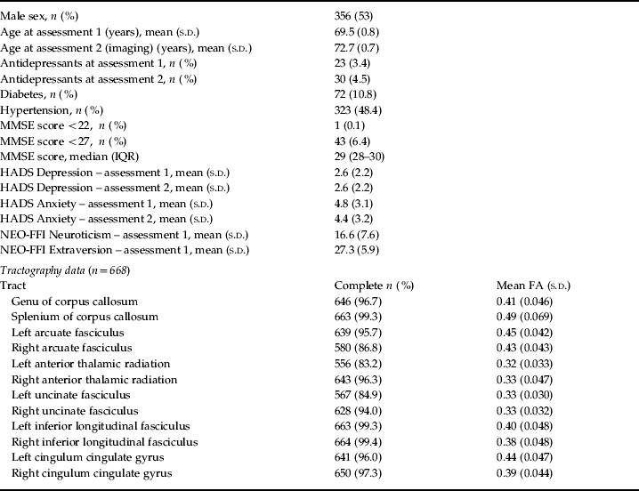
A total of 668 individuals from the LBC1936 provided useable tractography data from the 740 who presented for MRI. As indicated in Table 1, the average age of the subjects was 72.7 (s.d.=0.7) years at the time of the imaging examination and 356 (53%) were male. The rates of antidepressant prescribing were lower than in the general Scottish population (see www.isdscotlandarchive.scot.nhs.uk/isd/6517.html), approximately 10% of the sample reported a diagnosis of diabetes and 48.4% reported a diagnosis of hypertension. MMSE scores (Folstein et al. Reference Folstein, Folstein and McHugh1975) showed that only one subject met a specific threshold for organic brain disorder whereas 6.4% of the sample met a sensitive threshold. At both assessments, the HADS-D score correlated moderately and positively with neuroticism (Pearson's r=0.35 at age 69.5, and r=0.35 at age 72.7) and negatively with extraversion (Pearson's r=−0.38 at age 69.5, and r=−0.34 at age 72.7). Table 1 also shows that the number of individual tracts that did not meet quality criteria (truncation or failed to follow expected path) ranged from 0.6% for the right inferior longitudinal fasciculus to 16.8% for the left anterior thalamic radiation.
Demographic and historical information
MMSE, Mini Mental State Examination; HADS, Hospital Anxiety and Depression Scale; NEO-FFI, Neuroticism, Extraversion and Openness Five Factor Inventory; FA, fractional anisotropy; s.d., standard deviation; IQR, interquartile range.
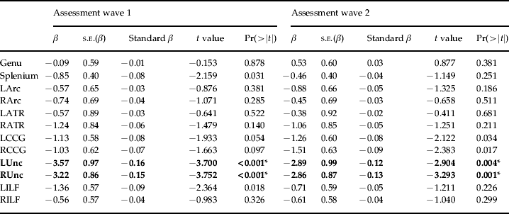
We first examined the relationship of the HADS-D subscale scores with FA in each tract (Table 2). Depressive symptoms measured at the time of the imaging assessment were significantly and negatively associated with FA in the left and right uncinate fasciculus (standardized β=−0.12, p=0.0038 in the left hemisphere; standardized β=−0.13, p=0.0011 in the right hemisphere). Depressive symptoms measured at the first assessment 3 years earlier were also significantly and negatively associated with bilateral uncinate FA, with similar effect sizes (standardized β=−0.16, p=0.0002 in the left hemisphere; standardized β=−0.15, p=0.0002 in the right hemisphere). The association between uncinate fasciculus FA and depressive symptoms survived FDR correction bilaterally at both assessments. There were no significant associations between HADS-A scores and FA in any tract (Supplementary Table S1).
Association between white-matter tract FA and symptoms of depression
FA, Fractional anisotropy; L, left; R, right; Arc, arcuate fasciculus; ATR, anterior thalamic radiation; CCG, cingulum cingulate gyrus; Unc, uncinate fasciculus; ILF, inferior longitudinal fasciculus; s.e., standard error. Bold type indicates effects that were statistically significant at both study assessments.
* False discovery rate (FDR) <0.05.
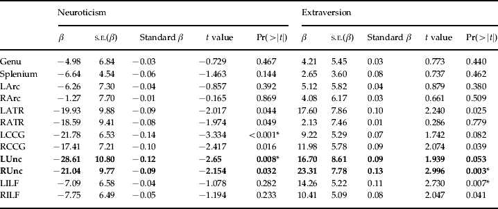
We next examined the relationship between neuroticism and extraversion, trait measures of liability to depression that were measured at the first assessment, and mean white-matter FA in each tract determined at the second assessment 3 years later (Table 3). There was a significant and negative association between neuroticism scores and FA in the uncinate fasciculus bilaterally (standardized β=−0.12, p=0.0083 in the left hemisphere; standardized β=−0.09, p=0.032 in the right hemisphere). Extraversion was significantly and positively correlated with uncinate fasciculus FA in the right hemisphere only (standardized β=0.13, p=0.0029), although the association with FA in the left uncinate fasciculus narrowly failed to reach conventional statistical significance (standardized β=0.09, p=0.053). Two of these associations, namely a negative association between left uncinate FA and neuroticism and a positive association between right uncinate FA and extraversion, remained significant after FDR adjustment across all 12 tracts assessed.
Association between white-matter tract FA, neuroticism and extraversion
FA, Fractional anisotropy; L, left; R, right; Arc, arcuate fasciculus; ATR, anterior thalamic radiation; CCG, cingulum cingulate gyrus; Unc, uncinate fasciculus; ILF, inferior longitudinal fasciculus. Bold text used to highlight effects on Unc.
* False discovery rate (FDR) <0.05.
We then reanalyzed the data after the graphical detection and subsequent removal of a single outlying observation with an FA value of <0.2, with no effect on any of our significant findings, all of which remained significant. We then adjusted for the effects of antidepressants prescribed at time 1 or time 2, the presence of a self-reported history of stroke, current alcohol use at the time of the scan, and whether the individual reported a history of past or current tobacco use. In each case the relationship between uncinate white-matter integrity remained in the same direction as reported in the whole sample (see online Supplementary material). However, the relationship between right uncinate white-matter integrity and neuroticism became non-significant.
We then assessed the effect of adding neuroticism or extraversion to a statistical model where tract FA and age and sex were already fitted as independent variables and depression was the dependent variable. The purpose of this analysis was to examine whether the addition of neuroticism/extraversion attenuated the effect of uncinate tract FA on depressive symptoms, indicating that neuroticism was more closely associated with depression and potentially mediating the association of tract FA with depressive symptoms. The addition of neuroticism led to a significantly improved model fit in both the left (see online Supplementary Table S2) and right hemispheres. The regression coefficient estimates for the effect of uncinate tract FA on depressive symptoms were somewhat attenuated in both the left hemispheres after the inclusion of neuroticism in the model (see Supplementary Table S2). Similarly, the addition of extraversion to a model where depressive symptoms was the dependent variable and right uncinate FA was the independent variable also improved the model fit whereas the effect of tract FA on depressive symptoms was also somewhat attenuated (see supplementary Table S2).
Mediation analysis supported our hypothesis that neuroticism and extraversion partly mediate the relationship between tract FA and depression. Specifically, Sobel tests showed a significant mediating effect of neuroticism between lower uncinate fasciculus FA and depressive symptoms at the first wave of assessment in both the left (test for mediation: Z=−2.38, p=0.017) and right (test for mediation: Z=−2.03, p=0.042) hemispheres. Extraversion also significantly partly mediated the relationship between right uncinate fasciculus FA and depressive symptoms in the right hemisphere (test for mediation: Z=−2.90, p=0.004) but not the left (test for mediation: Z=−1.91, p=0.056). In each case, the mediating effect of neuroticism was partial, as the (albeit attenuated) effect of white-matter integrity on depressive symptoms remained significant in each case.
Discussion
Using data from the LBC1936 we found that there were significant negative associations between depressive symptoms and white-matter integrity in the bilateral uncinate fasciculus. This relationship was highly significant when the depressive symptoms were rated either at the time of the imaging assessment or 3 years earlier. Subsequently we found that the personality traits of neuroticism and extraversion also have significant associations with white-matter integrity in the uncinate, consistent with their risk-conferring effects on depression. The association between depressive symptoms and white-matter integrity was slightly and significantly attenuated after the inclusion of neuroticism or extraversion in the model. Sobel analyses confirmed that neuroticism and extraversion partially mediated the relationship between white-matter integrity and depression. These associations were significant after adjustment for age and sex, after correction for multiple testing, and were robust to planned sensitivity analyses that took into account previous antidepressant treatment or stroke. These findings suggest that the effect of reduced uncinate white-matter integrity on depressive symptoms is conferred partially through effects on extraversion and neuroticism that have been shown to increase liability to depression in other studies.
No significant associations were found between depressive symptoms and white-matter integrity in any other tract, nor were there any associations between anxiety rating scale scores and white-matter integrity. By contrast, there was a significant negative association (surviving FDR correction) between left cingulum white-matter integrity and neuroticism. Similarly, there was a significant positive association between left ILF integrity and extraversion. To the best of our knowledge, neither of these associations has been reported previously in the literature. The absence of a relationship with anxiety is superficially surprising, as anxiety frequently accompanies depressive symptoms in the clinical disorder. The absence of a relationship between white-matter integrity and anxiety implies a more specific relationship with depressive symptoms, and suggests that anxiety symptoms are associated with a smaller detectable signal or that their neural correlates are more detectable as changes in gray-matter structure or brain function.
The uncinate fasciculus connects the prefrontal cortex to limbic and emotional regions of the temporal lobe, such as the amygdala and anterior temporal cortex. These areas are implicated in the encoding of rewarding or punishing stimuli and in normal emotional processing and regulation in both the animal (Cardinal et al. Reference Cardinal, Parkinson, Hall and Everitt2002) and human (Phillips et al. Reference Phillips, Drevets, Rauch and Lane2003) literature. Abnormalities in these tracts have been found in studies of unipolar depression (Taylor et al. Reference Taylor, MacFall, Gerig and Krishnan2007; Cullen et al. Reference Cullen, Klimes-Dougan, Muetzel, Mueller, Camchong, Houri, Kurma and Lim2010; Dalby et al. Reference Dalby, Frandsen, Chakravarty, Ahdidan, Sorensen, Rosenberg, Videbech and Ostergaard2010) and people at high familial risk of major mental illness (Huang et al. Reference Huang, Fan, Williamson and Rao2011). There is also a growing body of literature showing abnormal frontotemporal activation and connectivity in depression (Anand et al. Reference Anand, Li, Wang, Wu, Gao, Bukhari, Mathews, Kalnin and Lowe2005a,b) and in a variety of other mental disorders (Lawrie et al. Reference Lawrie, Buechel, Whalley, Frith, Friston and Johnstone2002; Chepenik et al. Reference Chepenik, Raffo, Hampson, Lacadie, Wang, Jones, Pittman, Skudlarski and Blumberg2010) for which neuroticism is an identified risk factor (Maier et al. Reference Maier, Minges, Lichtermann, Heun and Franke1994; van Os & Jones, Reference van Os and Jones2001; Angst et al. Reference Angst, Gamma and Endrass2003; Barnett et al. Reference Barnett, Huang, Perlis, Young, Rosenbaum, Nierenberg, Sachs, Nimgaonkar, Miklowitz and Smoller2011).
Few studies have directly examined the relationship of white-matter tract FA to neuroticism, although recently a negative association between harm avoidance and measures of tract integrity was reported in several long white-matter tracts (Westlye et al. Reference Westlye, Bjornebekk, Grydeland, Fjell and Walhovd2011). Structural and functional abnormalities of frontotemporal brain regions have, however, been linked to specific genetic risk factors for depression and other mental illnesses. These studies include several examining the role of the 5-HTTLPR polymorphism (Ball et al. Reference Ball, Hill, Freeman, Eley, Strelau, Riemann, Spinath, Angleitner and Plomin1997), in which the s allele confers reduced frontotemporal functional connectivity (Pezawas et al. Reference Pezawas, Meyer-Lindenberg, Drabant, Verchinski, Munoz, Kolachana, Egan, Mattay, Hariri and Weinberger2005), increased neuroticism (Barnett et al. Reference Barnett, Huang, Perlis, Young, Rosenbaum, Nierenberg, Sachs, Nimgaonkar, Miklowitz and Smoller2011) and an increased risk of depression (Caspi et al. Reference Caspi, Sugden, Moffitt, Taylor, Craig, Harrington, McClay, Mill, Martin, Braithwaite and Poulton2003). The current findings extend that literature to imply that the personality traits of neuroticism and extraversion may mediate the effects of reduced frontotemporal connectivity on the risk of depressive symptoms.
Although no relationship between left ILF FA and depressive symptoms was found in the current study, left ILF FA was significantly and positively related to extraversion. The ILF connects occipital brain areas with the temporal lobe whereas the cingulum connects the medial prefrontal cortex to the hippocampus. There is evidence that the integrity of these tracts is also associated with liability to depression (Sheline et al. Reference Sheline, Price, Vaishnavi, Mintun, Barch, Epstein, Wilkins, Snyder, Couture, Schechtman and McKinstry2008), bipolar disorder (Chaddock et al. Reference Chaddock, Barker, Marshall, Schulze, Hall, Fern, Walshe, Bramon, Chitnis, Murray and McDonald2009; Zanetti et al. Reference Zanetti, Jackowski, Versace, Almeida, Hassel, Duran, Busatto, Kupfer and Phillips2009; Sprooten et al. Reference Sprooten, Sussmann, Clugston, Peel, McKirdy, William, Moorhead, Anderson, Shand, Giles, Bastin, Hall, Johnstone, Lawrie and McIntosh2011) and schizophrenia. It is also possible that reductions in tract integrity in these regions may have important consequences for cognitive function in non-demented adults (Kantarci et al. Reference Kantarci, Senjem, Avula, Zhang, Samikoglu, Weigand, Przybelski, Edmonson, Vemuri, Knopman, Boeve, Ivnik, Smith, Petersen and Jack2011), a hypothesis that could be addressed in the current cohort at a later imaging assessment.
The current study is the largest investigation to date of depressive symptoms and white-matter tract FA as a proxy for integrity in any non-clinical sample. It should be noted that <10 people in the current study met the specific HADS-D threshold for clinical depression. These thresholds have been largely determined in healthcare-seeking populations, so the positive predictive value is likely to be higher in the current sample where the prevalence of depression is likely to be lower. The association of depressive symptoms with white-matter FA in a non-clinical cohort is likely to have broader applicability to clinical depression for several reasons. First, the symptoms of clinical depression are identical to those captured by the items of most depression-rating scales, albeit without the usual 2-week duration criterion needed to make a clinical diagnosis. However, the choice of a 2-week duration criterion for depression is arbitrary and has limited validity, with shorter duration of symptoms predicting depression in both affected subjects and co-twins (Kendler & Gardner, Reference Kendler and Gardner1998). Depressive symptoms measured at a single episode also show similar heritability estimates to clinically defined syndromal depression (McGue & Christensen, Reference McGue and Christensen1997) and higher estimates of heritability when measured repeatedly over time (McGue & Christensen, Reference McGue and Christensen2003). Furthermore, there is evidence that major and subthreshold ‘minor’ depression share a common neural basis (Kumar et al. Reference Kumar, Jin, Bilker, Udupa and Gottlieb1998). Overall, these findings suggest that scale-measured depressive symptoms are associated with trait liability to depression and are not merely measures of current mental state.
A further limitation of this study is the relatively small effect sizes that were found for the relationships between white-matter integrity, depression and the personality traits of neuroticism and depression. All scales for personality and depressive symptoms have strengths and limitations and each only approximates the measurement of the underlying latent variables. The estimates given here may therefore underestimate the true relationships, which cannot be measured in an error-free way. Finally, in the present sample, neuroticism and extraversion were correlated with one another (Pearson's r=−0.37) and it is possible that both measures may be measuring a common underlying trait. Future studies should explore the facets of each personality measure that are independently associated with white-matter integrity, and also perhaps mediate the association with symptoms of depression.
The current study is the first large-scale investigation showing an association between frontotemporal white-matter tract FA, depression and trait liability to clinical mood disorder. These findings are not due to differences in age, sex or the secondary effects of antidepressant treatment, and suggest that variation in frontotemporal interactions may underlie depressive symptoms at a population level, and may also apply to clinical populations.
Supplementary material
For supplementary material accompanying this paper visit http://dx.doi.org/10.1017/S003329171200150X.
Acknowledgments
Funding from the following bodies is acknowledged: The Disconnected Mind Project (www.disconnectedmind.ed.ac.uk/) funded by Age UK and the UK Medical Research Council (MRC), the Row Fogo Trust, the Carnegie Trust for the Universities of Scotland, the Scottish Funding Council (SFC), which funds the SINAPSE Collaboration (www.sinapse.ac.uk), and The Centre for Cognitive Ageing and Cognitive Epidemiology (G0700704/84698; www.ccace.ed.ac.uk/), part of the cross-council Lifelong Health and Wellbeing Initiative (LLWH). Funding from the Biotechnology and Biological Sciences Research Council (BBSRC), the Engineering and Physical Sciences Research Council (EPSRC), the Economic and Social Research Council (ESRC) and the MRC is also gratefully acknowledged. Personality, medical and cognitive testing was performed at the Wellcome Trust Clinical Research Facility, Edinburgh (www.wtcrf.ed.ac.uk/), and MRI was performed at the Brain Research Imaging Centre, University of Edinburgh (www.sbirc.ed.ac.uk/). M.L. is a Royal Society of Edinburgh/Lloyds TSB Foundation for Scotland Personal Research Fellow. J.H. is supported by a Scottish Senior Clinical Fellowship. A.M.M. is supported by the Brain and Behavior Research Fund (formerly NARSAD) through an Independent Investigator Award and by the Health Foundation through a Clinician Scientist Fellowship.
Declaration of Interest
A.M.M. and S.M.L. report having received research support from Pfizer Pharmaceuticals and consultancy fees from Roche Pharmaceuticals. J.H. reports having received research funding from Pfizer Pharmaceuticals.