Ventricular septal defects represent the commonest congenital cardiac malformation, accounting for almost one-fifth of all defects.1 Of these lesions, seven-tenths are located in the area of the membranous septum, with various extensions to open towards the inlet, outlet or apical components of the right ventricle. These are the perimembranous defects, with those opening towards the inlet excavating beneath the septal leaflet of the tricuspid valve. Defects opening directly beneath both the aortic and pulmonary valves are defined as being doubly committed and juxta-arterial, or supracristal. These defects are quite rare in western countries, albeit more frequent in asian countries. Ventricular septal defects can also be entirely located within the muscular portion of the septum, and these account for around one-sixth of patients seen in postnatal life. The defects can, of course, be multiple, and the worst example of multiple lesions is the so-called “swiss cheese” septum.
Acquired ventricular septal defects are rare. They can be seen in patients after myocardial infarction, or as residual defects subsequent to attempted surgical closure of a congenital defect.
Surgical closure of a congenital ventricular septal defect was performed for the first time by Lillehei and associates in 1954.2 Since that time, surgical closure has come to be regarded as the gold standard for treatment, albeit that it remains associated with morbidity and mortality,3–10 postoperative discomfort, the need for sternotomy, and a residual scar. Complications due to significant residual leaks are reported in up to one-twentieth of cases,3–7 while iatrogenic atrioventricular block can occur in around 1 to 8% of cases.3–9 Reoperations because of indications other than residual leakage are needed in a further 2% of subjects.3–7 The occurrence of post-pericardiotomy syndrome, arrhythmias, infections, and respiratory or neurological complications are also reported.3–7 Mortality may occur in a proportion of patients,3–7 albeit that this would be most unexpected in the current era. The risk for all these events, nonetheless, is increased in small infants, in patients whit multiple defects, associated lesions or when additional surgery is required in patients with residual defects.3–5 It should also be remembered that negative long term effects on developmental and neurocognitive functions have been reported in children who underwent bypass surgery.10 It is hardly surprising, therefore, that various attempts have been made over the years to develop less invasive techniques so as to reduce the impact of morbidity, mortality, and psychological stress.
In this review, we report and discuss data about the state of the art concerning interventional closure of ventricular septal defects, assessing the situation for muscular and perimembranous congenital defects, and in the acquired defects seen after previous surgery or subsequent to myocardial infarction.
Historical review
It was in 1988 that Lock and et al.11 reported the first human experience of transcatheter closure of muscular defects. They closed such defects in 7 patients using the Rashkind double umbrella device. Since then, various devices have been used, such as the Clamshell or CardioSeal device,12, 13 the Sideris buttoned device,14 and Gianturco coils.15 The rate of success of such procedures was between 77 and 100%, albeit that residual shunting was reported in between 35% and 100%.12–14, 16–20 Furthermore, the procedure was difficult when using these devices, and complications were encountered with some frequency.12–14, 16–20 The more recent introduction of the Amplatzer family of devices has markedly widened the application of transcatheter techniques for closure of these defects.21
This is particularly true for perimembranous defects. Due to the proximity of these defects to the aortic and the atrioventricular valves, devices designed for other applications did not fit perfectly when used in this setting.13 Only very recently, with the introduction of the specially designed eccentric Amplatzer device, has general closure of these defects become feasible.22 Substantially less experience has been reported for percutaneous closure of post-infarction ventricular septal defects, and it was not until 1998 that successful percutaneous closure of such a defect was first reported.23
Present state and clinical indications
Congenital defects
Indications for closure are symptoms of heart failure, and/or signs of left heart volume overload. In patients, and in particular in children, with left atrial and ventricular overload, closure may be needed in order to prevent pulmonary arterial hypertension, ventricular dilation, arrhythmias, aortic regurgitation, and development of double chambered right ventricle. Even subjects with small defects, with neither symptoms of cardiac failure nor overload, may also need closure if they experience endocarditis.
Large defects give signs and symptoms of cardiac failure in early infancy, and they have to be treated surgically in the first months of life. Defects of moderate size may also be responsible for failure to thrive, respiratory infections, and diastolic left heart overload. These defects may be suitable for percutaneous closure if they are located within the muscular septum, or if they are perimembranous. Surgical repair is currently the only option for doubly committed or supracristal defects, for perimembranous defects associated with prolapse of aortic valve and aortic regurgitation, and for any defect associated with malalignment of the muscular outlet septum, or straddling and overriding atrioventricular valves.
Concerning the limits in term of weight at which percutaneous closure is possible, we think that muscular defects can be treated ideally in patients weighing greater than 6 kilograms. Closure can be achieved, however, even at weights of less than 3 kilograms. The decision to perform percutaneous closure in these latter cases must be carefully weighed, given the challenging nature of the technique. Those with perimembranous defects can ideally be considered suitable candidates for closure once their weight is more than 8 kilograms.
Acquired defects
Rupture of the ventricular septum occurs in a small proportion of myocardial infarctions, and remains associated with very high morbidity and mortality.24 Such defects are usually observed within one week of the initial myocardial infarction.24 Without closure, the mortality exceeds 90%. Even with surgical intervention, the early mortality is from 20 to 60%.24–26 The clinical course is characterized by sudden haemodynamic deterioration, even in patients that appear clinically stable. Ideally, better results are obtained in subjects more than 14 days after the initial infarction. An attempt can be made earlier in the face of clinical deterioration.
Dehiscence of a patch placed fro surgical closure of a congenital defect is reported in from 1 to 6% of cases.3–7 If the residual defect is haemodynamically significant, further surgery could be necessary. In these cases, the risk of mortality or morbidity are increased.3, 7
Issues of technique and equipment
All procedures are performed under general anaesthesia, with fluoroscopic and transoesophageal echocardiographic control. Routine right and left catheterizations are performed, one or more left ventricular angiographies are obtained in axial projections for best evaluation of the size and position of the defect, in addition to echocardiographic views. Full heparinization, using 100 international units per kilogram, is given routinely. Patients receive a dose of cephalosporin during catheterization, and two further doses at 8 hour intervals.
Congenital defects
a) Muscular defects
The Amplatzer muscular ventricular septal defect occluder is a self expandable device made of Nitinol, consisting of two flat discs having a diameter 8 millimetres larger than a central connecting waist. It is the diameter of the waist that determines the size of the device, and it is available in sizes from 4 to 18 millimetres. Polyester fabric is incorporated within the Nitinol wire mesh of both discs and the connecting waist. The device is secured to a delivery cable, and is inserted into a delivery sheath ranging from 6 to 9 French in size.21, 27–32
Procedure and technique of implantation.
Vascular access is achieved through the right femoral artery and the right internal jugular vein in case of apical or mid-muscular defects. In cases of muscular defects opening between the outlets, right femoral venous access is used. Angiography is performed using 35 degrees left atrial oblique plus 35 degrees cranial view. The defect is crossed from the left side by using a Right Judkins catheter and a soft Terumo guide wire. The wire is advanced to the pulmonary trunk, where it is snared with an Amplatz Gooseneck snare (Microvena Corporation), and exteriorized out of the right internal jugular vein or femoral vein, thus establishing an artero-venous circuit. When the tip of the sheath is placed in the mid cavity of the left ventricle, the dilator and the wire are gently removed and the sheath should be deaired and flushed. Sometimes, the long sheath can kink. In order to avoid this problem we use a Flexor sheath which is braided and has a Teflon inner lining that permits smooth advancement of the device. Depending on the results of both angiographic and echocardiographic interrogation, an occluder 1 to 2 millimetres larger than the maximum size of the defect is chosen. The device is attached to the delivery cable, loaded, introduced and advanced into the sheath. The left disc is extruded in the left ventricular cavity, making sure it is not engaged in the tension apparatus of the mitral valve. The entire system is then pulled back onto the left ventricular surface of the defect; and the sheath is pulled back, allowing the right ventricular disc to open. Angiography and echocardiography are repeated in order to verify the correct position of the device, with the discs on the left and right sides of the septum, respectively, and the central waist within the muscular septum. The device is then released by unscrewing the microscrew. A final angiogram is performed approximately 15 to 20 minutes later to assess the position of the device and any possible residual shunting. The electrocardiogram is carefully monitored during all the procedures and manoeuvres.
b) Perimembranous defects
Device and delivery system.
The Amplatzer membranous ventricular septal defect occluder has two discs of unequal size. The aortic rim of the asymmetric left ventricular disc exceeds the dimensions of the connecting waist by only 0.5 millimetres, so as to avoid impingement on the aortic valve, whereas the apical end is 5.5 millimetres larger than the waist. This apical end of the left ventricular disc contains a platinum marker to facilitate correct orientation during implantation. The right ventricular disk is symmetrical, and it exceeds the diameter of the connecting waist by 2 millimetres throughout its circumference. The device is available in sizes from 4 to 18 millimetres, and requires delivery sheaths from 7 to 9 French. The delivery system consists of a delivery cable and a pusher catheter having a sharp curvature of 180 degrees inferiorly. This allows correct orientation of the left ventricular disc during implantation. It has a flattened part of the socket that matches the flat portion of the microscrew, in order to force the larger part of the left ventricular disc to be oriented downwards so that it points to the left ventricular apex.33–36
Early in our experience, when only the muscular ventricular septal defect occluder was available, we used this device in selected cases, these patients being judged to have at least 5 millimetres distance between the superior rim of the defect and the leaflets of the aortic valve. Since the specially designed occluder became available, we have used it routinely, even in patients with defects only 2 millimetres distant from the aortic valve.
Procedure and technique of implantation (Figs. 3, 4 and 5).

Figure 1. Closure of a muscular ventricular septal defect. Cine angiograms in the left anterior oblique and anteroposterior projections. Top right: angiogram shows a single mid-muscular ventricular septal defect; Top left: cine angiogram demonstrating snaring of the guide wire passed from the left ventricle up to the left pulmonary artery; Low right: the long sheath is placed at the level of the outflow tract of the left ventricle. The device is inside the delivery sheath; Low left: opening of the left disk of the device. The right disk is inside the delivery sheath.

Figure 2. Closure of a muscular ventricular septal defect. Cine angiograms in the left anterior oblique projection. Right: the right atrial disk is opened and the device is fully configured across the defect. The device is still attached to the delivery cable; Middle: the device is unscrewed; Left: left ventriculography showing complete closure.

Figure 3. Closure of a perimembranous defect. Cine angiograms in the hepatoclavicular projection. Top left: angiogram demonstrates a single perimembranous ventricular septal defect and left ventricular overload; Top right: cine angiogram demonstrating snaring of the guide wire passed from the left ventricle up to the left pulmonary artery; Bottom left: the delivery sheath is in the ascending aorta over the arteriovenous circuit; Bottom right: cine image showing the position of the delivery sheath in the apex of the left ventricle. The guide wire of the arteriovenous circuit is still in place.

Figure 4. Closure of a perimembranous ventricular septal defect. Cine angiograms in the hepatoclavicular projection. Top left: cine image showing the position of the delivery sheath in the apex of the left ventricle; Top right: the device is fully configured but it is still attached to the delivery system; Bottom left: left ventricular angiogram after device release, showing good device position and trivial intraprothesic residual shunt; Bottom right: aortography showing no aortic regurgitation.

Figure 5. Echocardiographic five-chamber views in a patient with a perimembranous ventricular septal defect RV: right ventricle; LV: left ventricle; LA: left atrium; Ao: aorta. Top left: Picture shows perimembranous ventricular septal defect (arrow). Colour Doppler shows left-to-right shunt. Left ventricular cavity appears enlarged; Top right: The long sheath is across the defect (arrow); Bottom left: the device is correctly placed (arrow); Bottom right: at colour Doppler examination no residual shunt is seen.
Vascular access is via the right femoral artery and vein. Angiography is performed using 60 degrees left atrial oblique plus 20 degrees cranial view. An angiogram of the ascending aorta is also performed in 50 degrees left atrial oblique view to check for aortic insufficiency. The size of the defect, and its relationship to the aorta, are confirmed. The defect is crossed from the left ventricle by using a Right Judkins or a Right Amplatzer catheter and a Terumo wire. The catheter is advanced to the pulmonary arteries or the superior or inferior caval vein. The Terumo wire is then replaced by the soft exchange noodle wire (AGA Medical Corporation, Golden Valley, MN). The noodle wire is snared with a goose neck snare, exteriorized from the femoral vein, and an arteriovenous circuit is created. The AGA braided sheath is advanced over the wire up to ascending aorta. Sometimes this manoeuvre is quite difficult. A “kissing” technique may be needed, using the tip of the sheath and the arterial catheter over the wire. Another technique consists in holding the guide wire circuit taut and pushing the sheath and the dilator over this quite rigid system. The dilator is then withdrawn approximately 10 centimetres, the sheath is slowly withdrawn, and the arterial catheter advanced, making a loop of the wire that is then pushed into the left ventricular apex. The sheath is advanced over the wire until it reaches the apex of the left ventricle and the wire is gently removed. The device, having been sized at equal to or 1 millimetre larger than the size of the defect, is secured on the delivery cable; and the flat part of the micro-screw is aligned with the flat part of the capsule of the pusher catheter. The device is advanced up to the tip of the sheath and the entire system is withdrawn to the left ventricular outflow tract. The left disc deployed, echocardiographic monitoring being of paramount importance at this stage to confirm normal function of both mitral and aortic valve. The platinum marker of the distal disc should point downwards. The proximal disc is then deployed on the right side of the septum and angiographic testing is done before releasing the device.
When it is difficult to achieve the position of the braided sheath towards the left ventricular apex; the sheath can be left in the ascending aorta and the left ventricular disc opened under the aortic valve while coming with the sheath from the aorta. Then the right ventricular disc is opened by advancing the delivery cable.
After 10 to 15 minutes, the left ventricular angiogram and aortogram are repeated to assess possible residual shunting or aortic regurgitation. Throughout the procedure, the electrocardiogram is carefully screened in order to assess the occurrence of abnormalities of atrioventricular conduction or tachyarrhythmias.
Acquired defects
a) Post-infarction defects
Device and procedure.
In most instances, the Amplatzer muscular device has been used for closing post-infarction defects. More recently, a new device specifically designed for closure of post-infarction defects has become available.25 This is the Amplatzer post-infarct muscular ventricular septal defect device. It is similar to the muscular device, albeit with two important differences. The length of the waist is larger, at10 millimetres, and the disks are 10 millimetres larger than the connecting waist. The device is available in sizes from 16 to 24 millimetres.
The principal steps of the procedure are similar to those used for closure of muscular defects (Fig. 6). There are two major differences. First, patients are usually in critical clinical conditions, often requiring inotropic support and intraaortic balloon pumping. Second, when possible, it is advisable to perform percutaneous coronary arterial revascularization before attempting closure of the ventricular septal defect. Vascular access is the same. The defect can be balloon sized to be sure that the guide wire is not trapped around a cord, or that the wire is not across the largest hole. The other steps are the same as for insertion of the muscular device.

Figure 6. Left ventricular angiography in the anteroposterior view in a patient with a post-infarction ventricular septal defect. Left: the device is unscrewed and fully configured across the ventricular septum; Right: left ventricular angiogram after device release, showing good device position and no significant intraprothesic residual shunt.
b) Residual post-operative ventricular septal defect
Device and procedure.
The steps are the same as for closure of muscular or perimembranous defects (Fig. 7). In cases of residual perimembranous defects, due to the presence of the patch, it can be quite difficult to direct the tip of the long sheath towards the left ventricular apex. In these cases it is possible to leave the sheath in the ascending aorta, advance the device up to the tip of the sheath, slowly withdraw the entire unit from the aorta in the left ventricular outflow tract, and then deploy the distal disc in the left ventricle rather quickly, thus avoiding a falling back into the right ventricle of the delivery system. Otherwise, a retrograde transaortic approach can be used when it is possible to use an Amplatzer muscular occluder. In these cases, the residual defect is crossed from the left ventricle and the wire is placed in the apex of the right ventricle. The long sheath is advanced from the aorta, through the defect, and into the right ventricular apex. The device is advanced in the long sheath and then the distal disc is opened in the right ventricle and withdrawn to the septum. The proximal disc is then opened, taking care to avoid any entrapment of the aortic valve with the disc of the device. The other steps are the same as previously described.

Figure 7. Cine angiograms in the left anterior oblique projection. Top left: angiogram shows a single residual post surgical defect. Notice the sternal metallic stitches; Top right: cine image showing the position of the delivery sheath that is not directed towards the apex of the left ventricle but it is directed towards the mitral valve; Bottom left: aortography showing no aortic regurgitation; Bottom right: left ventricular angiogram after device release, showing good device position and no significant intraprothesic residual shunt.
Acute and long-term outcomes
Congenital defects
Muscular ventricular septal defect.
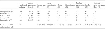
Pooling data from literature, we calculate a mean success rate of 96%, with 95% confidence intervals from 88 to 100% (Table 1). In our own series32 of 30 patients with muscular defects, the procedure was successfully performed in all, confirming the encouraging results cited in the literature.21,27–32 Complications in closure are reported in 2.8%, with 95% confidence interval from 0 to 8.5%. Holzer and coworkers30 reported the results of a multicentric trial involving 14 tertiary referral centres in the United States of America. In all, 75 patients were treated, with a total of 59 (45%) adverse events, with 8 out of 75 (10.7%) major procedure related complications, including embolization of the device, cardiac perforation, stroke, and 2 deaths (2.7%). Many of these problems are likely to be related to the learning curve, or lack of experience by the operator.30 In our experience,32 we had a very low incidence of complications, We suffered 2 transient disturbances of rhythm, and 1 embolization of the device followed by transcatheter retrieval and successful implantation of a second device. We had no deaths, strokes, or other complications, neither immediately nor during follow-up. In other studies from the literature, the rate of complications is very low (Table 1).
Table 1. Literature of percutaneous closure of muscular ventricular septal defect.

Perimembranous defects (Table 2).
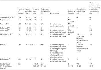
Table 2. Data from literature of perimembranous ventricular septal defect percutaneous closure.

It is only very recently that closure of perimembranous defects has become commonplace. At the beginning of our experience, we used the muscular occluder in 10 selected patients, having at least 5 millimetres distance between the superior rim of the defect and the aortic valve. The device was successfully deployed by using a retrograde approach in all. Similar good results with the use of a muscular device for properly selected patients with perimembranous defects were reported by Arora et al.37 When the membranous occluder became available, indications were expanded also to patients having only 1 to 2 millimetres distance between the defect and the aortic valve.33,34
Pooling data from the literature, we calculate the mean rate of successful closure at 96.3%, with 95% confidence interval from 93 to 99%. In our series,32 among the 84 patients who had the device successfully implanted, complete closure was 97% at 6 months, as in previous reports.33–35, 37–40
The most common morphological variation is the presence of an aneurysm of the ventricular septum. We found this feature in one-third of our patients. Usually, we tried to close the true anatomical hole with the more appropriate device, as judged from case to case. Sometimes, when the redundant tissue of the aneurysm was relatively small, the device could cover the hole along with the aneurysm. In cases of very large aneurysms, the device was implanted within the aneurysm itself, with the aim of closing the true anatomical hole, and not to place the device at the “entrance” on the left ventricular side, in order to avoid insertion of a dangerously oversized device. Studies reported in the literature show that major acute complications occur in 2.2% of cases, with 95% confidence interval from 0–4 to 6%.
The most important complications are embolization of the device, haemolysis, aortic regurgitation, and disturbances of conduction. In our series, embolization occurred in 2 cases, but we could retrieve the device and successfully implant a second device in both. Transient haemolysis occurred in 2 out of 35 cases in the phase one trial carried out in the United States of America,38 and in 2 out of 84 of our patients.32 Trivial aortic and tricuspid regurgitation related to insertion of the device occurred in only 3 cases in our series. Complete heart block was the most important complication we encountered. It occurred acutely, albeit transiently, during the procedure in 1 patient, within 48 hours after the procedure in 2, one of these being permanent, 4 months afterwards in permanent fashion in another, and then after more than 6 months, but again permanently, in a final patient, Implantation of a pacemaker was required in 3 of or 84 patients (3.5%). No instances of complete heart block were reported by Thanoupoulos et al.35 in 10 children, by Hijazi33 in 6 cases, or by Bass et al.34 in 25 cases. Complete atrioventricular block, however, was reported in 2 out of 35 patients (5.7%) by Fu and associates,38 in 2 out of 100 subjects by Holzer et al.,40 and in 1 out of 12 patients (8%) during catheter manipulation leading to abandonment of the procedure by Pedra et al.39 Also in our experience, the procedure was aborted in 2 patients after the occurrence of transient complete atrioventricular block during catheter manoeuvres. From data published in the literature, we calculate the incidence of complete atrioventricular block needing implantation of a pacemaker to be 1.7%, with 95% confidence intervals from 0–2 to 6%.
The occurrence of complete heart block has to be regarded as the major issue in percutaneous closure of perimembranous defects. The proximity of the conduction tissues to the rims of the perimembranous defect explains how, sometimes, a simple catheter or wire manipulation across the defect may cause heart block. If heart block occurs after placement of the device, it is likely that that expansion of the device against the conducting tissue plays a major role. The use of oversized devices, therefore, should be avoided. Complete heart block may also occur, however, when the discs appear nicely flat on both sides. In our patients, an oversized device could have been possibly used in 1, but certainly not in the 2 others. Other mechanisms can be considered. The device may give rise to an inflammatory reaction or formation of scarring in the conduction tissue. Steroid therapy may be useful, as was observed previously in some cases of complete atrioventricular block occurring subsequent to surgery. As yet, there is no data relating to the mechanisms of complete atrioventricular block occurring after percutaneous closure of perimembranous defects. Large studies are needed to clarify the real impact of arrhythmic problems in these patients, and the mechanism of these events.
Acquired defects
Post-infarction defects.
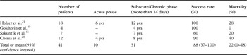
A few studies have reported experiences using Amplatzer devices in these subjects25, 26, 29, 41, 42 (Table 3).
Table 3. Post myocardial infarction ventricular septal defect transcatheter closure. Data from literautre

These papers from the literature25, 29, 41, 42 show that, in the acute phase, morbidity and mortality are quite high, and rate of successful closure is reduced. In the chronic phase, in contrast, that is 14 days after myocardial infarction, results are very encouraging, with low morbidity, mortality and a high rate of complete closure. Pooling all data from the literature, we calculate the mean success rate at 88%, with 95% confidence intervals from 57 to 100%, with a mortality of 22%, and 95% confidence intervals from 0 to 48%.
Residual post-operative defects.
Transcatheter closure is an appealing option in these subjects. In our study,32 we reported a series of 16 subjects in whom a successful and complete closure was obtained. Mean hospital stay was 2.5 plus or minus 1 days. At a mean follow-up of 12 plus or minus 3 months, all subjects but one had a stable result. In one subject, the patch dehisced again, and the patient was finally sent for repeated surgical closure.
Place in clinical management
We think that, at present, percutaneous closure can be regarded as a valuable alternative to surgery for treatment of muscular defects in subjects whose weight is more than 6 kilograms. Concerning perimembranous defects, percutaneous closure can be considered a real alternative to surgery in patients weighting more than 8 kilograms. For patients with reduced weight, or with other anatomical defects, a surgical approach is needed. It is important to emphasise in closing that children with ventricular septal defects are usually symptomatic early in life, when a percutaneous approach is not feasible. Hence, surgical closure remains the first option in a large majority of subjects. Concerning patients with residual post-surgical defects, and those with chronic post-infarction defects, the percutaneous approach can be considered the first option. In cases with acute post-infarction defects, the choice of method of closure is highly debated. It depends on multiple factors, including the experience of the centre in surgical closure. In terms of what we have reported about percutaneous closure, this can be considered as routine in only a few experienced centres among the tertiary centres of paediatric cardiology. Experience in small numbers of patients has to be discouraged.
Future outlook
In the future, efforts have to be made to reduce the dimensions of devices in order to apply this technology to even smaller babies, and to reduce the size of the introducers used. Eccentric devices for closing perimembranous defects have provided excellent rates of closure, but the occurrence of atrioventricular block remains a concern. Should it be possible to make somewhat “softer” devices, this could be advantageous in reducing traumatic injury to the conduction tissues. Post-infarction defects represent another field where innovation is needed. The largest Amplatzer device for closure of such defects is 24 millimetres, and this is sometimes not enough to close the holes. Further-more, the Amplatzer device is probably too traumatic, stiff and heavy for necrotized and ischaemic tissues. Another field where, in the future, we will see a great development is the so called hybrid approach. In hybrid procedures, surgery and transcatheter approaches are used together in order to reduce or eliminate bypass time.
There are already small series reporting the hybrid approach performed in the operating theatre. Amin et al.42 and Bacha et al.43 have proposed the so called perventricular approach. This method of closure can be done in small infants. After the chest and the pericardium are opened, and under transoesophageal echocardiographic control, a needle is used to puncture the right ventricular free wall. A short guidewire is then passed through the needle and the defect. Over the wire, a short sheath is advanced to the left ventricular cavity. Finally, an Amplatzer muscular device of appropriate size is delivered in the usual way. Bacha et al.43 have reported a series of 12 infants in whom this approach has been used successfully. Operating rooms with catheterization facilities have to be implemented, and materials and devices have to be modified in a proper way in order to deal with this different approach. In the future, resorbable devices will probably be developed in order to minimize the presence of foreign materials within the body.
Conclusions
Nowadays, and in experienced hands, percutaneous closure is a safe and effective procedure. In selected patients, percutaneous closure of muscular and perimembranous defects can be considered a real alternative to the standard surgical approach, with the advantage of a significantly reduced rate of complications.