Introduction
Artificial insemination (AI) using frozen–thawed bull semen is a commonly used technique for the reproduction of dairy and beef cattle. It has been reported that improvement in frozen–thawed semen quality, such as motility, malformation, and concentration of spermatozoa in semen, was positively correlated with pregnancy rate (Brito et al., Reference Brito, Silva, Rodrigues, Vieira, Deragon and Kastelic2002). Semen collected from bulls is diluted, cooled, and frozen for long-term storage until insemination into the female genital tract. All processing steps of semen cryopreservation may induce damage to the plasma membrane and cellular structure of spermatozoa (Hammerstedt et al., Reference Hammerstedt, Graham and Nolan1990; Watson, Reference Watson2000; Silva & Gadella, Reference Silva and Gadella2006). The evaluation of spermatozoon characteristics by laboratory assay, therefore, is a very important step in achieving a high pregnancy rate by AI using frozen–thawed semen. There have been several reports on criteria for evaluation of various spermatozoon characteristics: motility, viability, morphological abnormality, and organelle functions (Linford et al., Reference Linford, Glover, Bishop and Stewart1976; Söderquist et al., Reference Söderquist, Janson, Larsson and Einarsson1991; Den Daas et al., Reference Den Daas, De Jong, Lansbergen and Van Wagtendonk-De Leeuw1998; Thomas et al., Reference Thomas, Garner, DeJarnette and Marshall1998). Most evaluation methods in these reports were subjective, however, because they were achieved by microscopic observation, which may vary depending on the observer. Objective and quantitative methods, therefore, should be used for evaluation of spermatozoon characteristics. It has been stated that results from any single laboratory assay will not effectively estimate the fertilizing potential of a semen sample (Graham & Mocé, Reference Graham and Mocé2005). Combined multiple assays are necessary, therefore, to estimate the characteristics of spermatozoa more accurately.
In the last few years, flow cytometry has been used as an objective tool for evaluating multiple characteristics of a large number of spermatozoa (Vincent et al., Reference Vincent, Underwood, Dolbec, Bouchard, Kroetsch and Blondin2012). Nagy et al. (Reference Nagy, Jansen, Topper and Gadella2003) demonstrated that triple staining by SYBR-14, propidium iodide (PI), and phycoerythrin-conjugated peanut agglutinin (PE–PNA) was an effective method for the simultaneous evaluation of viability and acrosomal integrity of bovine spermatozoa. In addition, Thomas et al. (Reference Thomas, Garner, DeJarnette and Marshall1998) showed that the mitochondrial membrane potential of spermatozoa could be assessed by flow cytometry using JC-1 as a probe. If these two methods could be combined, we would be able to evaluate three variables (viability, acrosomal integrity and mitochondrial membrane potential) of spermatozoa simultaneously and therefore possibly obtain more detailed information about each spermatozoon. A combination of these reagents cannot be used for flow cytometry, however, because these stains use the same excitation band (488 nm) and the broad-emission spectral properties of JC-1 (green, 510–520 nm, and red-orange, 590 nm) overlap with SYBR-14 (517 nm). There is also difficulty in distinguishing JC-1 from PI (617 nm) and PE–PNA (580 nm) by flow cytometry. Celeghini et al. (Reference Celeghini, de Arruda, de Andrade, Nascimento and Raphael2007) reported the simultaneous evaluation of viability, acrosome integrity, and mitochondrial membrane potential using fluorescence microscopy. However, their method cannot be applied to flow cytometry because they used PI and JC-1. Hallap et al. (Reference Hallap, Nagy, Jaakma, Johannisson and Rodriguez-Martinez2005) reported that spermatozoa having a high mitochondrial membrane potential (double staining with SYBR-14 and MitoTracker Deep Red (MTDR)) showed high motility. The excitation laser band for MTDR (640 nm) is different from that of SYBR-14, PI, and PE–PNA (488 nm) and MTDR is known as a highly specific probe for mitochondria (Martínez-Pastor et al., Reference Martínez-Pastor, Mata-Campuzano, Álvarez-Rodríguez, Alvarez, Anel and De Paz2010) MTDR is, therefore, a suitable candidate for evaluating mitochondrial membrane potential simultaneously with the triple staining method mentioned above.
In the present study, we aimed to develop an objective simultaneous evaluation procedure for plasma membrane integrity, acrosomal integrity, and mitochondrial membrane potential of spermatozoa using flow cytometry after staining with SYBR-14, PI, PE–PNA, and MTDR.
Materials and Methods
Semen
Frozen semen, previously diluted with egg yolk-Tris-glycerol (6%) extender and packed in 0.5-ml straws, was derived from the same ejaculates of five Holstein bulls donated from Genetics Hokkaido Association (Sapporo, Japan), were used for this study. The semen was thawed at 37°C for 45 s in water and expelled into a 1.5-ml tube. The thawed semen was used for different staining, as follows. Dead spermatozoa used in Experiment 1 were prepared by thawing at 37°C in water and refreezing in liquid nitrogen twice.
Double staining for evaluation of mitochondrial membrane potential
Staining solution was prepared as described in a previous study (Hallap et al., Reference Hallap, Nagy, Jaakma, Johannisson and Rodriguez-Martinez2005). In brief, 100 μl of MTDR (final concentration 10 nM; M22426, Life Technologies, Carlsbad, CA, USA), 1 μl of SYBR-14 (final concentration 100 μM; L-7011 LIVE/DEAD Sperm Viability Kit, Molecular Probes, Eugene, OR, USA) and 800 μl of Dulbecco's phosphate-buffered saline without calcium and magnesium (DPBS) were mixed (staining solution). Then, 100 μl of semen was mixed with staining solution and warmed at 37°C for 10 min in the dark.
Triple staining for evaluation of viability and acrosomal integrity of spermatozoa
Staining solution was prepared as described in a previous study (Nagy et al., Reference Nagy, Jansen, Topper and Gadella2003). Briefly, 1 μl of SYBR-14, 2.5 μl of PE–PNA (final concentration 2.5 μg/ml; GTX01509, GeneTex, Irvine, CA, USA), 5 μl of PI (final concentration 12 μM; L-7011, LIVE/DEAD Sperm Viability Kit, Molecular Probes), and 900 μl of DPBS were mixed (staining solution). Then, 100 μl of semen was mixed with staining solution and warmed at 37°C for 10 min in the dark.
Quadruple staining for simultaneous evaluation of viability, acrosomal integrity, and mitochondrial membrane potential of spermatozoa
The same volume and types of fluorescent dye as in the triple staining along with 100 μl of MTDR solution were added to 800 μl of DPBS (staining solution). Then, 100 μl of semen was mixed with the staining solution and warmed at 37°C for 10 min in the dark.
Analysis by flow cytometry
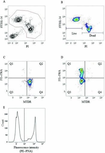
After staining, 10 μl of 10% (v/v) formaldehyde (final concentration 0.1%) was added to all samples (1000 μl) to immobilize the living spermatozoa in the staining solution, as described in a previous study (Harrison & Vickers, Reference Harrison and Vickers1990). Subsequently, 100 μl of stained sample was mixed with 400 μl of DPBS and subjected to flow cytometry. Sperm suspensions were run through a flow cytometer (FACS Verse™, BD Biosciences, San Jose, CA, USA). SYBR-14, PI, and PE–PNA were excited using a 488-nm excitation laser and detected by an FITC filter (527/32 nm), PE filter (586/42 nm), and Per-CP-Cy5.5 filter (700/54 nm), respectively. MTDR was excited at 640 nm and detected by an APC filter (660/10 nm). Flow cytometric gating of spermatozoa was performed as reported by Hallap et al. (Reference Hallap, Nagy, Jaakma, Johannisson and Rodriguez-Martinez2005) and Nagy et al. (Reference Nagy, Jansen, Topper and Gadella2003). The gating of quadruple staining was performed as described in Fig. 1. Briefly, particles stained with SYBR-14 or PI were judged as spermatozoa (Fig. 1 A). Spermatozoa were divided into two groups (live and dead) by PI emission (Fig. 1 B) and then each group was gated by PE–PNA and MTDR (Fig. 1 C, D). Fluorescence data of all events were collected and 10,000 gated events were counted. Triplicate measurements per sample were conducted and the average was used as a value of the sample.
Gating procedure and judgement for quadruple staining analysis by flow cytometry. Items stained with SYBR-14 and propidium iodide (PI) were distinguished as spermatozoa and gated from all events (area in the line; A). Gated spermatozoa were divided into live and dead clusters (B) followed by classification into four groups (Q1–4) by acrosome integrity and mitochondrial membrane potential in live (C) and dead (D) spermatozoa. Q1: damaged acrosome with low mitochondrial membrane potential, Q2: damaged acrosome with high mitochondrial membrane potential, Q3: intact acrosome with low mitochondrial membrane potential, and Q4: intact acrosome with high mitochondrial membrane potential. PE–PNA: phycoerythrin-conjugated peanut agglutinin, MTDR: MitoTracker Deep Red. The judgement of each spermatozoon characteristic by flow cytometry depended on fluorescence intensity. Spermatozoa with low fluorescence intensity (<103) was judged as PE–PNA negative (intact acrosome) and spermatozoa with high fluorescence intensity (≥103) was judged as PE–PNA positive (damaged acrosome) (E).

Analysis by fluorescence microscopy
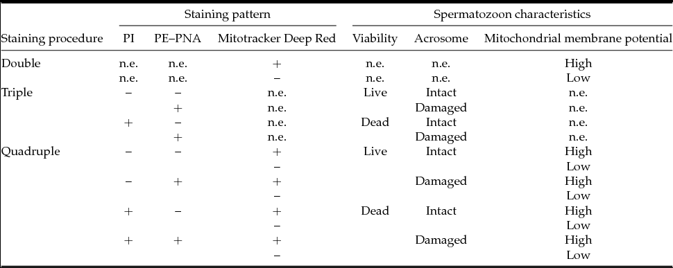
After staining and immobilization, an 8-μl sample was loaded onto a slide, coverslipped, and evaluated immediately under a fluorescence microscope (ECLIPSE Ci, Nikon, Tokyo, Japan) equipped with a B-2A filter (excitation 450–490 nm and emission >520 nm) and a G2-A filter (excitation 510–560 nm and emission >590 nm) at ×400 magnification. Microscopic examination was mainly conducted by using a B-2A filter. A G2-A filter was used for the evaluation of plasma membrane integrity when PI emission was not clear. Two hundred spermatozoa per slide were examined and classified based on the fluorescence emitted from each probe (Table 1). Three slides per sample were examined and the average was used as the value of the sample.
Staining patterns and evaluation of spermatozoon characteristics by each staining method

+, fluorescence positive; –, fluorescence negative; n.e., not evaluated.
Evaluation of spermatozoon characteristics
Spermatozoon characteristics estimated by flow cytometry are described in Table 1. Briefly, spermatozoa stained with PI are categorized as dead as plasma membrane damage allows PI to penetrate the cells. When the acrosome was stained with PE–PNA, it was classed as damaged. When the spermatozoon midpiece was stained with MTDR, the spermatozoa was classified as having a high mitochondrial membrane potential.
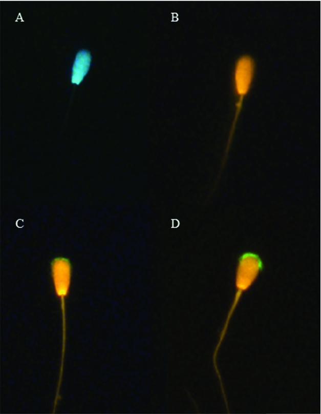
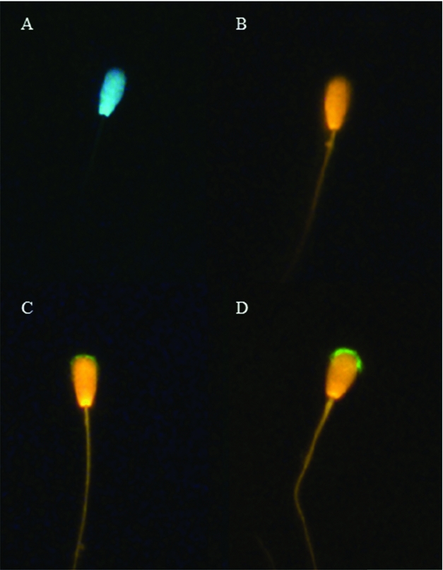
Spermatozoon characteristics evaluated by fluorescence microscopy are expressed in Fig. 2. Spermatozoa stained with PI were evaluated as dead in the same way as by flow cytometry. When the acrosomal region of the spermatozoon was stained with PE–PNA, it was classified as damaged. Some spermatozoa stained with PE–PNA intermediately (Fig. 2 C), they were also judged as being spermatozoon with a damaged acrosome.
The photographs of spermatozoon triple staining taken by a fluorescence microscopy. The head of spermatozoon stained with SYBR-14 and acrosomal region not stained with PE–PNA (A) were judged as a live spermatozoon with an intact acrosome. The head of spermatozoon stained with PI but acrosomal region not stained with PE–PNA (B) was judged as dead spermatozoon with an intact acrosome. The heads of spermatozoon stained with PI and acrosomal region stained intermediately (C) and completely with PE–PNA (D) were both judged as a dead spermatozoon with a damaged acrosome.
Experimental design
In Experiment 1, frozen–thawed semen was mixed with 0, 25, 50, 75 or 100% dead spermatozoa. These samples were subjected to triple staining. After staining, half of the sample was evaluated by flow cytometry and the other half by fluorescence microscopy; the obtained results were then compared. Semen derived from five bulls was used for this experiment.
In Experiment 2, frozen–thawed semen derived from a bull was subjected to double, triple, or quadruple staining. Then, the results of the spermatozoon characteristics estimated by quadruple staining were compared with those of double or triple staining samples. The experiment was repeated four times on independent samples.
Statistical analysis
Statistical analysis was performed using JMP 9.0.2 software (SAS, NC, USA). The correlation between each characteristic of spermatozoa estimated by flow cytometry and by fluorescence microscopy was analyzed by linear regression analysis. The percentages of spermatozoon characteristics examined using different equipment and different staining procedures were compared by Student's t-test. Differences with a P-value <0.05 were recognized as significant.
Results
Experiment 1
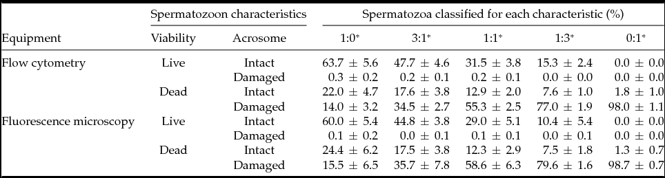
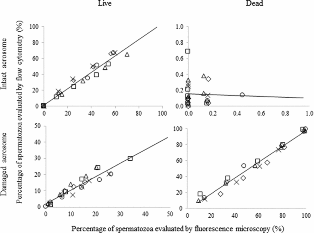
The viability and acrosomal integrity of spermatozoa evaluated by flow cytometry and fluorescence microscopy were significantly correlated (r > 0.9, P < 0.01), except for the live spermatozoa with a damaged acrosome (P = 0.866), as shown in Fig. 3. The percentages of each characteristic evaluated by flow cytometry and fluorescence microscopy are shown in Table 2. There were no significant differences in all characteristics (live spermatozoa with an intact acrosome, live spermatozoa with a damaged acrosome, dead spermatozoa with an intact acrosome, and dead spermatozoa with a damaged acrosome) evaluated by the two types of equipment (P > 0.05). The percentages of live spermatozoa with a damaged acrosome were low among the samples with different mixed ratios of dead spermatozoa.
Characteristics of bovine spermatozoa, the mixture of thawed semen and dead spermatozoa, evaluated by flow cytometry and fluorescence microscopy after triple staining

Values are mean ± standard deviation (five bulls/group).
*Mixed ratio of frozen–thawed semen and dead spermatozoa.
Scatter plots and regression lines of percentages of spermatozoa evaluated by flow cytometry and fluorescence microscopy. Semen from five bulls was used and data from the same bull are indicated by the same symbol.

Experiment 2
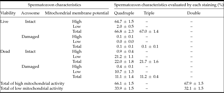
The viability, acrosomal status, and mitochondrial membrane potential of spermatozoa evaluated by flow cytometry after quadruple staining and by the other staining procedures are shown in Table 3. There were no significant differences in all characteristics evaluated by quadruple staining and the other procedures (P > 0.05). By quadruple staining, more than 95% of the live spermatozoa having an intact acrosome showed high mitochondrial membrane potential. In addition, more than 95% of dead spermatozoa having an intact acrosome showed low mitochondrial membrane potential.
Spermatozoon characteristics evaluated by flow cytometry using different staining procedures

Values are mean ± standard deviation (four replicates).
Discussion
In the present study, the results of spermatozoon characteristics evaluated by flow cytometry and fluorescence microscopy were similar, except for live spermatozoa with a damaged acrosome. High correlation may be due to the criteria of spermatozoa evaluation. The fluorescence intensity value obtained by flow cytometry indicated two obvious peaks, those evaluated as negative and positive. The PE–PNA positive peak, however, had a broad base towards the low intensity (103–104) as shown in Fig. 1 E, and this small peak might be a subpopulation of those spermatozoa observed as intermediately stained and judged as positive under fluorescence microscopy. Evaluation of spermatozoa using two types of equipment was therefore able to access spermatozoa characteristics by the same criteria and provide similar results. The exception may have been caused by the small population size (<1%) of live spermatozoa with a damaged acrosome in semen. The number of spermatozoa evaluated by flow cytometry was about 50 times greater than that by fluorescence microscopy, for which we examined about 200 spermatozoa (Celeghini et al., Reference Celeghini, de Arruda, de Andrade, Nascimento and Raphael2007; Somfai et al., Reference Somfai, Bodó, Nagy, Papp, Ivancsics, Baranyai, Gocza and Kovacs2002). Although we evaluated a large number of spermatozoa, quite a small population of this type of spermatozoa may indicate that spermatozoa die immediately after damage to the acrosome.
In the present study, the viability, acrosomal integrity, and mitochondrial membrane potential of spermatozoa could be evaluated accurately by quadruple staining without interference between fluorescent dyes. The present results mean that most spermatozoa with damage to the plasma membrane had an impaired mitochondrial membrane potential, even though about two-third of them had an intact acrosome. Mitochondria produce ATP, which is required for housekeeping of the plasma membrane of spermatozoa (Silva & Gadella, Reference Silva and Gadella2006). In the present study, most live spermatozoa had a high mitochondrial membrane potential, while the dead spermatozoa showed a low one. Low mitochondrial membrane potential indicates a lack or decrease in ATP production, which may become a cause of spermatozoon death without acrosomal damage. Mitochondrial activity is crucial and correlates with the fertilization ability of spermatozoa (Amaral et al., Reference Amaral, Lourenço, Marques and Ramalho-Santos2013). In further studies, the relationship between fertility and spermatozoon characteristics as evaluated by flow cytometry using quadruple staining should be performed.
A staining method to estimate the viability of spermatozoa, acrosomal integrity, and mitochondrial functions simultaneously under a fluorescence microscope using four fluorescent dyes (Hoechst 33342, PI, FITC-PSA, and JC-1) has been reported previously (Celeghini et al., Reference Celeghini, de Arruda, de Andrade, Nascimento and Raphael2007). In this previous study (Celeghini et al., Reference Celeghini, de Arruda, de Andrade, Nascimento and Raphael2007), only fewer numbers, hundreds of spermatozoa, could be evaluated subjectively. Conversely, the method developed in the present study enables the objective estimation of more than 10,000 sperm by flow cytometry and in a short time. This result means that the characteristics of spermatozoa can be evaluated more accurately and faster by our procedure.
In conclusion, quadruple staining using SYBR-14, PI, PE–PNA, and MTDR for flow cytometry can evaluate the plasma membrane integrity, acrosomal integrity, and mitochondrial membrane potential of bovine spermatozoa simultaneously. The procedure can be applied to the quality control of bovine frozen–thawed semen.
Acknowledgements
This study was supported by a Grant-in-Aid for Scientific Research from the Japan Society for the Promotion of Science to M. Nagano (No. 25450441).