Introduction
In the 1950s scientists at Hoffmann-La-Roche (Switzerland) discovered that some of the canthaxanthin (CX) added to chicken feed was deposited in the egg yolk (Paust, Reference PAUST1991). This was the beginning of CX research which brought the egg yolk pigmentation issue to a new level, and created much interest in the egg industry as well as in breeder nutrition. In laying hens, carotenoid deposition in egg yolk varies widely with CX being deposited at 30 to 45% efficiency, zeaxanthin 25% and astaxanthin 14% (Hencken, Reference HENCKEN1992; Schiedt, Reference SCHIEDT1998) and carotenoid excretion being about 70% for astaxanthin and zeaxanthin and 45 to 50% for CX. Grashorn and Steinberg (Reference GRASHORN and STEINBERG2002) showed that 1 mg CX per kg diet resulted in 2.5 mg CX per kg egg yolk. Forty percent of the deposition rate of CX was independent of dietary supplementation level. It is interesting that, in laying hens, 50% of the total body zeaxanthin and 80% of the CX are located in the ovary (Nys, Reference NYS2000). During the rapid growth of the ova at the onset of lay, xanthophylls are deposited in the egg yolk in a dose-dependent manner (Hinton et al., Reference HINTON, FRY and HARMS1974). Increased egg pigmentation due to CX dietary supplementation was observed by Pinchasov et al. (Reference PINCHASOV, DAVID and ZOHARI1992). Commercial eggs in Germany contain about 206 µg CX/egg (Grashorn, Reference GRASHORN2005). Inclusion of very high doses of CX (0.04%) into the breeder diet was associated with extremely high CX concentration (426.1 µg/g) in the egg yolk (Haq et al., Reference HAQ, BAILEY and CHINNAH1996). CX transfer to the yolk from the diet was also reported by Galobart et al., Reference GALOBART, BARROETA, BAUCELLS and GUARDIOLA2001). A good nutritional status of the parent birds is crucial to the transfer to the egg of an adequate, balanced supply of nutrients required for normal development of the embryo (Wilson, Reference WILSON1997). In this respect it is important that CX was effectively transferred from the egg yolk to the developing embryo. As a result, CX concentration in the liver, yolk sac membrane and plasma at d 16 of embryonic development, in 1-d-old and 7-d-old chicks was significantly increased in proportion to its concentration in the egg yolk (Surai et al., Reference SURAI, SURAI, STEINBERG W., WAKEMAN, SPEAKE and SPARKS2003). Similar observations were previously reported, with CX being a part of a tested carotenoid mixture (Surai and Speake, Reference SURAI and SPEAKE1998) as well as with gull embryos (Surai et al., 2001).
It is important to note that, after hatching, the CX that accumulated in the chicken's body during embryonic development is depleted. Even so at d 7 posthatch there was an elevated CX concentration in the liver and plasma of chicks hatched from eggs enriched with this carotenoid. These results show the importance of the maternal diet on the body composition of the chick during early postnatal development. Similar results have been obtained with other carotenoids fed to the breeding birds (Karadas et al., Reference KARADAS, PAPPAS, SURAI and SPEAKE2005). Previously it was shown that CX was detectable in the plasma and liver of birds hatched from CX-enriched eggs, while there was no CX in plasma or liver of the control birds (Haq et al., Reference HAQ, BAILEY and CHINNAH1995).
In the wild, CX is incorporated into gull egg yolk (Royle et al., Reference ROYLE, SURAI, MCCARTNEY and SPEAKE1999; Reference ROYLE, SURAI and HARTLEY2001; Surai et al., 2001; Surai, Reference SURAI2002), and dietary CX supplementation of gulls increased deposition of this carotenoid in their eggs (Blount et al., Reference BLOUNT, SURAI, NAGER, HOUSTON, MOLLER, TREWBY and KENNEDY2002a). In fact, CX was found in the egg yolk of wild gulls at concentrations of about 5 µg/g and was increased almost five fold after CX supplementation (Blount et al., 2002b). In the tissues of adult gulls in the wild, CX concentration was reported to be 1.53; 0.59; 0.58; 0.38; 0.23; 0.16 and 0.11 µg/g in the liver, fat, kidney, breast muscle, heart, lung and pancreas respectively (Surai et al., Reference SURAI, ROYLE and SPARKS2000), while in newly hatched gulls CX concentrations in the liver, heart and leg muscle were 21.2; 5.7 and 4.7 µg/g respectively (Surai et al., 2001). CX was also found to be one of the most common carotenoids in the liver, plasma and gut content of wild mallard (Anas platyrhynchos) ducklings (Butler and McGraw, Reference BUTLER and MCGRAW2010). CX has been determined as a colouring agent of beaks and legs of zebra finches (McGraw and Toomey, Reference MCGRAW and TOOMEY2010) and plumages of yellow-crowned bishop and southern red bishop (Prager et al., Reference PRAGER, JOHANSSON and ANDERSSON2009).
Antioxidant defences of the chicken egg and possible roles of canthaxanthin
The chicken egg consists of yolk, white (albumen) and shell (Figure 1). Practically all lipids are located in the egg yolk, where a range of fat soluble antioxidants are located (Surai, Reference SURAI2002). Water soluble compounds (e.g. vitamin C) are not accumulated in the egg yolk and antioxidant protection of the this area is afforded mainly by vitamin E and carotenoids. Other compounds possessing antioxidant activities (e.g. coenzyme Q, glutathione, etc.) present in the egg are only in very low concentrations and therefore play a minor role. It has been shown that increased vitamin E (Surai, Reference SURAI1999) or carotenoid concentrations (Surai, Reference SURAI2002) in the egg yolk are associated with increased resistance to lipid peroxidation. However, long chain polyunsaturated fatty acids in the layer diet could substantially change the fatty acid profile of the egg yolk and have detrimental effects on antioxidant defences (Cherian and Sim, Reference CHERIAN and SIM1997; Pappas et al., Reference PAPPAS, ACAMOVIC, SPARKS, SURAI and MCDEVITT2005) thereby increasing embryonic mortality (Pappas et al., Reference PAPPAS, ACAMOVIC, SPARKS, SURAI and MCDEVITT2006). During egg incubation vitamin E and carotenoids are effectively transferred from the egg yolk to the developing embryo and maximum concentrations of these compounds in the liver of newly hatched chicks are considered to be an adaptive mechanism to protect tissues from the oxidative stress of hatching (Surai, Reference SURAI1999; Reference SURAI2002; Surai et al., Reference SURAI, NOBLE and SPEAKE1996). It is interesting to note a positive correlation between carotenoid and vitamin E concentration in the egg yolk of wild passerine birds (Hargitai et al., Reference HARGITAI, MATUS, HEGYI, MICHL, TÓTH and TÖRÖK2006). Furthermore, there is a strong correlation between various carotenoids at both the inter- and intra-specific levels in wild bids (Cohen and McGraw, Reference COHEN and MCGRAW2009).
Antioxidant system in the egg.

During embryonic development other antioxidant compounds are synthesized in various tissues. These include glutathione, ascorbic and uric acids, as well as antioxidant enzymes (Figure 1) such as glutathione peroxidase (GSH), catalase (CA) and superoxide dismutase (SOD) (Surai, Reference SURAI2006). There is tissue specificity associated with the synthesis of such antioxidants and, in combination with vitamin E and carotenoids, this contributes to an integrated antioxidant system in the developing embryo and postnatal chicken (Surai, Reference SURAI2006). This system is responsible for the protection of polyunsaturated fatty acids in biological membranes from damaging effects of free radicals and toxic products of their metabolism.
Since CX possesses antioxidant properties in vitro and in vivo (Surai, Reference SURAI2002) and is effectively transferred from the diet to the egg yolk and further to the developing embryo, it can be considered as an effective modulator of the antioxidant system of the egg and the embryo.
The role of natural antioxidants within the maternal diet in maintaining egg quality and chicken hatch should not be underestimated (Rocha et al., Reference ROCHA, LARA, BAIAO, VASCONCELOS, BARBOSA, POMPEU and FERNANDES2010). In recent years this topic received much attention from researchers in both evolutionary and population biology. It has been suggested that there are different strategies in the allocation of natural antioxidants, in particular carotenoids, in males and females. Male birds allocate carotenoids to their plumage to indicate their health status (Blount et al., Reference BLOUNT, METCALFE, BIRKHEAD and SURAI2003) making them more successful in attracting a mate, whereas females invest carotenoids mainly into the egg to build up the antioxidant defences of the developing embryo (Surai, Reference SURAI2002; Monaghan et al., Reference MONAGHAN, METCALFE and TORRES2009). Indeed, maternal effects mediated by egg quality are important sources of offspring phenotypic variation, and can influence the course of evolutionary processes. Mothers allocate a diverse range of antioxidants to the egg that protect the embryo from oxidative stress. Furthermore, the immunomodulating properties of carotenoids may be of importance in progeny (Constantini and Moller, 2008). Analysis of yolk samples from clutches of wild birds, that were subsequently partially cross-fostered, revealed a positive effect of yolk antioxidant capacity on embryonic development and chick growth (Rubolini et al., Reference RUBOLINI, ROMANO, BONISOLI ALQUATI and SAINO2006). Thus, egg antioxidants are important mediators of maternal effects in wild bird populations, especially during the critical early post-hatching phase. A comparative analysis of lipid-soluble antioxidants (carotenoids and vitamins A and E), which was conducted in the eggs of 112 species of bird has shown that species with larger clutch sizes deposited higher yolk concentrations of the three antioxidants (Biard et al., Reference BIARD, GIL, KARADAŞ, SAINO, SPOTTISWOODE, SURAI and MØLLER2009).
A further area of interest is the effect of maternal nutrition, including natural antioxidants, on gene expression in progeny. Recently the effect of breeder diet fortified with vitamins and minerals on gene expression in the intestine of progeny was investigated (Rebel et al., Reference REBEL, VAN HEMERT, HOEKMAN, BALK, STOCKHOFE-ZURWIEDEN, BAKKER and SMITS2006). Unfortunately the authors did not report the egg composition before and after dietary fortification. From the data reported, it can be expected that such fortified diets leads to substantially increased levels of vitamin E in the egg and slightly increased levels of vitamins A, D, B1 and B2 as well as selenium. Gene expression patterns in the intestine were measured at 3 d and 14 d of age using intestinal cDNA-microarray methodology. Between the two groups, 11 genes were found to be differently expressed both at 3 d and 14 d of age. Genes that are differently expressed will affect intestinal cell turnover, proliferation and development, as well as metabolism and feed absorption. Taking into account the results presented by Koutsos et al. (Reference KOUTSOS, CALVERT and KLASING2003), which showed the maternal effect of carotenoids on their concentration in 4-week-old chickens; it is possible to suggest that various antioxidants, including carotenoids, could affect gene expression during embryonic development. This may result in better antioxidant defences related to higher hatchability and improved chick viability post-hatch. The authors showed that the concentration of carotenoids in the egg yolk had a profound effect on the immune system development post-hatch. They suggested that carotenoid depletion in the egg yolk may compromise the capacity for appropriate immune responses later in the bird's life (Koutsos et al., Reference KOUTSOS, GARCÍA LÓPEZ and KLASING2007). Although this hypothesis needs further clarification it is clear that this effect of maternal diet is seen beyond newly hatched chicks, and could be an example of maternal nutritional programming responsible for various changes in post-natal life. The prevailing nutritional environment during foetal development exerts powerful and long-lasting effects upon physiology and metabolism (Langley-Evans, Reference LANGLEY-EVANS2009). Indeed, in mammals there are several genes described which are specifically regulated by carotenoids, including CX (Zhang et al., Reference ZHANG, COONEY and BERTRAM1992; Bertram, Reference BERTRAM1999; Stahl et al., Reference STAHL, ALE-AGHA and POLIDORI2002). It is a matter of time before such genes are identified in chickens.
Testing canthaxanthin in chickens
To address some features of CX activity in breeder chickens a study was conducted at the Scottish Agricultural College (SAC) (Surai et al., Reference SURAI, SURAI, STEINBERG W., WAKEMAN, SPEAKE and SPARKS2003) using 320 female broiler breeder birds, sourced from the same rearing flock. Four treatments were compared, a control diet containing < 2 mg/kg total xanthophylls, and then test diets supplemented with either 3, 6, 12 and 24 mg/kg of a commercial CX product, (CAROPHYLL® Red 10% (Roche Vitamins (UK) Ltd, Heanor, UK). The trial data showed that inclusion of CX into the maternal diet caused a significant dose-dependent response in terms of its accumulation in the egg yolk. Egg yolk colour gradually changed from pale yellow in the control group to dark red in the laying hens fed CX at 24 mg/kg. These results are in agreement with previous observations from a similar trial using a carotenoid mixture (Surai and Speake, Reference SURAI and SPEAKE1998).
The most important finding of this study was a positive effect of CX on vitamin E in the developing chicks. First of all, CX improved assimilation of γ-tocopherol from the diet and its transfer to the egg yolk. As a result, an increased γ-tocopherol concentration was observed in the liver of the embryo at d 16 of the development. However, at later stages of the development, these differences disappeared, probably due to a tocopherol-binding protein (Surai and Speake, Reference SURAI and SPEAKE1998). Secondly, increased CX concentration in the egg yolk and embryonic tissues was associated with higher α-tocopherol concentrations in the liver and plasma of 1-d-old chicks. This is probably a reflection of antioxidant interactions during embryonic development. Indeed, if the CX that accumulated in tissues scavenged free radicals, fewer radicals would be available to react with vitamin E. This could spare some vitamin E and lead to increased availability and concentrations. There is also a possibility that CX may take part in vitamin E recycling. In this scenario vitamin E concentration would also be increased. In fact, a possibility of vitamin E recycling by ϐ-carotene has been reported earlier (Bohm et al., Reference BOHM, EDGE, LAND, McGARVEY and TRUSCOTT1997). The synergistic effect of CX on vitamin E was seen in 7-d-old chicks, when vitamin E concentrations in the liver and plasma were significantly elevated as a result of maternal diet supplementation with CX. In fact α-tocopherol concentration in the chick liver decreased more than 10-times in the first week. In such an extreme situation, increased CX concentration in the chick liver slowed down the depletion of vitamin E from the liver, which could be explained by the same mechanisms of CX/vitamin E interactions.
As a result of enrichment of the chick liver with CX and vitamin E due to the maternal dietary supplementation, tissue susceptibility to lipid peroxidation significantly decreased in 1-d-old and 7-d-old chicks. The protective effect CX against lipid peroxidation may be mediated via its effect on antioxidant enzymes (Palozza et al., Reference PALOZZA, CALVIELLO, EMILIA DE LEO, SERINI and BARTOLI2000).
The enhancement of the antioxidant system in the developing chick as a result of supplementing the maternal diet presents great opportunities for poultry producers. Postnatal development of the chicken is a crucial time for the maturation of major physiological systems, including the immune system, as well as a time of high risk of peroxidation (Surai, Reference SURAI2002). Therefore, increased supplementation of the maternal diet with carotenoids, in particular CX, could help maintain antioxidant system efficiency and increase chick viability.
The idea of exploiting the beneficial effects of dietary CX supplementation in breeder diets was further developed by Zhang et al. (Reference ZHANG, ZHANG, DING, BAI, HERNANDEZ, YAO and Q. ZHU2011). In this study, 270 Chinese Three-Yellow breeder hens were randomly divided into two groups consisting of 135 birds each (five replicates of 27). The breeder hens were fed either a basal diet or one supplemented with 6 mg of CX/ kg for 24 weeks. At the end of the experiment, all hatching eggs laid over five consecutive days from each group were collected and incubated. For each breeder group, 100 newly hatched chicks (five replicates of 20) were reared under environmentally controlled conditions for 21 d. The results from this trial showed that CX supplementation gave a significant increase in the yolk colorimetric score on the Roche Yolk Color Scale (P<0.001), confirming an effective CX transfer from the diet to the egg yolk. Egg yolk enrichment with CX was associated with a significant improvement of the antioxidant status of the egg yolk (P<0.05), whereby malondialdehyde (MDA) content of the egg yolk decreased from 139.83 nmol/g down to 86.92 nmol/g (P=0.023). At the same time total antioxidant capacity (TAC) of the egg yolk increased from 1.87 U/g up to 3.16 U/g (P<0.001). These important improvements in the antioxidant status of yolk could be due to CX transfer or improvements of vitamin E status (Surai et al., Reference SURAI, SURAI, STEINBERG W., WAKEMAN, SPEAKE and SPARKS2003). One important finding was the improvement of the serum TAC in the breeder hen (P<0.029). This could be an important element in an improvement of protection of laying birds from commercially-relevant stresses, including fluctuations in temperature and feed quality (Surai, Reference SURAI2002).
The chicks that hatched from eggs laid by breeder hens fed the CX diet demonstrated a higher pigmentation colorimetric score of their shank skin (P<0.05), ascribed to CX accumulation (Surai et al., Reference SURAI, SURAI, STEINBERG W., WAKEMAN, SPEAKE and SPARKS2003) and the antioxidant capacity of the newly hatched chicks was significantly increased (P<0.05). There was a significant decrease in MDA (from 4.28 down to 2.61 nmol/ml, P<0.001) in the plasma of day-old chicks, thought to be a result of increased CX and vitamin E concentration in plasma (Surai et al., Reference SURAI, SURAI, STEINBERG W., WAKEMAN, SPEAKE and SPARKS2003). It is of great importance to see increased SOD activity (from 98.4 to 144.7 U/ml, P=0.031) in plasma, and these findings are in agreement with previous observations indicating a stimulatory effect of CX on SOD (Palozza et al., Reference PALOZZA, CALVIELLO, EMILIA DE LEO, SERINI and BARTOLI2000). It is interesting to underline that maternal supplementation with CX has a positive effect on TAC of newly hatched chicks, which increased by 33% (from 13.8 to 18.3 U/ml, P= 0.052).
In a field trial, Robert et al. (Reference ROBERT, PANHELEUX- LE BASTARD, HAMELIN and BOULARD2008) studied the effect of CX in ROSS breeders on the anti-oxidant status of their progeny. They observed that the status of sera in was significantly higher and the TBARS level significantly lower 1-day old chicks from hens fed 6 ppm CX in the breeder feed. According to these authors, the results indicate that maternal supplementation with CX (6 ppm) enhances antioxidant capability and depresses oxidative stress in chicks. Similar to finding from previous work (Surai et al., Reference SURAI, SURAI, STEINBERG W., WAKEMAN, SPEAKE and SPARKS2003), maternal effects of CX supplementation has been seen in post-natal chicken development. Indeed shank pigmentation score (a reflection of CX accumulation) and MDA in plasma were significantly lower in 7 day-old chicks hatched from the CX-enriched eggs. The experimental chicks also showed a lower mortality (0 vs. 4%) during the first 21 d post-hatch. These findings support the hypothesis that CX supplementation of the maternal diet enhances the protective capacity of tissues against oxidative stress in vivo, which might be beneficial for poultry producers.
The next step in examining the beneficial effects of CX in the maternal diet was provided by an experiment conducted in Brazil in the Poultry Science Laboratory of the Department of Zootechnics at the Federal University of Santa Maria (UFSM); Rosa et al., Reference ROSA, SCHER, SORBARA, BOEMO, FORGIARINI and LONDERO A.2012). Three hundred and sixty female pullets and 36 roosters were placed in an open-sided house allocated into 12 pens. At 42 weeks of age, the breeder hens and roosters were distributed into two experimental groups with similar body weight and uniformity, and from weeks 46 to 66, one group received 6 ppm of CX, supplemented in the diet, and the other group received the diet without addition of CX (control diet). Those fed CX had significantly better egg laying rates during certain periods, but overall there was no difference between the two treatment groups. Two weeks after the start of the supplementation the pigmentation measured with the colour fan were 9.33 in the control group and 14.67 in the CX group (P<0.0001) and this difference in yolk coloration remained stable throughout the experiment. These results are in agreement with previous observations (Surai et al., Reference SURAI, SURAI, STEINBERG W., WAKEMAN, SPEAKE and SPARKS2003; Zhang et al., Reference ZHANG, ZHANG, DING, BAI, HERNANDEZ, YAO and Q. ZHU2011). In concurrence with previous observations, a reduction of TBARS was observed in yolks from stored hatching eggs produced by breeders fed CX. The reduction in TBARS was observed in eggs submitted for analysis on the same day they were produced (P= 0.0214) and in eggs stored for four (P<0.0002), eight (P<0.0003) and twelve days (P<0.0001).
The most important finding of this study was the positive effect of CX supplementation of the maternal diet on fertility, hatchability and embryonic mortality. In fact, CX in comparison to the control improved fertility (92.1 vs. 91.0%, P<0.02) and hatchability (93.7 vs. 91.3%, P=0.0003) and reduced embryonic mortality (3.7 vs. 5.5%, P<0.003). As a result, hatching rate was significantly (86.2 vs. 83.0%, P=0.0001) improved. As to the different stages of embryo mortality, CX was most efficacious for its prevention in the first 48 hours (1.04 vs. 1.8%, P= 0.008) as well as between day 15 and 21 of incubation (1.44 vs. 2.07, P=0.017). It can be postulated that CX supplementation improved breeder fertility (P=0.0171) by improving survival and storage of spermatozoa within the reproductive tract of the hen. Indeed, according to Breque et al. (Reference BREQUE, SURAI and BRILLARD2003), long term storage of spermatozoa is supported by a complex antioxidant defence system present within the oviduct that protects spermatozoa against lipid peroxidation. CX may play an important role in that system. As for the positive effect of CX on hatchability, this can be explained by participation in antioxidant defences during embryonic development (Surai, Reference SURAI2002). It is interesting to mention that another carotenoid with a similar structure astaxanthin (AX) fed to breeders was shown to be transferred to the egg yolk and had a significant protective effect against decreasing hatchability due to egg storage at 21ºC (Saito and Kita, Reference SAITO and KITA2011). The hatchability of eggs stored at 21ºC was significantly lower than that at 10ºC, and lowered hatchability was restored by maternal intake of AX. Elevated levels of dietary AX from 5 to 20 ppm gradually restrained the decrease in hatchability at 21ºC. And it is interesting to note an additional benefit to use CX supplementation of the breeder diet as an opportunity to identify low-producing breeders by observing high coloration of shanks (Pinchasov et al., Reference PINCHASOV, DAVID and ZOHARI1992). Indeed, there is a distribution of carotenoids between different tissues in the body and if egg production rate is decreasing more CX is available for shank coloration.
Consistent with the improved antioxidant capacity due to dietary CX addition and aforementioned results, it is possible to postulate that:
• CX added via maternal feed is efficiently deposited in egg yolk and consequently in embryo liver
• CX significantly decreased detectable MDA (TBARS) in:
• Egg yolks (on storage or up to seven days incubation)
• 16 d old liver embryos
• 1 d to 7 d old chicks
• TBARS is lower in eggs with CX stored up to 12 days
• TBARS increases after 7 days of incubation, losing statistical significance - possibly due to utilisation of CX as an antioxidant during embryo development
• Improvement in antioxidant status in eggs obtained by addition of CX via maternal feed produces a beneficial effect on embryo development resulting in healthier chicks (first quality chicks).
Сonclusions
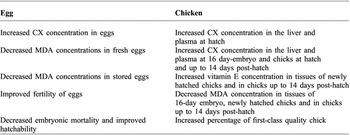
Antioxidant defences of chicken eggs are based mainly on vitamin E and carotenoids and their deposition and concentrations in the egg yolk is dependent on dietary provision. CX is well absorbed from the feed and effectively transferred to the egg yolk and further to the developing embryo. Increased CX concentration in embryonic tissues is associated with increased resistance to oxidative stress. Since oxidative stress is an important element of increased embryonic mortality during the last week of incubation it is highly likely that dietary CX could support chicken viability after hatch. Research has shown that CX supplementation of breeder diets significantly increased anti-oxidative status in the egg yolk and significantly increased hatching rate of chicken eggs. Taken together, the aforementioned results clearly indicate that CX provides a great deal of benefits for chicken eggs, embryos and chickens during early postnatal development (Table 1).
Main benefits of CX for eggs and chickens.