INTRODUCTION
The trematode Schistosoma mansoni is the major causative agent of schistosomiasis, a human disease affecting around 200 million people worldwide (Mascie-Taylor and Karim, Reference Mascie-Taylor and Karim2003). During its life-cycle, the freshwater snail Biomphalaria glabrata acts as intermediate host, being essential for asexual reproduction of the parasite. While numerous investigations have been performed on the parasite's invasion of its snail host, the molecular mechanisms of this process are still not completely understood (Loker and Bayne, Reference Loker and Bayne1986; Yoshino and Boswell, Reference Yoshino and Boswell1986; Loker et al. Reference Loker, Adema, Zhang and Kepler2004). Different invasion strategies of the parasite have been discussed (El-Ansary, Reference El-Ansary2003; El-Ansary and Al-Daihan, Reference El-Ansary and Al-Daihan2006), including molecular mimicry which implies that the parasite covers itself with host-like antigens to escape from defence mechanisms of the host (Damian, Reference Damian1989). In this context, schistosome glycoconjugates, such as glycoproteins and glycolipids, have gained increasing interest, since it is known that S. mansoni synthesizes a complex set of different, in part unique, carbohydrate structures throughout its life-cycle. It is generally accepted that these molecules play an important role in the immunology of schistosomiasis. This holds true, in particular for glycoconjugates of S. mansoni eggs which are mainly responsible for the pathological consequences of human schistosomiasis (Hokke and Deelder, Reference Hokke and Deelder2001; Pearce, Reference Pearce2005).
Previous studies have demonstrated that glycolipids and glycoproteins of schistosomes are dominated by fucosylated carbohydrate epitopes including F-LDN [Fuc(α1-3)GalNAc(β1-4)GlcNAc(β1-)], LDN-F [GalNAc(β1-4)[Fuc(α1-3)]GlcNAc(β1-)], F-LDN-F [Fuc(α1-3)GalNAc(β1-4)[Fuc(α1-3)]GlcNAc(β1-)], LDN-DF [GalNAc(β1-4)[Fuc(α1-2)Fuc(α1-3)]GlcNAc(β1-)] and Lewis X [Gal(β1-4)[Fuc(α1-3)]GlcNAc(β1-)] (van Remoortere et al. Reference Van Remoortere, Hokke, Van Dam, Van Die, Deelder and Van Den Eijnden2000; Wuhrer et al. Reference Wuhrer, Dennis, Doenhoff, Lochnit and Geyer2000b; Kantelhardt et al. Reference Kantelhardt, Wuhrer, Dennis, Doenhoff, Bickle and Geyer2002; Wuhrer et al. Reference Wuhrer, Kantelhardt, Dennis, Doenhoff, Lochnit and Geyer2002; Robijn et al. Reference Robijn, Wuhrer, Kornelis, Deelder, Geyer and Hokke2005; Wuhrer and Geyer, Reference Wuhrer, Geyer, Maule and Marks2006). Although carbohydrate-based molecular mimicry has been postulated as an invasion strategy of S. mansoni at the level of its intermediate host, B. glabrata, already some years ago (Dissous et al. Reference Dissous, Grzych and Capron1986), the relevant carbohydrate epitopes were not defined in that study. Recently, we could show, however, that B. glabrata synthesizes a broad battery of glycoprotein-N-glycans comprising at least 2 carbohydrate determinants that cross-react with glycoconjugates from S. mansoni eggs (Lehr et al. Reference Lehr, Geyer, Maass, Doenhoff and Geyer2007). Corresponding glycan epitopes included terminal Fuc(α1-3)GalNAc(β1-4)[±Fuc(α1-3)]GlcNAc(β1-) moieties, i. e., F-LDN and F-LDN-F units which have been reported to occur in both glycoproteins and glycolipids (Wuhrer et al. Reference Wuhrer, Dennis, Doenhoff and Geyer2000a, Reference Wuhrer, Dennis, Doenhoff, Lochnit and Geyerb; Wuhrer et al. Reference Wuhrer, Kantelhardt, Dennis, Doenhoff, Lochnit and Geyer2002; Robijn et al. Reference Robijn, Wuhrer, Kornelis, Deelder, Geyer and Hokke2005). In addition, N-glycans comprising a Xyl(β1-2)-substituted chitobiose core region could be identified which is in so far relevant as this type of structural modification has been also described for schistosome egg glycoprotein glycans (Khoo et al. Reference Khoo, Chatterjee, Caulfield, Morris and Dell1997; Hokke et al. Reference Hokke, Deelder, Hoffmann and Wuhrer2007).
In the present study, we have investigated the occurrence of carbohydrate epitopes common to non-infected and infected adult B. glabrata snails as well as sporocysts and miracidia, i. e. the snail-pathogenic larval stages of schistosomes. During invasion of the snail body, the miracidium strips off some epithelial cells, while others remain attached (Chiang and Caulfield, Reference Chiang and Caulfield1988). Thus, the glycan structures of the miracidial glycocalyx and, especially, the carbohydrate epitopes being present on the surface of the miracidium itself may be of prime importance during the invasion process. So far, investigations on miracidia of S. mansoni have been mainly focussed on histological studies on actin filament organization as well as the fine structure of the miracidium (Pan, Reference Pan1980), whereas investigations regarding the surface distribution of carbohydrate structures are still scarce (Köster and Strand, Reference Köster and Strand1994). Likewise, studies performed on non-infected and infected B. glabrata mainly described the histological changes to be observed in the course of schistosome infection (de Souza et al. Reference De Souza, Cunha Rde and Andrade1995; Lemos, Reference Lemos1999; Lemos and Andrade, Reference Lemos and Andrade2001; Borges and Andrade, Reference Borges and Andrade2003). In order to characterize resultant alterations also at the level of distinct carbohydrate determinants, this study has been primarily focussed on tissue-expressed glycan antigens in B. glabrata before and after infection with S. mansoni.
MATERIALS AND METHODS
Tissue samples
Adult species of laboratory-bred Biomphalaria glabrata susceptible to schistosome infection were either employed in an uninfected state or used after 6 weeks of infection with Schistosoma mansoni. Three snails have been used for each immunohistochemical staining procedure. The snails were fixed in toto for 12 h using a 4% paraformaldehyde solution (Fluka, Taufkirchen, Germany) in an aliquot of the water used for snail culturing. Snail shells were then carefully removed, and the fixation procedure was continued for a further 6 h. The snails were subsequently washed in phosphate-buffered saline (PBS; 137 mm NaCl, 2·7 mm KCl, 8 mm Na2HPO4, 1·5 mm KH2PO4, pH 7·4). To examine representative snail tissues (foot, anterior tissue, hepatopancreas), the snail body was cut into small pieces and embedded using 3% agarose (Serva, Heidelberg, Germany) solution in PBS to simplify paraffin embedding. Sections of 1 μm thickness were prepared and mounted on SuperFrost glass slides (Roth, Karlsruhe, Germany). Paraffin embedding and the preparation of serial sections were kindly performed in the laboratory of Professor R. M. Bohle (Institute of Pathology, University Hospital Giessen-Marburg, Germany).
Miracidia of S. mansoni were obtained by hatching from eggs derived from highly infected hamsters (Grevelding et al. Reference Grevelding, Sommer and Kunz1997). After hatching, the miracidia were fixed using 0·1 m PEM-buffer (0·1 mPIPES, 2 mmEGTA, 1 mmMgSO4, pH 6·8) (Ogbadoyi et al. Reference Ogbadoyi, Ersfeld, Robinson, Sherwin and Gull2000) containing 4% paraformaldehyde. Fixed miracidia could be stored at 4°C in fixation buffer until use.
Antisera and antibodies
Polyclonal rabbit antiserum was raised against soluble egg antigens (SEA) of S. mansoni as described earlier (Dunne et al. Reference Dunne, Agnew, Mohda and Doenhoff1986). For the detection of specific carbohydrate epitopes, the monoclonal antibody (mAb) M2D3H (Bickle and Andrews, Reference Bickle and Andrews1988), interacting specifically with Fuc(α1-3)GalNAc(β1-4)[±Fuc(α1-3)]GlcNAc(β1-) (F-LDN or F-LDN-F) carbohydrate units (Kantelhardt et al. Reference Kantelhardt, Wuhrer, Dennis, Doenhoff, Bickle and Geyer2002) and mAb G8G12, reacting with the Gal(β1-4)[Fuc(α1-3)]GlcNAc(β1-) (Lewis X) motif (Wuhrer et al. Reference Wuhrer, Dennis, Doenhoff, Lochnit and Geyer2000b), as well as different mAbs recognizing LDN, F-LDN, LDN-F and LDN-DF carbohydrate epitopes were used (van Remoortere et al. Reference Van Remoortere, Hokke, Van Dam, Van Die, Deelder and Van Den Eijnden2000; Robijn et al. Reference Robijn, Wuhrer, Kornelis, Deelder, Geyer and Hokke2005). These antibodies were kindly provided by Dr Q. Bickle (London School of Hygiene and Tropical Medicine, Department of Infectious and Tropical Diseases, London, UK) and Professor A. M. Deelder and Dr C. H. Hokke (Department of Parasitology, Leiden University Medical Centre, The Netherlands), respectively. As secondary antibodies, rabbit-anti-mouse and swine-anti-rabbit antibodies, conjugated with horse-radish peroxidase, as well as goat-anti-mouse and swine-anti-rabbit fluorescein isothiocyanate (FITC)-conjugated antibodies were obtained from DakoCytomation (Hamburg, Germany). Normal sera from rabbits and mice were purchased from Sigma (Taufkirchen, Germany).
Immunohistochemical staining of snail sections
Antibody treatment, as well as incubation with antisera, was performed according to a modified protocol described (van Leeuwen, Reference Van Leeuwen1986; Bankfalvi et al. Reference Bankfalvi, Navabi, Bier, Bocker, Jasani and Schmid1994). After removal of the paraffin wax and ethanolic rehydration of the sections, blocking of non-specific binding was performed with a 3% bovine serum albumin (BSA; PAA Laboratories, Cölbe, Germany) solution in Tris-buffered saline (TBS; 25 mm Tris-HCl, 0·1 m NaCl, pH 7·5), containing 0·01% Triton-X-100 (Sigma). Double negative controls were performed with PBS instead of the first antibody and with normal sera from rabbits and mice. Incubation with antibodies and antisera took place in a humid chamber overnight at 4°C. The following antibody dilutions were applied: anti-SEA serum, 1:5000; mAb M2D3H, 1:100; mAbs recognizing LDN, F-LDN and LDN-F, 1:10 each; mAb anti-LDN-DF, 1:100; mAb G8G12, 1:200. The secondary antibodies (coupled to horseradish peroxidase) were used and diluted 1:100 in TBS, containing 0·1% BSA and 0·1% Triton-X-100. After 1·5 h incubation at room temperature, slides were washed 4 times with TBS for 10 min. Subsequently, slides were developed using a 2·5 mm solution of diaminobenzidine (DAB; Sigma) in TBS, containing 20 μl of a 30% H2O2 solution per 10 ml of staining solution (Merck, Darmstadt, Germany). The staining process was optically controlled and stopped after 3 min by a water passage. Staining of nuclei was achieved by using a standardized staining protocol with haemalaunic solution (Romeis, Reference Romeis1989). Sections were drained by incubation in 70, 80, 90 and 100% ethanolic solutions in TBS (2 min each) followed by a short storage in Roti-Histol and covered using Roti-Histokitt (Roth).
Staining of miracidia
Staining of miracidia was performed using a modified protocol of Pinheiro and co-workers (Pinheiro et al. Reference Pinheiro, Maldonado and Lanfredi2004) for staining on whole mounts. Glass chamber slides (Nunc, Wiesbaden, Germany) were covered overnight at 4°C with 0·01% aqueous poly-L-lysine solution (Sigma) and subsequently dried for 6 h at 37°C. Fixed miracidia were carefully sedimented by short centrifugation (500 rpm, 3 min) and washed twice with PBS. Approximately 50 miracidia were transferred in 100 μl of PBS to each chamber and allowed to adhere overnight at 4°C. PBS was carefully removed and miracidia were incubated for 1 h at room temperature in microtubule stabilizing buffer (4 m glycerol, 100 mm PIPES, 1 mm EGTA, 5 mm MgCl2, pH 6·8) as described (Gordon et al. Reference Gordon, Howard and Compton2001). Non-specific binding sites were blocked with 3% BSA solution in TBS, containing 0·1% Triton-X-100. Adhered miracidia were incubated with antibodies or antisera, diluted as indicated below, overnight at 4°C. Normal sera of rabbits and mice as well as PBS were used as negative controls. The following antibody dilutions were applied: anti-SEA serum, 1:5000; mAb M2D3H, 1:100; mAbs recognizing LDN, F-LDN and LDN-F, 1:10 each; mAb anti-LDN-DF, 1:100; mAb G8G12, 1:200. After incubation, the chambers were washed 3 times for 10 min with TBS and incubated with respective secondary antibodies (FITC-conjugated, 1:20) for 1 h at room temperature. Miracidia were carefully washed 3 times with PBS and covered with Vectashield H-1000 fluid (Vector Laboratories, Burlingame, USA) to stabilize the FITC-fluorescence.
Documentation of antibody binding
The stained snail tissue sections and miracidia were analysed and staining was documented using the automated digital microscope for Life Sciences LEICA DM 6000 B (Leica, Wetzlar, Germany). Photographs were taken in both cases first from control incubations, applying in part the phase-contrast technique in order to localize the object, when fluorescence was not measurable. Tissue sections and miracidia were then examined for antibody staining using either the excitatory light wavelength of 488 nm for FITC fluorescence or normal light field for the localization of brown precipitates. The evaluation of the microscopic photographs was performed using the microscope software LEICA FW 4000. In order to ease localization of the respective tissue areas within the snail body, a simplified snail model (provided by courtesy of Professor Dr E. S. Loker and Dr Si-Ming Zhang, University of New Mexico, Albuquerque, USA; http://biology.unm.edu/biology/esloker/pi/lokerindex.htm) is displayed in the upper left corner of the respective figures.
RESULTS
Detection of cross-reacting carbohydrate epitopes in different tissues of uninfected snails
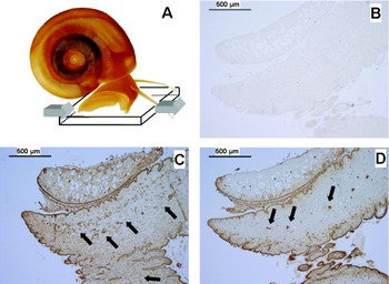
As documented in Fig. 1, foot tissue sections of uninfected snails demonstrated a distinct binding of both anti-SEA serum as well as mAb M2D3H, confirming the presence of Fuc(α1-3)GalNAc(β1-4)[±Fuc(α1-3)]GlcNAc(β1-) (F-LDN or F-LDN-F) carbohydrate epitopes (Fig. 1C,D) which is marked in both cases by arrows. In comparison with mAb M2D3H, however, anti-SEA serum led to a more intense labelling of the foot tissue. Incubation with monoclonal antibodies directed against Lewis X, LDN, LDN-F or LDN-DF sugar epitopes revealed no binding (data not shown). Likewise, control experiments employing either normal rabbit serum (NRS) or normal mouse serum (NMS) yielded at most staining of nuclei under the conditions used. Therefore, only the result obtained after NRS incubation is shown (Fig. 1B). Since these controls proved to be negative in all experiments performed, they are not explicitly addressed in the text further on, but are included in each figure.
Immunohistochemical staining of uninfected Biomphalaria glabrata foot tissue for carbohydrate epitopes. (A) Model illustrating the analysed tissue section. (B) Negative control obtained by incubation with normal rabbit serum (NRS). (C) Reaction of foot tissue with polyclonal rabbit antiserum raised against schistosome soluble egg antigens (SEA). (D) Staining with mAb M2D3H recognizing Fuc(α1-3)GalNAc(β1-4)[±Fuc(α1-3)]GlcNAc(β1-) (F-LDN or F-LDN-F) carbohydrate moieties.

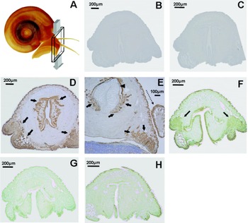
Cross-sections of anterior tissue of uninfected snails revealed strong binding of anti-SEA serum (Fig. 2D) as well as mAb M2D3H as marked by arrows (Fig. 2E). Intriguingly, the middle part of this tissue was devoid of antibody staining and could be shown to contain mainly muscle fibres by the Goldner staining technique applied in parallel (data not shown). Obviously, these muscle fibres do not express corresponding carbohydrate epitopes. The included arrow head marks part of the radula and the dashed arrow points to a cross-section of the tentacle (Fig. 2E). Antibodies reacting either with LDN (Fig. 2G) or LDN-DF (Fig. 2H) failed to stain anterior tissue, whereas the anti-F-LDN mAb employed in this study revealed the same binding pattern as mAb M2D3H (Fig. 2F).
Immunohistochemical staining of uninfected Biomphalaria glabrata anterior tissue for carbohydrate epitopes. (A) Model illustrating the analysed tissue section; (B and C) Negative controls obtained by incubation with NRS and normal mouse antiserum (NMS), respectively. (D–F) Tissue staining (marked by arrows) obtained with polyclonal anti-SEA serum (D), mAb M2D3H (E) and mAb anti-F-LDN (F). (G and H) Results obtained after incubation with monoclonal antibodies specific for GalNAc(β1-4)GlcNAc(β1-) (LDN) and GalNAc(β1-4)[Fuc(α1-2)Fuc(α1-3)]GlcNAc(β1-) (LDN-DF) carbohydrate moieties, respectively. The arrow-head and the dashed arrow in (E) mark part of the radula and a cross-section of a tentacle.

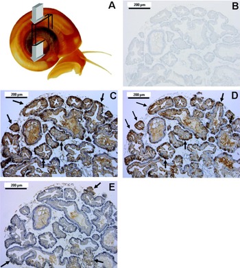
Cross-sections of uninfected hepatopancreas tissue revealed a pronounced binding of anti-SEA serum and mAb M2D3H (Fig. 3C,D), whereas the Lewis X-specific mAb G8G12 led to weaker, but clearly detectable staining (Fig. 3E). These results could be confirmed by immunohistochemical staining of longitudinal sections of the uninfected hepatopancreas with anti-SEA serum and mAb M2D3H, revealing the presence of single M2D3H-positive hepatopancreas cells (data not shown). Monoclonal antibodies recognizing LDN, LDN-F or LDN-DF carbohydrate epitopes did not yield any immunohistochemical staining of uninfected hepatopancreas tissue.
Immunohistochemical staining of uninfected hepatopancreas cross-sections for carbohydrate determinants. (A) Model illustrating the analysed tissue section. (B) Negative control incubation with NMS. (C–E) Tissue staining with polyclonal anti-SEA serum (C), mAb M2D3H interacting with Fuc(α1-3)GalNAc(β1-4)[±Fuc(α1-3)]GlcNAc(β1-) (F-LDN or F-LDN-F) carbohydrate moieties (D) and mAb G8G12 recognizing Gal(β1-4)[Fuc(α1-3)]GlcNAc(β1-) (Lewis X) carbohydrate entities (E). Arrows mark tissue areas of high antibody binding.

Detection of cross-reacting carbohydrate epitopes in hepatopancreas tissue of infected snails
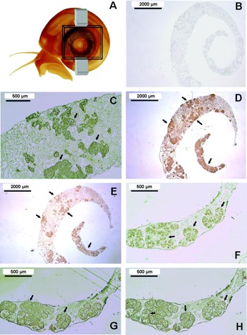
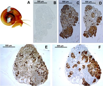
In comparison to uninfected snails, infected snail tissue displayed, in part, a more pronounced, but also different immunohistochemical staining behaviour. Since the snails had been infected already for a long time prior to analysis, only daughter sporocysts were observed in their hepatopancreas, whereas their foot tissue regions appeared to be free of any parasites. Consequently, antibody staining of this area yielded similar results as in the case of uninfected snails (data not shown). In contrast, striking differences were observed with regard to infected hepatopancreas. As shown in Fig. 4, staining of longitudinal sections with anti-SEA serum (Fig. 4C), mAb M2D3H (Fig. 4D) and mAb G8G12 (Fig. 4E), recognizing Lewis X determinants, revealed a strong binding of the applied antibodies to the daughter sporocysts. It is interesting to note, however, that incubation with anti-SEA serum and mAb M2D3H resulted simultaneously in an intense staining of the surrounding tissue which was less pronounced in the case of the anti-Lewis X mAb. In order to obtain more detailed information, additional carbohydrate-specific monoclonal antibodies were employed. Similar to mAb M2D3H, incubation with the anti-F-LDN mAb yielded a clear staining of both sporocysts as well as surrounding tissue (data not shown). In contrast, mAbs recognizing LDN, LDN-F and LDN-DF (Fig. 4F-H) displayed a different binding pattern in so far as antibodies reacting with LDN-F (Fig. 4G) and LDN-DF (Fig. 4H) yielded an intense staining of the inner part of the sporocysts and, in addition, also of ring-like structures surrounding these sporocysts which are marked by arrows. Antibodies recognizing the unfucosylated LDN epitope bound mainly to the inner part of the sporocysts, while the surrounding tissue remained unaffected (Fig. 4F).
Immunohistochemical staining of longitudinal sections of infected hepatopancreas tissue for carbohydrate determinants. (A) Model illustrating the analysed tissue section. (B) Negative control incubation with NMS. (C–H) Tissue staining achieved by incubation with polyclonal anti-SEA serum (C), mAb M2D3H interacting with Fuc(α1-3)GalNAc(β1-4)[±Fuc(α1-3)]GlcNAc(β1-) (F-LDN or F-LDN-F) carbohydrate moieties (D), mAb G8G12 reacting with Lewis X entities (E) and monoclonal antibodies recognizing GalNAc(β1-4)GlcNAc(β1-) (LDN) (F), GalNAc(β1-4)[Fuc(α1-3)]GlcNAc(β1-) (LDN-F) (G) and GalNAc(β1-4)[Fuc(α1-2)Fuc(α1-3)]GlcNAc(β1-) (LDN-DF) carbohydrate motifs (H). Arrows mark tissue areas exhibiting high antibody binding.

In agreement with these data, immunohistochemical staining of infected hepatopancreas cross-sections revealed again a preferential binding of anti-SEA serum (Fig. 5C) and mAb M2D3H (Fig. 5D) to sporocysts. Likewise, incubation with the Lewis X-specific mAb G8G12 resulted in substantial staining of the parasites (Fig. 5E). A combined incubation of these tissue sections with mAbs against LDN-F and LDN-DF confirmed that these epitopes are primarily located within the sporocysts (Fig. 5F). In contrast to anti-SEA serum, mAb M2D3H and mAb G8G12, however, the surrounding tissue remained unstained in this case.
Immunohistochemical staining of cross-sections from 2 snail samples (B–D and E+F) of infected hepatopancreas tissue for carbohydrate determinants. (A) Model illustrating the analysed tissue section. (B) Negative control incubation with NMS. (C–F) Tissue staining with polyclonal anti-SEA serum (C), mAb M2D3H recognizing Fuc(α1-3)GalNAc(β1-4)[±Fuc(α1-3)]GlcNAc(β1-) (F-LDN or F-LDN-F) carbohydrate moieties (D), the Lewis X-specific mAb G8G12 (E) or mAbs recognizing GalNAc(β1-4)[Fuc(α1-3)]GlcNAc(β1-) (LDN-F) and GalNAc(β1-4)[Fuc(α1-2)Fuc(α1-3)]GlcNAc(β1-) (LDN-DF) motifs (F).

Surface staining of miracidia as whole mounts
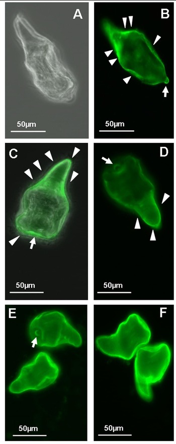
In a first set of experiments, miracidia were incubated with polyclonal rabbit antiserum raised against soluble egg antigens (SEA) of S. mansoni. Subsequent treatment with fluorescently labelled secondary antibodies revealed strong binding of anti-SEA antibodies to plate-like areas at the miracidial surface (arrow heads in Fig. 6B), indicating a wide distribution of the recognized antigenic determinants. Intriguingly, antibody staining was also observed at the terebratorium (arrows in Fig. 6B–E) which is highly involved in the invasion process of miracidia entering the snail body. Control incubation with NRS yielded no staining, thus confirming the lack of non-specific secondary antibody binding and auto-fluorescence of the preparations. In order to localize the miracidium, a superimposition of pictures registered by phase contrast and fluorescence detection is shown in this case (Fig. 6A).
Immunofluorescence staining of miracidia. (A) Negative control incubation with NMS. In order to localize the miracidium, a superimposition of pictures registered by phase contrast and fluorescence detection is shown. (B) Anti-SEA antibody-mediated immunofluorescence detection of antigens located at the miracidial surface (arrow-heads) and at the terebratorium (arrow). (C and D) Immunofluorescence detection of Fuc(α1-3)GalNAc(β1-4)[±Fuc(α1-3)]GlcNAc(β1-) (F-LDN or F-LDN-F) carbohydrate epitopes at the miracidial surface by mAb M2D3H. Arrow-heads mark areas of surface staining which appeared to be more intense in the posterior part of the miracidium, whereas staining of the terebratorium is marked by arrow. In (C) a superimposition of pictures obtained by phase contrast and fluorescence detection is shown, demonstrating an intense staining of the posterior part of the larvae. (E and F) Immunofluorescence detection of GalNAc(β1-4)[Fuc(α1-2)Fuc(α1-3)]GlcNAc(β1-) (LDN-DF) epitopes (E) and GalNAc(β1-4)[Fuc(α1-3)]GlcNAc(β1-) (LDN-F) carbohydrate entities (F). Staining of the terebratorium is marked by arrow (E).

In order to obtain more detailed information on the occurrence of individual carbohydrate epitopes, monoclonal antibodies were employed. The results revealed distinct binding of mAbs directed against fucosylated LDN carbohydrate epitopes. Staining with mAb M2D3H clearly confirmed the presence of F-LDN and/or F-LDN-F carbohydrate epitopes at the miracidial surface (arrow heads in Fig. 6C,D) including the terebratorium. Interestingly, increased fluorescence intensity was observed at the posterior part of the miracidium which is illustrated best by superimposition of the pictures obtained by phase contrast and fluorescence detection (Fig. 6C). Incubation with mAb anti-F-LDN revealed again the same binding pattern as mAb M2D3H (data not shown).
Incubation with monoclonal antibodies interacting with LDN-DF (Fig. 6E) and LDN-F (Fig. 6F) carbohydrate epitopes resulted in strong, mostly uniform staining of the miracidial surface and the terebratorium, whereas monoclonal antibodies recognizing Lewis X and LDN units revealed no binding to the surface of miracidia (data not shown).
DISCUSSION
Localization of carbohydrate epitopes in uninfected snail tissue
Previous histological studies on B. glabrata have been either focussed on the snail tissue itself or on changes occurring in the context of schistosome infection (Pan, Reference Pan1980; Jeong et al. Reference Jeong, Lie and Heyneman1983; de Souza et al. Reference De Souza, Cunha Rde and Andrade1995; Sire et al. Reference Sire, Rognon and Theron1998; Lemos, Reference Lemos1999; Lemos and Andrade, Reference Lemos and Andrade2001; Nyame et al. Reference Nyame, Yoshino and Cummings2002) whereas the occurrence and distribution of distinct glycan epitopes within the snail have not been investigated in detail so far. Recently, we could demonstrate, however, that B. glabrata haemolymph glycoproteins exhibit a wide range of N-glycans which share carbohydrate structures cross-reacting with glycoconjugates of S. mansoni (Lehr et al. Reference Lehr, Geyer, Maass, Doenhoff and Geyer2007), thus supporting the hypothesis of carbohydrate-based molecular mimicry postulated earlier (Dissous et al. Reference Dissous, Grzych and Capron1986; Damian, Reference Damian1989; Dissous and Capron, Reference Dissous and Capron1989). The aim of this study was, therefore, to use polyclonal rabbit antibodies raised against S. mansoni soluble egg antigens (SEA) and monoclonal mouse antibodies recognizing defined carbohydrate epitopes for allocation of serologically cross-reacting glycan structures in both uninfected and infected snail tissue as well as sporocysts and miracidia, i. e., the snail-pathogenic larvae of S. mansoni.
As shown recently, polyclonal anti-SEA serum recognizes at least 2 different types of carbohydrates epitopes, Fuc(α1-3)GalNAc(β1-4)[±Fuc(α1-3)]GlcNAc(β1-), i. e., F-LDN or F-LDN-F, as well as core-linked Xyl(β1-2) units (Lehr et al. Reference Lehr, Geyer, Maass, Doenhoff and Geyer2007). The immunohistochemical data presented in this study clearly demonstrated an interaction of anti-SEA antibodies with distinct areas within the foot, anterior tissue and the hepatopancreas of uninfected snails, indicating the presence of cross-reacting antigenic determinants. Intriguingly, these tissues are important areas of host-parasite interaction during invasion, since the miracidium enters the snail via the foot tissue, whereas further development of the parasite takes place in the hepatopancreas. Thus, common antigenic determinants could contribute to the parasite's survival within its intermediate host.
In agreement with these findings, mAb M2D3H interacting specifically with Fuc(α1-3)GalNAc(β1-4)[±Fuc(α1-3)]GlcNAc(β1-) (F-LDN or F-LDN-F) carbohydrate units (Kantelhardt et al. Reference Kantelhardt, Wuhrer, Dennis, Doenhoff, Bickle and Geyer2002), i.e., only one type of structure bound by anti-SEA serum, displayed a similar, but weaker, binding to these tissues which could be confirmed by a second monoclonal antibody similarly reacting with F-LDN moieties. This finding is remarkable since F-LDN and F-LDN-F carbohydrate motifs represent major glycan epitopes of schistosome egg glycoconjugates (Wuhrer et al. Reference Wuhrer, Dennis, Doenhoff, Bickle, Lochnit and Geyer1999; Robijn et al. Reference Robijn, Wuhrer, Kornelis, Deelder, Geyer and Hokke2005). In contrast, unfucosylated lacdiNAc (GalNAc(β1-4)GlcNAc(β1-); LDN) or difucosylated LDN units (GalNAc(β1-4)[Fuc(α1-2)Fuc(α1-3)]GlcNAc(β1-); LDN-DF) were not detected in uninfected snail tissue by this approach, although our previous studies on individual haemolymph glycoprotein-N-glycans from uninfected snails revealed, in fact, certain amounts of sugar chains with unfucosylated LDN units (Lehr et al. Reference Lehr, Geyer, Maass, Doenhoff and Geyer2007). The latter finding is in agreement with results obtained by Western blot analyses of tissue extracts of uninfected B. glabrata performed by Nyame and co-workers (Nyame et al. Reference Nyame, Yoshino and Cummings2002) which similarly demonstrated the presence of glycoproteins with LDN antigens. Obviously, glycoconjugates expressing this epitope do not occur or are only expressed in small quantities in hepatopancreas tissue, thus preventing staining by the corresponding antibody.
Application of the Lewis X-specific mAb G8G12 (Wuhrer et al. Reference Wuhrer, Dennis, Doenhoff, Lochnit and Geyer2000b) yielded weak staining of uninfected hepatopancreas tissue. This finding is also remarkable since N-glycans carrying a Lewis X epitope have not been identified in our previous structural analysis of haemolymph glycoproteins. Possibly, such glycans are only expressed in a tissue-specific manner in the hepatopancreas of the snail or occur primarily in O-linked glycoprotein glycans or glycolipids of B. glabrata which have not been studied so far. From the standpoint of molecular mimicry, however, the pre-existence of Lewis X structures in the uninfected hepatopancreas might be of relevance with regard to the survival of the parasite, since sporocysts formed after infection are shown in this study to abundantly express this carbohydrate motif.
Localization of carbohydrate epitopes in infected snails
Immunohistochemical studies revealed no differences between foot tissue of uninfected and long-term infected adult B. glabrata snails. In contrast, striking changes were observed for their hepatopancreas. In comparison with uninfected snails, treatment with polyclonal anti-SEA antibodies and mAb M2D3H resulted in increased staining of this tissue and, in particular, of the daughter sporocysts, indicating an enhanced expression of F-LDN and/or F-LDN-F carbohydrate determinants which have been also detected in uninfected hepatopancreas tissue. The question remains open, however, as to whether this observation is due to increased biosynthesis of these molecules by the sporocysts themselves or to acquisition and presentation of host-derived antigens, since previous studies have shown that sporocysts are, in fact, able to acquire and process molecules of their host (Bayne et al. Reference Bayne, Boswell, Loker and Yui1985; Bayne et al. Reference Bayne, Loker and Yui1986; Spray and Granath, Reference Spray and Granath1990). Since corresponding glycan structures are also present at the surface of miracidia, it appears to be more likely, however, that these glycoconjugates are of parasitic origin. The enhanced staining of the host tissue surrounding the sporocysts might be due to a secretion of these antigens by the parasite which has been also suggested by Nyame and co-workers (Nyame et al. Reference Nyame, Yoshino and Cummings2002).
In contrast, Lewis X carbohydrate epitopes were preponderantly detected on sporocysts, whereas the surrounding tissue was much less stained by the respective antibody. Hence, it can be assumed that this glycan motif is primarily synthesized by the parasite, possibly in order to prepare itself for a future invasion of its definitive host, since Lewis X epitopes have been demonstrated to represent major determinants of cercarial glycolipids (Wuhrer et al. Reference Wuhrer, Dennis, Doenhoff, Lochnit and Geyer2000b) and have been also detected at the surface of mechanically transformed schistosomula (Köster and Strand, Reference Köster and Strand1994). The staining of sporocysts with the Lewis X-specific mAb G8G12 is remarkable because immunohistochemical studies performed by Nyame and co-workers provided no evidence for the presence of Lewis X carbohydrate antigens in both in vitro-derived mother sporocysts and in vivo-derived mother and daughter sporocysts (Nyame et al. Reference Nyame, Yoshino and Cummings2002). One possible explanation for this finding might be that these authors used a different, commercially available anti-Lewis X IgM mAb recognizing primarily human CD 15, whereas in our study a monoclonal antibody which was generated against glycoconjugates of S. mansoni was employed. The importance of the kind of antibody for this type of study is underlined by the observation that the Lewis X-specific mAb 128-4F9-A (van Remoortere et al. Reference Van Remoortere, Hokke, Van Dam, Van Die, Deelder and Van Den Eijnden2000), which has been used in parallel, resulted only in very weak staining of the sporocysts (data not shown), even though this monoclonal antibody has been obtained from spleen cells of mice infected with S. mansoni. This is in agreement with data reported by van Remoortere and co-workers, who demonstrated that different isolates of anti-Lewis X mAbs may differ in their recognition pattern of glycoconjugates in individual developmental stages of S. mansoni (van Remoortere et al. Reference Van Remoortere, Hokke, Van Dam, Van Die, Deelder and Van Den Eijnden2000).
GalNAc(β1-4)GlcNAc(β1-) (LDN), GalNAc(β1-4)[Fuc(α1-3)]GlcNAc(β1-) (LDN-F) and GalNAc(β1-4)[Fuc(α1-2)Fuc(α1-3)]GlcNAc(β1-) (LDN-DF) motifs were found to be specifically expressed on hepatopancreas located sporocysts. Intriguingly, monoclonal antibodies recognizing LDN-F and LDN-DF stained, in addition, a ring-like structure surrounding the sporocysts which might be due to the excretion or secretion of parasite-expressed antigens, whereas LDN-specific monoclonal antibodies solely bound to sporocysts without staining the above mentioned ring-like structure.
Localization of carbohydrate epitopes in miracidia
Polyclonal anti-SEA serum led to intense staining of the entire surface, including the terebratorium of the miracidium, thus demonstrating a wide distribution of the recognized antigens. In principle, similar results were obtained with mAb M2D3H recognizing F-LDN and/or F-LDN-F carbohydrate determinants as well as a second anti-F-LDN antibody (data not shown), although the staining intensity was lower in both cases due to the fact that only one type of glycan epitope was registered.
The occurrence of cross-reacting carbohydrate antigens at the terebratorium is in so far interesting as this structure is utilized by the larva to invade the snail body. Possibly, invasion of the snail host is facilitated by masking this part of the miracidium by antigens occurring, at least in part, also in the uninfected foot tissue. A similar twist of epitope distribution has also been reported for cercariae which express the Lewis X carbohydrate motif primarily at their oral suckers (van Remoortere et al. Reference Van Remoortere, Hokke, Van Dam, Van Die, Deelder and Van Den Eijnden2000). In addition to cross-reacting carbohydrate epitopes, however, the miracidial surface exhibited also parasite-specific glycan motifs like LDN-F and LDN-DF, both of which were also found to be uniformly distributed at the miracidial surface including the terebratorium, whereas Lewis X and unfucosylated LDN glycan motifs could not be detected.
In conclusion, our study clearly revealed the presence of cross-reacting carbohydrate epitopes in both uninfected B. glabrata as well as snail-pathogenic larvae of S. mansoni, thus supporting the concept of carbohydrate-based molecular mimicry as an invasion strategy of this parasite at the level of its intermediate host.
The authors thank Professor Dr A. M. Deelder and Dr C. H. Hokke (Department of Parasitology, Leiden University Medical Centre, The Netherlands) for kindly providing monoclonal antibodies as well as Dr Q. Bickle (London School of Hygiene and Tropical Medicine, Department of Infectious and Tropical Diseases, London, UK) for mAb G8G12 and the mAb M2D3H producing hybridoma cell line. We acknowledge Professor Dr R. M. Bohle (Institute of Pathology, University Hospital Giessen-Marburg) and his laboratory for embedding of snail samples and preparation of serial tissue sections and thank Dr R. D. Dennis (Institute of Biochemistry, Faculty of Medicine, University of Giessen) for his help and scientific support. This study was financially supported by the Deutsche Forschungsgemeinschaft (SFB 535, project A15).