Introduction
Andrássy (Reference Andrássy2009a) created the genus Aporcelinus with nine species, two new and seven transferred. Since its proposal, the genus has received new incorporations (Andrássy, Reference Andrássy2009b, Reference Andrássy2012; Álvarez-Ortega and Peña-Santiago, Reference Álvarez-Ortega and Peña-Santiago2013; Nguyen et al., Reference Nguyen2016a, Reference Nguyenb; Peña-Santiago and Abolafia, Reference Peña-Santiago and Abolafia2016; Varela-Benavides and Peña-Santiago, Reference Varela-Benavides and Peña-Santiago2018) and its taxonomic position has been a matter of some controversy (Vinciguerra et al., Reference Vinciguerra, Orselli and Clausi2014). To date, the genus Aporcelinus contains 26 valid species plus two species inquirendae. Available molecular data are limited to only three of its representatives, which is a serious handicap to clarifying the evolutionary relationships with other members of the family Aporcelaimidae Heyns, 1965.
Fortunately, two soil samples, one from Kansas and another from Florida, received in 2011 for nematological inspection by the Nematology Laboratory at the Plant Pest Diagnostics Center, California Department of Food and Agriculture, yielded fresh specimens of two putative new, non-described Aporcelinus species. Both species are morphologically and molecularly characterized here.
Materials and methods
Sampling, extraction and morphological identification
Nematodes were extracted from soil samples by sieving and a sucrose centrifugation technique, somewhat modified (specific density = 1.18), according to Barker (Reference Barker, Barker, Carter and Sasser1985), relaxed and killed by heat, fixed in 4% formaldehyde, and processed to anhydrous glycerin following the technique of Siddiqi (Reference Siddiqi1964). The specimens were mounted on permanent glass slides for observation under a light microscope.
Specimens were examined and measured using an Olympus BH-2 light microscope equipped with differential interference contrast (DIC). Morphometrics included de Man's indices and standard measurements. The location of the pharyngeal gland nuclei was expressed according to Loof and Coomans (Reference Loof and Coomans1970) and the spicule terminology was according to Peña-Santiago et al. (Reference Peña-Santiago, Abolafia and Álvarez-Ortega2014). Some of the best-preserved specimens were photographed with a Nikon Eclipse 80i microscope equipped with DIC and a Nikon DS digital camera. Digital images were edited using Adobe® Photoshop® CS. Drawings were made using a camera lucida attached to the microscope. After their examination and identification, one specimen of the new species Aporcelinus paolae sp. n. preserved in glycerin was selected for examination by scanning electron microscopy (SEM) following the protocol of Álvarez-Ortega and Peña-Santiago (Reference Álvarez-Ortega and Peña-Santiago2016). The nematode was hydrated in distilled water, dehydrated in a graded ethanol and acetone series, critical point-dried, coated with gold, and observed under a Zeiss Merlin microscope.
DNA extraction, polymerase chain reaction (PCR) and sequencing
DNA was extracted from a single individual using the proteinase K protocol. Nematode material was transferred to an Eppendorf tube containing 30 μl double distilled water, 3 μl polymerase chain reaction (PCR) buffer (Qiagen, Hilden, Germany) and 2 μl proteinase K (600 μg ml−1) (Qiagen). The tubes were incubated at 65°C (1 h) and then at 95°C (15 minutes). PCR and sequence protocols were as described by Álvarez-Ortega et al. (Reference Álvarez-Ortega, Subbotin and Peña-Santiago2013a). The primers used for amplification of the D2-D3 expansion segments of 28S rRNA gene were the D2A (5′-ACAAGTACCGTGAGGGAAAGTTG-3′) and the D3B (5′-TCGGAAGGAACCAGCTACTA-3′) primers (Subbotin et al., Reference Subbotin2006).
PCR products were purified using the QIAquick PCR purification Kit (Qiagen) and used for direct sequencing. The sequencing reactions were performed at Davis Sequencing (Davis, CA, USA). The sequences obtained were submitted to the GenBank database under accession numbers MK007552 and MK007553.
Phylogenetic analyses
The newly obtained sequences were aligned with the other 58 D2-D3 expansion segments of 28S rRNA gene sequences available in GenBank using ClustalX 1.83 (Thompson et al., Reference Thompson1997). Outgroup taxa were chosen according to the results of previous published data (Holterman et al., Reference Holterman2008; Álvarez-Ortega et al., Reference Álvarez-Ortega, Subbotin and Peña-Santiago2013b). Sequence alignments were manually edited using GenDoc 2.6.002 (Nicholas et al., Reference Nicholas, Nicholas and Deerfield II1997). Bayesian inference (BI) and maximum-likelihood (ML) analyses of the sequence dataset were performed at the CIPRES Science Gateway (Miller et al., Reference Miller, Pfeiffer and Schwartz2010), using MrBayes 3.2.6 (Ronquist et al., Reference Ronquist2012) and RAxML 8.2.10 (Stamatakis, Reference Stamatakis2014), respectively. The best fit model of DNA evolution was obtained using jModelTest 2.1.10 (Darriba et al., Reference Darriba2012) with the Akaike information criterion (AIC). The Akaike-supported model, the base frequency, the proportion of invariable sites, and the gamma distribution shape parameters and substitution rates in the AIC were then used in phylogenetic analyses. BI analysis under the general time reversible model with a proportion of invariable sites and a gamma-shaped distribution (GTR + I + G) was initiated with a random starting tree and run with the four Metropolis-coupled Markov chain Monte Carlo (MCMC) for 2 × 10⁶ generations. ML analysis was implemented under the same nucleotide substitution model as in the BI, with 1000 bootstrap replications. The topologies were used to generate a 50% majority rule consensus tree. Posterior probabilities (PP) and bootstrap support (BS) over 70% are given on appropriate clades. The trees were visualized using the program FigTree v1.4.3, and drawn with Adobe Acrobat XI Pro 11.0.1.
Results
Aporcelinus floridensis sp. n. (figs 1 and 2)
Material examined
Twelve females from one location, in variable (in general acceptable) states of preservation.
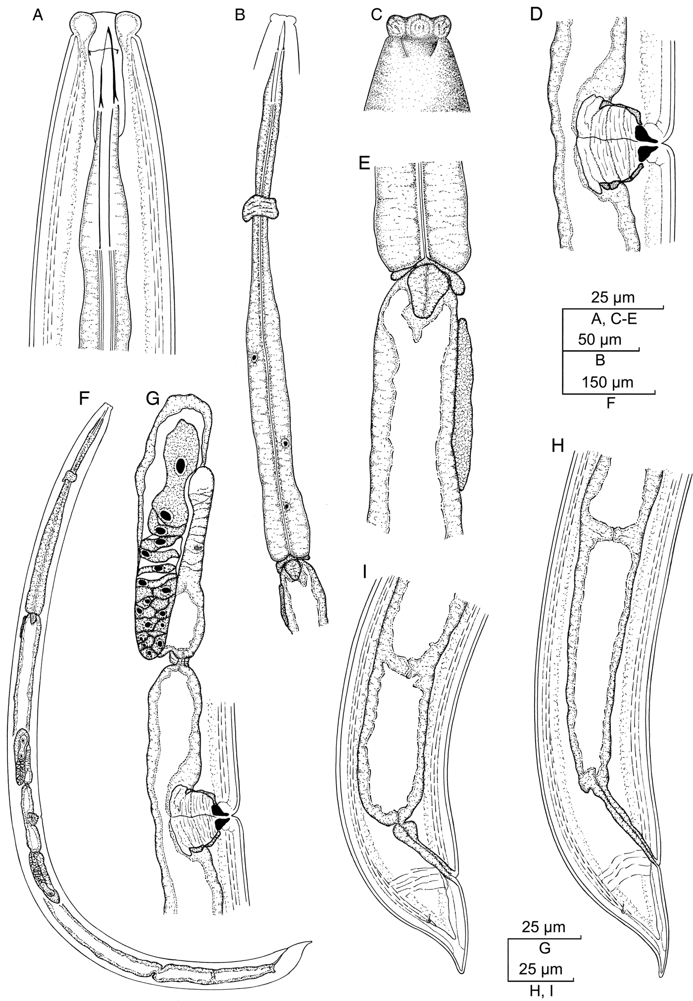
Aporcelinus floridensis sp. n. (female, line drawing). (A) Anterior region in lateral median view; (B) neck region; (C) lip region in lateral surface view; (D) vagina; (E) pharyngo-intestinal junction; (F) entire body; (G) anterior genital branch; (H, I) posterior body region.

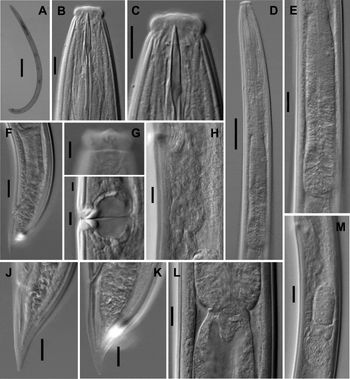
Aporcelinus floridensis sp. n. (female, light micrographs). (A) entire body; (B, C) anterior region in lateral median view; (D) neck region; (E) pharyngeal expansion; (F) caudal region and prerectum; (G) lip region in surface lateral view; (H) uterus; (I) vagina; (J, K) caudal region; (L) pharyngo-intestinal junction; (M) posterior genital branch. (Scale bars: A = 200 μm; B, C, H, J–L = 10 μm; D = 50 μm; E, F, M = 20 μm; G, I = 5 μm)

Morphometrics
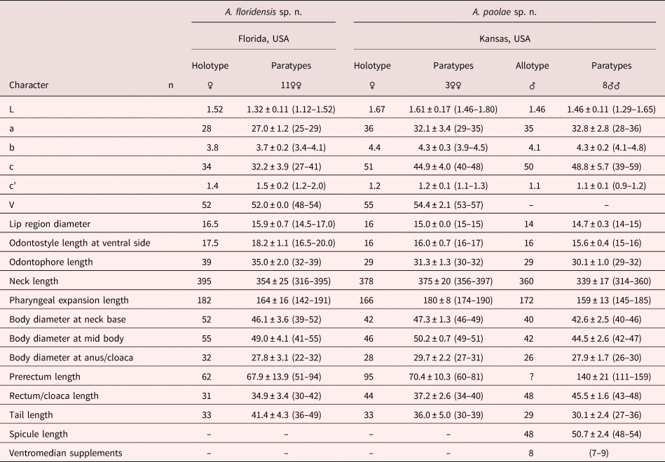
See table 1.
Morphometrics of Aporcelinus floridensis sp. n. and A. paolae sp. n. Measurements in μm (except L, in mm), and in the form of mean ± standard deviation (range).

Description
Female. Moderately slender (a = 25–29) nematodes of medium size, 1.12–1.52 mm long. Habitus curved ventrad upon fixation, in general, C-shaped. Cuticle two-layered, 2.0–2.5 μm thick at anterior region, 2.5–3.5 μm in mid-body and 3.0–4.5 μm on tail; outer layer thin and bearing fine but conspicuous transverse striation throughout the body; inner layer thicker and more refractive than the outer layer. Lateral chord 6–10 μm broad or 13–21% of mid-body diameter. Body pores often obscure under light microscope. Lip region offset by a distinct constriction, 2.9–3.4 times as wide as high and 32–37% of body diameter at neck base; labial and cephalic papillae slightly protruding; oral field visibly elevated, suggesting the existence of perioral liplets. Amphid fovea funnel-shaped, its opening 8–10 μm broad or 52–63% of lip region diameter. Cheilostom nearly as long as wide, with no specialization. Odontostyle strong, slightly longer (1.1–1.2 times) than lip region diameter, 5.8–7.8 times longer than wide and 1.15–1.56% of total body length; aperture 9.0–10.5 μm or 51–56% of its total length. Guiding ring simple but distinct and somewhat plicate, located at 8.0–10.5 μm or 0.5–0.6 times the lip region diameter from the anterior end. Odontophore rod-like, 1.6–2.2 times the odontostyle long. Pharynx consisting of a slender but muscular anterior section enlarging very gradually in the posterior, the expansion 4.9–6.7 times as long as wide, 2.8–4.0 times the corresponding body diameter and occupies 43–48% of total neck length; gland nuclei in general obscure in the specimens examined: DN = 66–69, S1N1 = 71–75, S1N2 = 78–82, S2N = 88–93. Nerve ring at 119–142 μm or 36–40% of total neck length from the anterior end. Pharyngo-intestinal junction surrounded by an asymmetrical ring-like structure, with a distinctly developed dorsal lobe; cardia conical, 17–23 × 10–15 μm; a dorsal cell mass is present at a variable distance behind the pharyngeal base. Genital system didelphic–amphidelphic, with both branches equally and well developed, 104–199 μm long or 9–14% of total body length: ovaries 60–190 μm long, often not reaching the oviduct–uterus junction; oviduct 54–78 μm long or 1.2–1.5 times the body diameter, and consisting of a slender portion and a small pars dilatata with visible lumen; a sphincter separates oviduct and uterus; uterus a short and simple tube-like structure 33–56 μm long or 0.8–1.2 times the corresponding body diameter, without sperm cells observed inside; vagina extending inwards 19–24 μm or 38–49% of body diameter, with pars proximalis 10–15 × 13–21 μm and convergent walls surrounded by weak musculature, pars refringens consisting of two drop-shaped to trapezoidal pieces 4.5–7.0 × 3.0–4.5 μm and a combined width of 6.5–10.0 μm, and pars distalis 4.5–6.5 μm long and visibly refractive; and vulva a transverse slit. Prerectum 1.7–3.2 and rectum 1.0–1.4 times the anal body diameter long. Tail conical with finely rounded tip, ventrally straight or slightly convex, dorsally first convex and then bearing a more or less conspicuous concavity and re-curved dorsad; inner cuticle layer reaching the tail tip, so that a hyaline portion is not perceptible; caudal pores two pairs at the middle of tail, one subdorsal, another sublateral.
Male. Unknown.
Molecular characterization
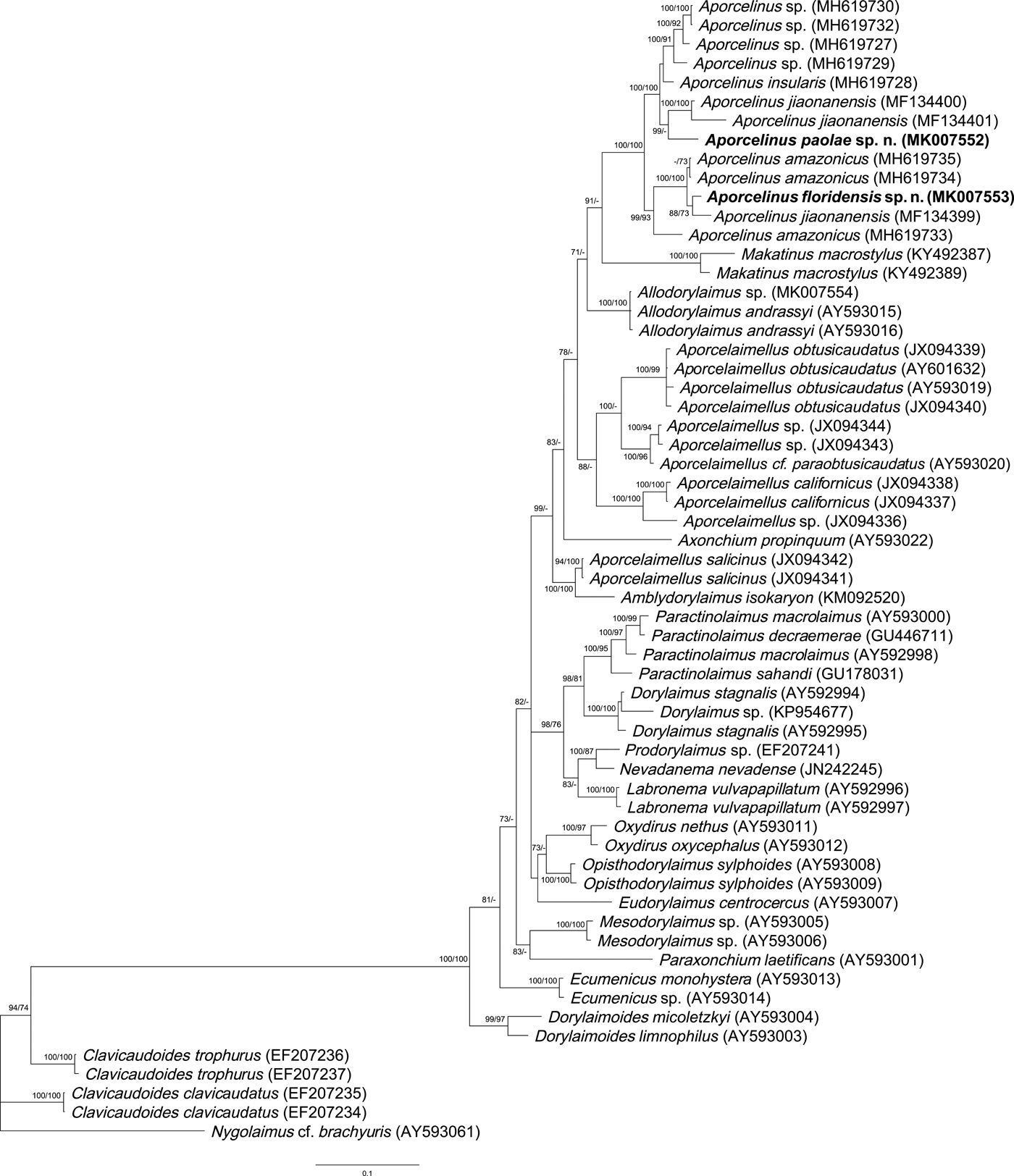
One sequence of the D2-D3 of 28S rRNA gene 747 bp long was obtained. The evolutionary relationships of the new species with several representatives of the order Dorylaimida are presented in fig. 3.
Bayesian 50% majority rule consensus tree as inferred from the D2-D3 expansion segments of 28S rRNA gene sequence alignments under the GTR + I + G model. Branch support of over 70% is given for appropriate clades and is indicated as posterior probability value in Bayesian inference analysis/bootstrap value from maximum-likelihood analysis. Newly obtained sequences are indicated by bold letters.

Diagnosis
This new species is characterized by its 1.12–1.52 mm long body, lip region offset by marked constriction and 14.5–17.0 μm broad with perioral liplets, odontostyle 16.5–20.0 μm at its ventral side and 1.1–1.2 times the lip region diameter, neck 316–395 μm long, pharyngeal expansion 142–191 μm long or 43–48% of total neck length, a dorsal cell mass present at variable distance behind the level of pharyngo-intestinal junction, uterus simple and 33–56 μm long or 0.8–1.2 times the corresponding body diameter, V = 48–54, female tail conical (36–49 μm, c = 27–41, c’ = 1.2–2.0) with finely rounded terminus and no hyaline portion, and male absent.
Relationships
In having medium general size (body length almost always > 1.0 mm), comparatively small odontostyle (up to 22 μm long) and female tail bearing a distinct dorsal concavity and ending in a finely rounded terminus, the new species resembles A. irritans (Cobb in Thorne & Swanger, 1936) Andrássy, 2009, A. jiaonanensis (Zhao & Zhao, 2010) Álvarez-Ortega & Peña-Santiago, 2013 (see recent description by Nguyen et al., Reference Nguyen2017) and A. neogranuliferus Peña-Santiago & Abolafia, 2016, a group of species with intricate taxonomy. It differs from A. irritans in its less angular and broader lip region (exceptionally under 15.5 vs 14–15 μm), shorter odontostyle (up to 20 vs 20–21 μm, 1.1–1.2 vs 1.4 times the lip region diameter), shorter female prerectum (up to 1.4 vs twice the anal body diameter long), and male absent (vs present); from A. jiaonanensis in its narrower lip region (up to 17 vs 17–20 μm wide), shorter odontostyle (up to 20 vs 20–23 μm long), comparatively shorter pharyngeal expansion (43–48 vs 47–55% of total neck length), shorter uterus (33–56 vs 56–97 μm long), comparatively longer female tail (c’ = 1.2–2.0 vs c’ = 0.7–1.3) without (vs with) hyaline terminal portion, male unknown (vs known), and in remarkable differences in partial sequences of D2-D3 of LSU rRNA gene; and from A. neogranuliferus, a very similar species, in its narrower lip region (14.5–17.0 vs 17.5–18.0 μm broad), slightly longer odontostyle (1.1–1.2 times vs equal to the lip region diameter) with larger aperture (51–56 vs 46–51% of total length), and longer female tail (36–49 vs 28–35 μm, c’ = 1.2–2.0 vs 0.8–1.0).
Type locality and habitat
Fort Pierce, Florida, USA, where the new species was collected in association with Ficus benjamina.
Type material
Female holotype, nine female paratypes deposited in the nematode collection of the University of Jaén, Spain. Two female paratypes deposited with the USDA Nematode Collection, Beltsville, MD, USA.
Etymology
The specific epithet refers to the geographical origin of the species.
Aporcelinus paolae sp. n. (figs 4–6)
Material examined
Four females and nine males from one location, in good state of preservation.
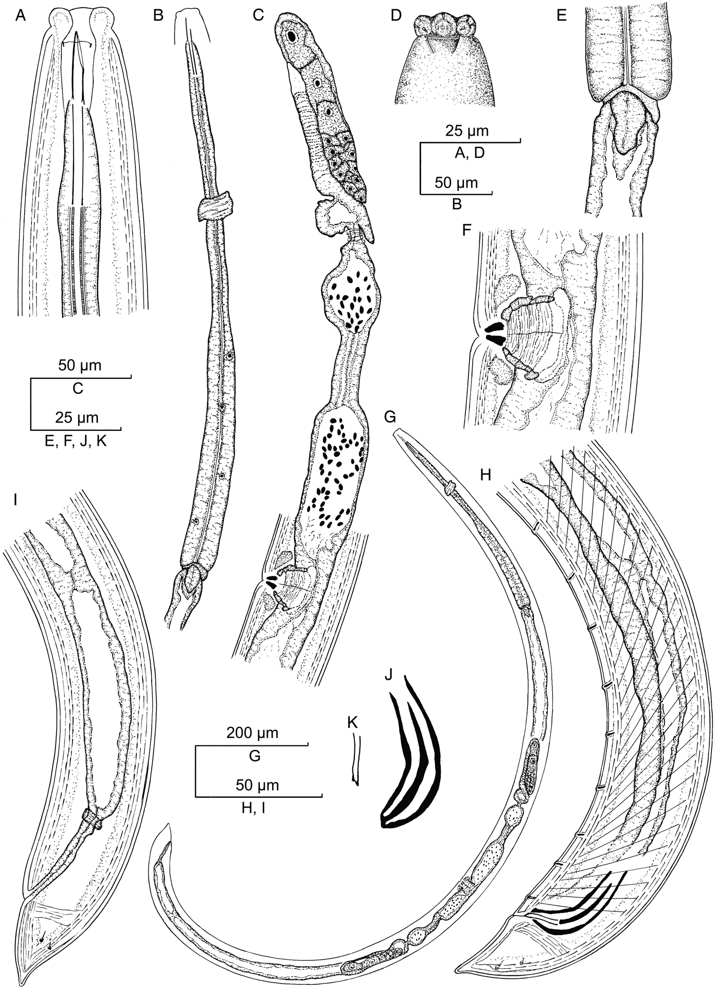
Aporcelinus paolae sp. n. (line drawing). (A) Anterior region in lateral median view; (B) neck region; (C) female, anterior genital branch; (D) lip region in lateral surface view; (E) pharyngo-intestinal junction; (F) vagina; (G) female, entire; (H) male, posterior body region; (I) female, posterior body region; (J) spicule; (K) lateral guiding piece.

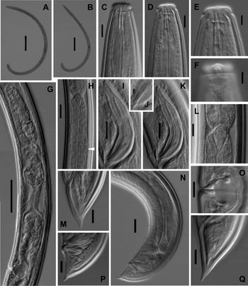
Aporcelinus paolae sp. n. (light micrographs). (A) female, entire; (B) male, entire; (C–E) anterior region in lateral median view; (F) lip region in lateral surface view; (G) female, anterior genital branch; (H) pharyngo-intestinal junction, arrow head pointing at dorsal cell mass; (I, K) spicule; (J) lateral guiding piece; (L) pharyngo-intestinal junction; (M) female, caudal region; (N) male, posterior body region; (O) vagina; (P, Q) male, caudal region. (Scale bars: A, B = 200 μm; C, D, I, K, L, M, O–Q = 10 μm; E, F = 5 μm; G = 50 μm; H, N = 20 μm; J = 2 μm)

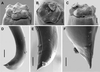
Aporcelinus paolae sp. n. (male, SEM). (A) Lip region in dorsal view; (B) lip region in frontal view; (C) lip region in lateral view; (D) posterior body region; (E) caudal region in subventral view; (F) caudal region in dorsal view. (Scale bars: A–C = 2 μm; D = 20 μm; E = 10 μm; F = 5 μm)

Morphometrics
See table 1.
Description
Adult. Moderately slender to slender (a = 28–36) nematodes of medium size, 1.29–1.80 mm long. Habitus visibly curved ventrad upon fixation, in general C-shaped. Cuticle two-layered, 2.0–2.5 μm thick at anterior region, 2.5–3.0 μm in mid-body and 2.5–4.0 μm on tail; outer layer thin and bearing fine but conspicuous transverse striation throughout the body; inner layer thicker and more refractive than the outer layer. Lateral chord 6–10 μm broad or 12–20% of mid-body diameter. Body pores often obscure, but two cervical pores are visible at both dorsal and ventral sides at level of odontostyle plus odontophore. Lip region moderately angular, offset by a distinct constriction, 3.2–3.5 times as wide as high and 31–37% of body diameter at neck base. Under SEM: lips mostly amalgamated, separated by marked interlabial depressions, each lip bearing two or three concentric incisures at its inner (oral field) part and five to six at its outer part; labial papillae button-like with a pore in the centre, with both inner and outer papillae equally sized and surrounded by a coarse annulus; cephalic papillae opening in a short transverse slit, which is not surrounded by a coarse annulus similar to that found in labial papillae; oral field poorly differentiated by a very weak elliptical (or somewhat hexagonal) incisure, bearing coarse radial striation ending at the base of the lips; oral opening a relatively short dorsoventral slit. Amphid fovea cup-shaped, its opening 7.0–8.5 μm broad or 48–56% of lip region diameter. Cheilostom nearly as long as wide, with no specialization. Odontostyle strong, nearly equal (1.0–1.1 times) to lip region diameter, 6.1–6.9 times longer than wide and 0.96–1.24% of total body length; aperture 7.5–9.0 μm or 47–54% of its total length. Guiding ring simple but distinct and somewhat plicate, located at 6.5–8.0 μm or 0.4–0.6 times the lip region diameter from the anterior end. Odontophore rod-like, 1.8–2.0 times the odontostyle long. Pharynx consisting of a slender but muscular anterior section enlarging very gradually in the posterior expansion that is 6.0–8.0 times as long as wide, 3.2–4.5 times the corresponding body diameter and occupies 43–53% of total neck length; most of the gland nuclei are obscure in the specimens examined: DN = 65–68, S2N = 89. Nerve ring at 114–147 μm or 33–39% of total neck length from the anterior end. Pharyngo-intestinal junction surrounded by an asymmetrical ring-like structure, with a distinctly developed dorsal lobe; cardia conical, 12–14 × 9–11 μm; a dorsal cell mass is present at variable distance behind the pharyngeal base.
Female. Genital system didelphic–amphidelphic, with both branches equally and very well developed, 231–277 μm long or 15–17% of total body length: ovaries 96–118 μm long, often not reaching the oviduct–uterus junction; oviduct 88–117 μm long or 1.7–2.3 times the body diameter, and consisting of a slender portion and a large pars dilatata with wide lumen and abundant sperm cells inside; a sphincter separates oviduct and uterus; uterus 128–164 μm long or 2.6–3.6 times the corresponding body diameter, tripartite, that is consisting of a short and wider proximal region, a narrower intermediate section with very narrow lumen, and a nearly spherical large distal part; vagina extending inwards 25–28 μm or 51–55% of body diameter, with pars proximalis 17–19 × 15–18 μm and somewhat sigmoid walls surrounded by weak musculature, pars refringens consisting of two drop-shaped to trapezoidal pieces 5–7 × 2–3.5 μm and a combined width of 6–7 μm, and pars distalis 4–5 μm long and visibly refractive; vulva a transverse slit. Prerectum 2.0–2.4 and rectum 1.1–1.6 times the anal body diameter long. Tail conical with finely rounded tip, ventrally convex, dorsally first convex and then bearing a more or less conspicuous concavity and re-curved dorsad; inner cuticle layer not reaching the tail tip, so that a hyaline portion up to 8 μm thick is perceptible; caudal pores two pairs at the middle of tail, one subdorsal, another sublateral.
Male. Genital system diorchic, with opposite testes. In addition to the ad-cloacal pair, situated at 7.5–11.0 μm from the cloacal aperture, there is a series of 7–9 irregularly spaced, 8–21 μm apart, ventromedian supplements, one (often) or two (rarely) of them located within the range of spicules, at 16–19 μm from the ad-cloacal pair. Spicules dorylaimoid, 4.8–5.5 times as long as wide, and 1.7–2.1 times longer than anal body diameter. Curvature 130–139°. Dorsal side regularly convex, ventral side lacking distinct hump and hollow. Head 1.3–1.6 times longer than wide, occupying 17–21% of total length, its dorsal side hardly longer than the ventral side, both sides nearly straight. Median piece 14–17 times longer than wide, occupying 25–29% of the spicule maximum width, reaching the terminal tip. Posterior end 3.0–4.0 μm wide. Lateral guiding pieces 10–11 μm long, 4.9–6.0 times longer than wide. Prerectum 3.7–6.0 and cloaca 1.4–1.9 times the corresponding body width long. Tail conical with finely rounded tip, ventrally straight or slightly curved ventrad, dorsally regularly convex; inner cuticle layer not reaching the tail tip, so that a hyaline terminal portion, up to 10 μm thick is perceptible; inner core of tail often bearing a dorsal, terminal indentation; caudal pores two pairs at the middle of tail, one subdorsal, another sublateral.
Molecular characterization
One sequence of the D2-D3 28S rRNA gene 754 bp long was obtained. The evolutionary relationships of the new species with several representatives of the order Dorylaimida are presented in fig. 3.
Diagnosis
This new species is characterized by its 1.29–1.80 mm long body, lip region offset by marked constriction and 14–16 μm broad, odontostyle 15–17 μm at its ventral side or 1.0–1.1 times the lip region diameter, neck 314–397 μm long, pharyngeal expansion 145–190 μm long or 43–53% of total neck length, a dorsal cell mass present at variable distance behind the level of pharyngo-intestinal junction, uterus tripartite and 128–164 μm long or 2.6–3.6 times the corresponding body diameter, V = 53–57, female tail conical (30–39 μm, c = 40–51, c’ = 1.1–1.3) with finely rounded terminus and variably re-curved dorsad, male tail conical (27–36 μm, c = 39–59, c’ = 0.9–1.2), ventrally straight and dorsally convex, spicules 48–54 μm long, and 7–9 irregularly spaced ventromedian supplements lacking hiatus.
Relationships
In having medium general size (body length > 1.0 mm), comparatively small odontostyle (< 22 μm long) and female tail bearing a distinct dorsal concavity and ending in a finely rounded terminus, the new species resembles A. irritans, A. jiaonanensis and A. neogranuliferus. It can be distinguished from A. irritans, a poorly known species, by its shorter odontostyle (15–17 vs 20–21 μm), more posterior vulva (V = 53–57 vs 50), and shorter tail (27–36 vs 39–42 μm, c’ = 0.9–1.2 vs c’ = 1.3–1.4); from A. jiaonanensis in its narrower lip region (14–16 vs 17–20 μm), shorter odontostyle (15–17 vs 20–23 μm), and longer uterus (128–164 vs 56–97 μm, 2.6–3.6 vs 1.0–1.5 times the corresponding body diameter); and from A. neogranuliferus in its narrower lip region (14–16 vs 17–18 μm), longer (128–164 vs 41–48 μm, 2.6–3.6 vs 0.6 times the corresponding body diameter) and complex (tripartite vs unipartite) uterus, more posterior vulva (V = 53–57 vs 49–53), presence of a distinct hyaline portion at tail end (inner cuticle layer not reaching vs reaching the tip), and males as frequent as females (vs male absent).
Type locality
Washington Marlatt, Kansas, USA, where the new species was collected in a prairie.
Type material
Female holotype, male allotype, two female and seven male paratypes deposited in the nematode collection of the University of Jaén, Spain. One female and one male paratype deposited with the USDA Nematode Collection, Beltsville, MD, USA.
Etymology
The new species is named after the first author's daughter, Paola.
Discussion
The two new taxa described herein together with A. irritans, A. jiaonanensis and A. neogranuliferus form a group of very similar species, with intricate taxonomy. Their separation may occasionally be problematic because, in general, it is based on small morphological differences. Aporcelinus paolae sp. n. and A. jiaonanensis are distinguishable from A. neogranuliferus and A. floridensis sp. n. by the morphology of their female genital tract (long and tripartite vs short and simple uterus, respectively), a significant difference indeed. Unfortunately, no information is available about the genital tract of A. irritans. Aporcelinus floridensis sp. n. and A. neogranuliferus differ from A. jianonanensis and A. paolae sp. n. in lacking (vs bearing) a perceptible hyaline terminal portion at tail, a subtle but appreciable and relevant difference. Unfortunately, again, there are no data on this feature in A. irritans. Other differences observed among these species (see relationships above) are morphometrical and small but significant enough to support a provisional separate status for them.
Molecular data of Aporcelinus species are still insufficient and are currently limited to only seven sequences belonging to three species (A. amazonicus (Andrássy, 2004) Andrássy, 2009 MH619733–35, A. insularis (Andrássy, 2004) Álvarez-Ortega & Peña-Santiago, 2013 MH619728 and A. jiaoanensis MF134399–401). Additionally, there are four sequences that are currently identified only to genus level (MH619727, MH619729–30 and MH619732). However, these molecular data should be considered with caution, because the available sequences for A. amazonicus and A. jiaoanensis show remarkable differences between them, and may represent more than one different species (fig. 3).
The inferred phylogenetic tree based on the D2-D3 of 28S rRNA gene sequences is presented in fig. 3. It shows that the new species are clustered together with other Aporcelinus sequences in a well-supported clade (posterior probabilities for BI: 100% and bootstrap support for ML: 96%). Besides, the results obtained suggest a close relationship between Aporcelinus and the genus Makatinus Heyns, 1965, another member of the family Aporcelaimidae. Finally, the genera Aporcelinus and Makatinus are clustered within a well-supported clade (PP: 91%), which includes other two Aporcelaimidae genera, Aporcelaimellus and Amblydorylaimus Andrássy, 1998, as well as representatives of the genus Allodorylaimus Andrássy, 1986, a taxon morphologically quite similar to Aporcelinus, and Axonchium Cobb, 1920, a genus belonging to the family Belondiridae Thorne, 1939. Therefore, these results confirm a close relationship between Aporcelinus and other Aporcelaimidae members, and justify the classification of the genus Aporcelinus under this family.
On the other hand, Andrássy (Reference Andrássy2009a) created the subfamily Aporcelaimellinae to accommodate the genera Aporcelaimellus, Makatinus and Aporcelinus, and justified this action because these three aporcelaimid genera differ from the other representatives of the family Aporcelaimidae in the structure of the cuticle. The molecular results herein support and justify Andrássy´s action. Nevertheless, further morphological and molecular studies, using additional DNA markers, are necessary to test and clarify the evolutionary relationship between Aporcelinus and Allodorylaimus, especially because the latest genus belongs to another dorylaimid family, Qudsianematidae Jairajpuri, 1965.
Author ORCIDs
S. Álvarez-Ortega 0000-0003-2077-5041.
Acknowledgements
The authors are grateful to Dr Cheryl Blomquist (Sacramento, CA, USA) who collected the soil samples from Kansas. SEM pictures were obtained with the assistance of technical staff and equipment of Centro de Instrumentación Científico-Técnica (CICT) at the University of Jaén (Spain).
Conflict of interest
None.