Palygorskite (PLG) is a hydrated octahedral magnesium aluminium silicate mineral with a 2:1 layer–ribbon crystal structure and a one-dimensional rod-like morphology (Ogorodova et al., Reference Ogorodova, Vigasina, Melchakova, Krupskaya and Kiseleva2015). Due to the nanotunnels and various types of water molecules in the structure of PLG (Scheme S1), the combination of PLG with dye molecules may form stable hybrid pigments compared with other clay minerals such as montmorillonite (Trigueiro et al., Reference Trigueiro, Pereira, Guillermin, Rigaud, Balme and Janot2018), halloysite (Zhuang et al., Reference Zhuang, Jaber, Rodrigues, Rigaud, Walter and Zhang2019), laponite (Lezhnina et al., Reference Lezhnina, Grewe, Stoehr and Kynast2012) and sepiolite (Chen et al., Reference Chen, Zhang, Zhuang and Jiang2019). As a typical example, Maya blue composed of PLG and indigo exhibits excellent stability towards acid, alkali and organic reagents (Doménech et al., Reference Doménech, Doménech-Carbó, del Río and de Agredos Pascual2009). Therefore, many new hybrid pigments having PLG as a host and other organic dyes as guests have recently been developed (Bujdák, Reference Bujdák2015), and the research on PLG-based hybrid pigments is mainly focused on the interaction between dye molecules and zeolitic water/structural OH2 in the nano-tunnels of PLG. However, there is a limited number of studies on the effects of the physically adsorbed H2O on the environmental stability of PLG-based hybrid pigments. In addition, the grinding of PLG-based hybrid pigments has an obvious effect on their environmental stability due to the change in the length and aggregation of PLG rod crystals with the increase in grinding time (Zhang et al., Reference Zhang, Wang, Mu, Wang and Wang2015a).
Coumarin derivatives are widely used as fluorescent probes (Bugaenko et al., Reference Bugaenko, Karchava, Yunusova and Yurovskaya2019), chemosensors (Song et al., Reference Song, Chen and Li2012) and antioxidants (Hoult & Paya, Reference Hoult and Paya1996) due to their efficient photochemical properties and remarkable therapeutic and medicinal properties. Fluorescent yellow X-10GFF (FY-10G), 3-(1,3-dimethyl-2-benzimidazolyl)-7-(dimethylamino) coumarin (Figure S2, in the supplementary material), a cationic coumarin derivative, is soluble in water and produces green fluorescence. FY-10 G might be used as a lasing medium in dye lasers due to its high lasing efficiency and photochemical stability (Anufrik & Tarkovsky, Reference Anufrik and Tarkovsky2010), but its optical properties are influenced by the local chemical environment (e.g. pH, temperature, solvent). Inspired by Maya blue, the incoporation of PLG has been shown to be a feasible strategy for effective reduction of the interaction of FY-10 G with other species (Wang et al., Reference Wang, Mu, Zhang, Zhang and Wang2016).
In contrast to our previous work (Zhang et al., Reference Zhang, Wang, Mu, Wang and Wang2015a; Wang et al., Reference Wang, Mu, Zhang, Zhang and Wang2016), in this study a novel strategy was developed for the preparation of a hybrid fluorescent pigment based on X-10GFF and PLG via semi-dry grinding. The effects of the key preparation factors, including the superficial adsorbed water content and grinding time, on the environmental stability of the hybrid pigment were evalulated systematically. We aimed to develop a facile method for the preparation of a stable hybrid fluorescent pigment with excellent resistance to acids, organic solvents and ultraviolet (UV) irradiation and to meet the requirements of the relevant applications.
Experimental
Materials
The PLG, obtained from Huida Technology Co. (Jiangsu, China), was crushed and purified with 2% H2SO4 to remove quartz and other impurities. The chemical composition of the purified PLG was as follows: SiO2 56.72%, Al2O3 11.63%, MgO 6.78%, Fe2O3 7.57%, K2O 1.44% and CaO 2.25%. Examination of the X-ray diffraction (XRD) traces of PLG before and after purification showed that the relative diffraction intensities of the typical peaks of PLG were enhanced significantly, while those of quartz were weakened (Fig. S1). The FY-10 G was supplied by Jin-Jiang Chemical Dyestuff Co., Ltd (Zhejiang, China). The remaining reagents were of analytical grade and used without further purification; all solutions were prepared with deionized water.
Preparation of FY-10G/PAL hybrid fluorescent pigments
A total of 0.35 g of FY-10 G was dissolved in 50 mL of deionized water and then 1.0 g of PLG was added. The mixture was shaken in a thermostatic shaker bath (THZ-98A) at 150 rpm and 30°C for 24 h to reach the adsorption equilibrium. Afterwards, the mixture was centrifuged at 4500 rpm for 10 min. The solid was washed six times with deionized water to remove free FY-10 G and was then divided into seven equal aliquots. Six of the equal aliquots obtained were dried in an oven at 60°C for 10, 15, 30, 45, 50 and 60 min. In order to determine the amount of water adsorbed physically, the remaining aliquot was dried at 105°C until constant weight. Subsequently, the hybrid fluorescent pigments containing a certain amount of physically adsorbed water were ground for various durations (0.5, 1.0, 1.5, 2.0, 2.5 and 3.0 h) using a high-energy mortar-type grinder (MG100, Beijing Grinder Instrument Co. Ltd) followed by drying for 4 h at 105°C. The final samples were denoted as FY-10G/PLG-x/y, where x and y represent the amount of the water adsorbed physically and grinding time, respectively. The maximum adsorption amount of FY-10 G onto PLG was 298 mg g–1, calculated using the method reported by Zhang et al. (Reference Zhang, Wang, Zhang, Liu and Wang2015b). The physically adsorbed water content was calculated according to the following equation:

where w 1 (g) is the weight of the wet FY-10G/PLG hybrid fluorescent pigments after drying at 60°C for 10, 15, 30, 45, 50 and 60 min, and w 0 (g) is the constant weight of FY-10G/PLG hybrid fluorescent pigments after drying at 105°C.
Evaluation of resistance to acid and organic solutions
The hybrid fluorescent pigments (0.1 g) were immersed in 4 mL of 1.0 mol L–1 HCl or ethanol and stirred at 150 rpm and 30°C for 24 h. The solid was separated from the solution by centrifugation at 4500 rpm, and 2 mL of the supernatant was diluted to 10 mL with the same solution. After that, the absorption spectrum of FY-10 G in the diluted supernatant was measured using a UV-visible (UV-Vis) spectrophotometer (SPECORD 200, ANALYTIK JENA AG, Jena, Germany).
Evaluation of UV light irradiation
A total of 1.0 g of the FY-10G/PLG sample was spread smoothly on a 4 cm foil box and irradiated for various durations (0, 24, 48 and 72 h) in a UV ageing test chamber (LX-UV, Xin Lang Electronic Inc., China), providing a UV-B spectrum (320 W m–2, 60°C). To evaluate the photostability of the hybrid fluorescent pigments, the absorption spectra of the samples before and after irradiation were tested in deionized water according to the method for the evaluation of the resistance to acid and ethanol described in the previous section. For comparison purposes, the absorption spectra of pristine FY-10 G powders before and after irradiation were also collected under the same conditions.
Characterization
The Fourier-transform infrared (FTIR) spectra of the samples were recorded on a Thermo Nicolet NEXUS™ spectrophotometer using the KBr pellet method. The morphology and microstructure of the samples were observed using transmission electron microscopy (TEM; TECNAI-G2-F30, FEI) and scanning electron microscopy (SEM; JSM-6701F, JEOL Ltd). The SEM images of the samples were obtained using a JSM-5600LV SEM (JEOL Ltd) following dispersal in ethanol and coating with a gold film. Powder XRD analyses were performed with Cu-Kα radiation using a PANalytical X'pert PRO diffractometer running at 40 kV and 30 mA, in the range 3–70°2θ at a scanning speed of 3° min–1. N2 adsorption–desorption isotherms were measured with a Micromeritics ASAP 2020M apparatus (Micromeritics, Norcross, Georgia, USA) at 77 K after outgassing the samples at 90°C for 10 h to remove any moisture or adsorbed contaminants. The specific surface area (S BET) of the samples was calculated by the Brunauer–Emmett–Teller (BET) method. Micropore surface area (S micro) and micropore volume (V micro) were obtained using the t-plot method. Total pore volume (V total) was estimated from the volume of liquid nitrogen held at the relative pressure P/P 0 = 0.95. Thermogravimetric (TG) and differential thermogravimetric (DTG) analyses were carried out using a STA 6000 (Perkin Elmer Instrument Co. Ltd, USA) apparatus to investigate the thermal stability of the FY-10G/PLG hybrid fluorescent pigments over the temperature range of 25–800°C at a heating rate of 10°C min–1 under a nitrogen atmosphere. UV-Vis-near-infrared (NIR) spectra in diffuse reflectance mode were collected in the 200–600 nm range on samples previously diluted with BaSO4 (1:3/1:20) using a Perkin Elmer Lambda 900UV–Vis–NIR spectrometer. Emission spectra were recorded with 440 and 420 nm excitation wavelengths on a Hitachi F7000 fluorescence spectrophotometer.
Results and discussion
Structural analysis
Figure 1 shows the FTIR spectra of FY-10G, PLG and FY-10G/PLG hybrid fluorescent pigments. The main characteristic absorption bands of the pristine FY-10 G (Fig. 1a) are observed at: (1) 1710 cm–1, related to the stretching of the carbonyl group; (2) 1619 and 1579 cm–1, attributed to the C=N vibration in the conjugated benzimidazole ring; (3) 1504 cm–1, associated with ring C=C stretching; (4) 1451 cm–1, attributed to −CH2 bending vibration; and (5) 1347 and 1264 cm–1, assigned to the C–O bond vibration modes with an electron-drawing carbonyl group (Obi et al., Reference Obi, Morino and Ichimura1999; Christie & Lui, Reference Christie and Lui2000; Gong & Liu, Reference Gong and Liu2001). After incorporation of PLG (Fig. 1c), the main characteristic absorption bands of FY-10 G are still observed in the range f 1000–1800 cm–1, indicating that FY-10 G had been loaded successfully on PLG. However, the absorption bands of FY-10 G at 1710, 1619, 1579, 1504, 1347 and 1264 cm–1 shifted to 1719, 1621, 1589, 1508, 1352 and 1267 cm–1, respectively (Fig. 1c). This phenomenon may be attributed to the host–guest interaction between PLG and FY-10 G molecules.
FTIR spectra of (a) FY-10G, (b) PLG and (c) FY-10G/PLG-95/1.5.

The absorption bands at 1694 and 1656 cm–1 are attributed to the H–O–H bending vibrations of adsorbed, zeolitic and coordinated water of PLG (Fig. 1b) (Augsburger et al., Reference Augsburger, Strasser, Perino, Mercader and Pedregosa1998; Ogorodova et al., Reference Ogorodova, Vigasina, Melchakova, Krupskaya and Kiseleva2015). However, the absorption band of PLG at 1694 cm–1 disappears and the absorption band at 1654 cm–1 shifts slightly to 1652 cm–1, accompanied by a decrease in the relative intensity after the incorporation of FY-10 G (Fig. 1c). This suggests that the adsorbed water and some of the zeolitic water in the surface and grooves of PLG are removed after the introduction of FY-10G. On the other hand, the main characteristic absorption bands of the hybrid fluorescent pigment do not display evident changes with variation of the physically adsorbed water and the grinding time (Figs S2, S3), indicating that the physically adsorbed water and the grinding time do not affect the structure of the hybrid fluorescent pigment.
The XRD patterns of FY-10G, PLG and FY-10G/PLG hybrid fluorescent pigments are shown in Fig. 2. The typical diffraction peaks of FY-10 G are not observed when FY-10 G is incorporated into PLG, indicating that FY-10 G molecules exist in an amorphous state in the host. After introduction of FY-10G, the d 110 spacing of the diffraction peaks of natural PLG decreases (Table S1), which may be ascribed to the host–guest interaction and the removal of water molecules (including surface-adsorbed water and zeolitic water). However, the positions of the typical diffraction peaks of the hybrid fluorescent pigments do not shift with the change of the physically adsorbed water and the grinding time, indicating that the physically adsorbed water and the grinding time do not affect the microstructure of PLG. A similar trend was also reported by Zhang et al. (Reference Zhang, Wang, Mu, Wang and Wang2015a).
XRD traces of (a) FY-10G, (b) PLG, (c) FY-10G/PLG-95/1.5, (d) FY-10G/PLG-20/1.5, (e) FY-10G/PLG-95/1.0 and (f) FY-10G/PLG-95/3.0.

Morphological analysis
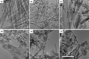
The effect of the physically adsorbed water and the grinding time on the surface morphology of FY-10G/PLG hybrid fluorescent pigments was observed using TEM and SEM (Figs 3, S4). The original PLG crystal bundles display good dispersion, and many individual nanorods 0.5–1.0 μm long can be observed (Fig. 3a). After loading of FY-10G, the PLG morphology does not change (Fig. 3b). However, the dispersion of PLG rod bundles is poor and they become shorter when the hybrid fluorescent pigments containing 95% physically adsorbed H2O are ground for 1.5 h (Fig. 3c). With the decrease in the physically adsorbed water content to 20%, the PLG rod bundles shorten considerably to form aggregations over the same grinding time (Fig. 3d). These phenomena may be attributed to the following: (1) the physically adsorbed water acts as a lubricant, decreasing the rubbing action among the PLG rod bundles and thus reducing the breakage of the PLG rods; and (2) the physically adsorbed water acts as a dispersion medium, promoting the dissociation of the crystal bundles under strong compression, shearing stress and looping stress. Therefore, the greater the physically adsorbed water content, the better the dispersion and the less the breakage of PLG rods.
TEM images of (a) PLG, (b) FY-10G/PLG-95/0 (without grinding), (c) FY-10G/PLG-95/1.5, (d) FY-10G/PLG-20/1.5, (e) FY-10G/PLG-95/1.0 and (f) FY-10G/PLG-95/3.0.

In our previous work, the grinding time influenced significantly the degree of dispersion and length of the PLG rods, both during dry grinding (Liu et al., Reference Liu, Wang and Wang2012) and during semi-dry grinding (Zhang et al., Reference Zhang, Wang, Mu, Wang and Wang2015a). A similar phenomenon is also observed in this work (Fig. 3e,f). After grinding for 1 h, the degree of aggregation of the PLG rod crystals evidently increases (Fig. 3e) and the rod length shortens compared with those without grinding (Fig. 3a,b). When the grinding time is prolonged to 3 h (Fig. 3f), the PLG rod crystals are significantly broken, accompanied by the formation of many submicron particles.
The above-mentioned phenomena are also demonstrated in Fig. S4. Hence, a larger amount of water adsorbed physically and an appropriate grinding time may effectively prevent the aggregation and breakage of the PLG crystal bundles and facilitate FY-10 G molecules to enter into the grooves of PLG crystals, which may be responsible for the stability of the FY-10G/PLG hybrid fluorescent pigments.
Thermal analysis
Figure 4 shows the TGA and DTG curves of the FY-10G, PLG and FY-10G/PLG hybrid fluorescent pigments. Major changes are observed in the TGA and DTG curves. In the DTG curves (inserts in Fig. 4), the mass losses observed at 59°C, 187°C and 393°C for FY-10 G and at 69°C, 187°C, 428°C and 584°C for PLG are mainly attributed to the decomposition of FY-10 G and the removal of the adsorbed water, zeolitic water, coordinated water and structural water, respectively. After incorporation of FY-10 G into PLG, the mass losses of the adsorbed water and zeolitic water at 69°C and 187°C do not change, but the mass loss at 350–700°C suggests that the incorporated FY-10 G molecules are loaded onto PLG via electrostatic interaction between the positively charged FY-10 G and negatively charged PLG (Wang et al., Reference Wang, Mu, Zhang, Zhang and Wang2016). According to the TGA curves (Fig. 4, main image), the mass loss of the hybrid fluorescent pigments (0.8%) at <70°C is related to the removal of the physisorbed H2O, which is similar to that of FY-10 G but lower than that of PLG (2.4%). This implies that FY-10 G molecules replace the physically adsorbed H2O and cover the external surface or groove of PLG in the process of grinding, which leads to the decrease in the physically adsorbed H2O content in the hybrid fluorescent pigment. This result is also consistent with the FTIR analysis and is also confirmed by the change in S BET, S micro, V total and V micro before and after the introduction of FY-10 G (Table S2). That is to say, the specific surface area and pore volume of the hybrid fluorescent pigment decrease due to the introduction of FY-10G. When the temperature varies from 70 to 280°C, the mass loss of the hybrid fluorescent pigments attributed to zeolitic H2O, the first structural –OH and small molecular compounds is lower than that of PLG. This implies that a proportion of the FY-10 G molecules replace partial zeolite water, which decreases the zeolite water content in the hybrid fluorescent pigments. This phenomenon is consistent with the results of Sanchez Del Rio et al. (Reference Sanchez Del Rio, Boccaleri, Milanesio, Croce, Van Beek and Tsiantos2009) and Giustetto et al. (Reference Giustetto, Wahyudi, Corazzari and Turci2011). However, the mass loss of the hybrid fluorescent pigments is greater than that of FY-10 G due to the removal of some of the zeolitic water. At temperatures of 280–480°C, the mass loss of FY-10 G exceeds 60%, whereas the mass loss of the hybrid fluorescent pigments is 10%, which is also lower than that of PLG. This may be due to the fact that the host–guest interaction of FY-10 G molecules with PLG enhances the thermal stability of FY-10G. At temperatures >480°C, the hybrid fluorescent pigments show greater mass loss compared with PLG due to the decomposition of FY-10G.
TGA and DTG curves of PLG, FY-10 G and FY-10G/PLG-95/1.5.

UV–Vis and fluorescence spectra
The UV-Vis diffuse reflectance spectrum of solid-state FY-10 G in Fig. 5 (insert) shows four main absorption peaks, namely three weak peaks in the UV region (249, 291 and 319 nm) and one broad peak in the visible region (an absorption from 370 to 550 nm). The PLG itself displays an absorption band with a maximum at ~250 nm, but no significant absorption in the visible region. After 32% of the FY-10 G (mass ratio of FY-10 G to PLG) is incorporated into PLG, the absorption spectra of the FY-10G/PLG hybrid fluorescent pigments are similar to those of FY-10 G (Fig. 5), and two weak peaks at 410 and 480 nm indicate that a small portion of the FY-10 G molecules aggregate in the PALG. These phenomena are also in accordance with Polette-Niewold et al. (Reference Polette-Niewold, Manciu, Torres, Alvarado and Chianelli2007). Moreover, the intensities of the absorption spectra increase with the increasing content of the physically adsorbed water. This may be related to the dispersion of the loaded FY-10G.
The effect of the physically adsorbed water content on the diffuse reflectance UV-Vis spectra of FY-10G/PLG hybrid fluorescent pigments (grinding time of 1.5 h).

Figure 6 shows the fluorescence spectra of pristine FY-10 G and FY-10 G loaded in PLG. The excitation and emission spectra of pristine FY-10 G in the deionized water are 440 and 500 nm (Fig. 6a, insert), respectively. When 32% of the FY-10 G is incorporated into PLG, the excitation spectrum of solid-state FY-10G/PLG hybrid fluorescent pigments is blue-shifted to 420 nm, while the emission spectrum shifts to 528 nm. The bathochromic shift of the emission bands in the PALG host might be explained in terms of the stereo effects associated with the pore structure and, in contrast with that in water, it reflects the greater-than-water polarity of the local environment surrounding the dye molecules (Yao et al., Reference Yao, Zhang, Shi, Gong, Zhang and Yang2001; Zhang et al., Reference Zhang, Cheng, Tang and Liao2003; Zhao et al., Reference Zhao, Li, He, Zhang, Huang and Zhang2005).
The effect of (a) the physically adsorbed water content and (b) grinding time on fluorescence spectra of FY-10G/PLG hybrid fluorescent pigments.

In addition, the intensities of the emission spectra increase with the increasing amount of physically adsorbed water (Fig. 6a) and when extending the grinding time from 0.5 to 1.5 h, but then decrease with further prolonging of the grinding time to 3 h (Fig. 6b). The reason for this behaviour may be that more physically adsorbed water and an appropriate grinding time effectively stop the aggregation and breakage of PLG rod bundles and improve the dispersion of FY-10 G loaded in PLG, which increases the intensity of the emission spectrum.
Environmental stability
The effect of the physically adsorbed water content
The effects of the physically adsorbed water content on the stability of the hybrid fluorescent pigments in various solutions are shown in Fig. 7. The absorption intensities of FY-10 G in the supernatant decrease with the increasing physically adsorbed water content following immersion in 1.0 mol L–1 HCl solution and ethanol for 24 h. These results indicate that a larger amount of physically adsorbed water is conducive to enhancing the stability of the hybrid fluorescent pigments in diluted acid and ethanol. This may be attributed to the following: (1) a larger amout of physically adsorbed water helps to prevent the aggregation and breakage of the PLG crystal bundles (Fig. 3c,d), which facilitates FY-10 G to enter into the external groove of PLG during the grinding process; and (2) the amount of physically adsorbed water has a significant effect on the pore characteristics of FY-10G/PLG hybrid fluorescent pigments (Fig. 8a). There are two maxima when the amount of physically adsorbed water is 95%: one at ~3.5 nm, which is attributed to the mesopores among the PLG layers, and another at 11.6 nm, which is related to the non-structural pores between crystal bundles of PLG (Zhang et al., Reference Zhang, Wang, Chen and Wang2010). As the amount of physically adsorbed water decreases from 65% to 13%, there is only one maximum, at ~3.5 nm, and its intensity decreases. This result indicates that the microstructure of PLG is retained well forlarger physically adsorbed water contents, and thus the hybrid pigments display good resistance to the external environment (e.g. acid and organic solvent).
The effect of the physically adsorbed water content on the absorption spectra of FY-10 G from FY-10G/PLG hybrid fluorescent pigments in 1.0 mol L–1 HCl solution and ethanol.

The effect of (a) the physically adsorbed water content and (b) grinding time on the pore-size distribution of the FY-10G/LG hybrid fluorescent pigments.

The resistance of FY-10G/PLG hybrid fluorescent pigments to alkaline media was also evaluated. There is no obvious effect of the physically adsorbed water content on the stability of the hybrid fluorescent pigments in 1.0 mol L–1 NaOH solution (Fig. S5a). However, the colour of the hybrid fluorescent pigments varies from yellow to brown, which is similar to the colour of PLG. This result is consistent with the results of our previous studies (Wang et al., Reference Wang, Mu, Zhang, Zhang and Wang2016), and it is ascribed to the fact that the ester group of the coumarin cycle of the dye molecule is attacked by the OH– ions in NaOH solution, or to the fact that the N+ linked to CH3 in the benzimidazole cycle reacts with the OH– ions.
The photostability of the FY-10G/PAL hybrid fluorescent pigments was tested through prolonged exposure to UV-B irradiation. The effects of the physically adsorbed water content and the irradiation time on the stability of the irradiated hybrid fluorescent pigments are shown in Fig. S6. The absorption intensities of FY-10 G in the supernatant decrease with the increasing physically adsorbed water content after being irradiated for 72 h. Therefore, the FY-10 G molecules loaded in the FY-10G/PLG hybrid fluorescent pigments with a more physically adsorbed water are difficult to elute due to the shielding effect of the groove of PLG on FY-10G. This trend is in accord with those seen after immersion of the hybrid fluorescent pigments in 1.0 mol L–1 HCl solution and ethanol. In addition, the maximum absorption peak at 419 nm of the hybrid fluorescent pigments shows a continuous decay in intensity as irradiation time increases (Fig. S6, insert), indicating that the FY-10 G molecules are gradually degraded. A similar phenomenon was reported by Yang et al. (Reference Yang, Tian, Xiao, Shang, Gong, Yao and Chen2001), and the photodegradation of the dye resulted from the formation of semi-oxidized dye free radical cations.
The effect of grinding time
The effect of grinding time on the stability of FY-10G/PLG hybrid fluorescent pigments is shown in Fig. 9. When the grinding time is <2 h, there is no significant difference in the absorption intensities of FY-10 G following immersion in 1.0 mol L–1 HCl solution for 24 h. The absorption intensities of FY-10 G in the centrifugate increase with the increase in grinding time from 2.5 to 3.0 h. A similar tendency is also observed in ethanol. These results indicate that a longer grinding time decreases the stability of the hybrid fluorescent pigments in acid and organic solvent. Zhang et al. (Reference Zhang, Wang, Mu, Wang and Wang2015a) reported that grinding time significantly influenced the environmental stability of methylene blue/PLG hybrid nanocomposites due to the disruption of the microstructure of PLG.
The effect of grinding time on the adsorption spectra of FY-10 G from FY-10G/PLG hybrid fluorescent pigments in 1.0 mol L–1 HCl solution and ethanol.

Figure 8b shows the effect of grinding time on the pore-size distribution of FY-10G/PLG hybrid fluorescent pigments. When the grinding time increases from 0.5 to 2.0 h, two maxima are observed: one sharp maximum at 3.5 nm and one broad maximum at 5.0, 8.5, 14.7 and 21.3 nm after grinding for 0.5, 1.0, 1.5 and 2.0 h, respectively. However, there is only a maximum at 3.5 nm after grinding for 2.5 and 3.0 h. The aforementioned results indicate that a longer grinding time decreases the pore-size distribution and pore volume due to damage to the PLG rods, and thus the stability of the hybrid fluorescent pigments is poor in acid and organic solvents. Therefore, it is essential to investigate the appropriate grinding time, because the grinding treatment on the hybrid pigments with an appropriate grinding time may enhance the interaction of dye molecules with PLG (Zhang et al., Reference Zhang, Wang, Mu, Wang and Wang2015a), which increases the stability of the hybrid fluorescent pigments and so extends the practical application of PLG-based hybrid pigments. Furthermore, the stability of the hybrid fluorescent pigments in 1.0 mol L–1 NaOH solution does not change with increasing grinding time (Fig. S5b).
The photostabilities of the FY-10G/PLG hybrid fluorescent pigments ground for various time were also tested through prolonged exposure to UV-B irradiation. There is no significant difference in the absorption intensities of FY-10 G derived from the hybrid fluorescent pigments following irradiation for 72 h when the grinding time is <2.0 h (Fig. S7). With further increases in the grinding time to 3 h, the absorption intensities increase. Thus, the stability of the hybrid fluorescent pigments depends on the microstructure of PLG during grinding. In addition, the maximum absorption peak at 419 nm in Fig. S7 (insert) does not shift with increasing irradiation time. Moreover, no change was observed in the absorption peak intensities when the irradiation time increases from 0 to 24 h, but it decreases with further increasing of irradiation time to 72 h. These results indicate that the FY-10 G molecules are degraded gradually with prolonged UV-B irradiation.
Conclusions
A FY-10G/PLG hybrid fluorescent pigment was prepared via facile semi-dry grinding. The FY-10 G molecules incorporated were mainly trapped on the external surface and grooves of PLG according to FTIR, TGA, specific surface area and pore volume analyses of PLG before and after the introduction of FY-10G. The introduction of PLG influenced the excitation and emission spectra of FY-10G, but no shifts were observed in the emission spectrum except for in terms of intensity with variation of the physically adsorbed water content and grinding time. The dispersion of PLG rod crystals and the pore sizes of the FY-10G/PLG hybrid fluorescent pigments are key factors that influence the intensities of their emission spectra and their resistance to dilute acid, organic solvent and UV irradiation. When the amount of physically adsorbed water exceeded 95% and the grinding time was <2 h, the resultant FY-10G/PLG hybrid fluorescent pigments displayed excellent resistance to dilute acid, organic solvent and UV irradiation. On the basis of the abovementioned results, the as-prepared FY-10G/PLG hybrid fluorescent pigments are promising for use in many fields, such as paint, printing and solid-state dye lasers.
Financial support
This work was supported financially by the Major Projects of the Natural Science Foundation of Gansu, China (18JR4RA001) and the Linze County Attapulgite Industry Development Open Project (LZKFKT-1901).
Supplementary material
To view supplementary material for this article, please visit https://doi.org/10.1180/clm.2021.12.