

INTRODUCTION: ROMANO-BRITISH CHILDHOOD
Human skeletal remains present the most direct form of evidence for studying past populations. Numerous palaeopathological studies of Romano-British adults have provided insight into population health, diet and lifeways, albeit with a bias towards urban cemeteries.Footnote 1 More recently, several studies have reported on health in both urban and rural contexts, yet the children did not receive emphasis.Footnote 2 Roman archaeology is no different from other branches of the discipline in its research concerns, therefore the childhood experience has not yet fully evolved as a dedicated subject in Roman scholarship. While stable isotope analysis of Romano-British children has received considerable attention,Footnote 3 the actual health status of children remains relatively unexplored and a comprehensive picture of growing up in Roman Britain is still lacking. As children are growing, they reflect adverse environmental conditions more readily than their parents.Footnote 4 Child health is therefore a very powerful indicator of overall population health and dynamics, making non-adult (0–17 years) palaeopathology a primary source of evidence for past lifestyles and living environments. The osteological term ‘non-adult’ is not without problems but allows us to label, observe and discuss immature individuals in the past without involuntarily attaching social age norms.Footnote 5 The cut-off age of 17 years is a product of the most accurate osteological ageing methods used and allows comparison between bioarchaeological datasets. Alongside age-at-death, a range of acquired conditions of ill-health can be observed in non-adult skeletons, which can provide information regarding cultural change, environmental pressures and life histories.
Literary evidence on Romano-British children is limited and, as so often in the study of Romano-British lifeways, we are forced to look to Rome for analogies and guidance. The classical literature, epigraphic and iconographic references provide a wealth of information on the Roman childhood experience.Footnote 6 However, particularly the written sources are based on individual accounts of a privileged minority, tainted with anecdotal undertones and with very little to no mention of the working classes, peasantry and slaves.Footnote 7 Inscriptions and imagery of Romano-British children are scarce and cannot fully communicate life histories of children from all orders of society within town and country.Footnote 8 It is difficult to trace the everyday realities of a child in Rome, let alone at the north-western fringes of the Empire, post-dating most of the ancient literature by several centuries. Yet, awareness of the life course of Roman children as described in important texts, such as Soranus’ second-century a.d. Gynaecology, is beneficial.Footnote 9 Soranus’ instructions on weaning practices,Footnote 10 believed to have been followed in Rome, may be detectable in the osteological record of Romano-British children. However, in the absence of historical sources, direct skeletal evidence of ill-health in the children is the most comprehensive means of exploring the childhood experience in Roman Britain. This study aims to provide an overview of childhood pathology across the province, giving new insights into growing up in both town and country. Most importantly, this analysis adds substantially to our current understanding of life in the understudied Romano-British countryside via the palaeopathology of the children.
CHILDREN FROM ROMAN ENGLAND: AN OVERVIEW OF HEALTH AND WELL-BEING
Acquired in childhood, cribra orbitalia and enamel hypoplasia have been addressed in skeletal populations across the Empire and in Rome itself, allowing insight into general health, nutritional status and, more specifically, the prevalence of malaria.Footnote 11 High prevalence of malaria favours individuals with genetic mutations of resistance, which can manifest as β-thalassaemia and other anaemias.Footnote 12 Cribra orbitalia and porotic hyperostosis are frequently observed in Italian Roman populations, suggesting high rates of acquired and genetic anaemic conditions.Footnote 13 Malaria is well recognised as an endemic disease in Classical Italy which impacted on morbidity and mortality to the extreme of yielding a ‘demography of malaria’, which may also have affected the southern areas of Roman Britain.Footnote 14 In Italy, correlations between high rates of marrow hypoplasia and malarious areas have been identified. In Roman Britain, however, cribrotic and enamel lesions as indicators of early childhood stress and a malarial threat were considerably less prevalent.Footnote 15
Important insights into child health in Roman Dorset are provided in several palaeopathological studies which highlight a decline in child health from the Iron Age through to the late Roman period.Footnote 16 Accounts of unprecedented detail of non-adult ill-health in Roman Britain stem from Lewis’ work on metabolic disease, trauma, tuberculosis and thalassaemia from 364 children (0–17 years) from third- to fifth-century Poundbury Camp, Dorchester, Dorset.Footnote 17 Elevated levels of metabolic disease at the site bear resemblance to those reported in post-medieval children.Footnote 18 The Poundbury Camp cohort included three non-adults who were likely to have suffered from thalassaemia, providing direct osteological evidence for immigrants from the Mediterranean in Roman Dorchester.Footnote 19 Particularly in the infants, high levels of blood-borne conditions and vitamin C and D deficiencies may be witness to early Christian fasting or ascetic practices adhered to by the women at the site.Footnote 20 We would also expect to see these lesions in children that suffered from thalassaemia major and subsequently could not feed.Footnote 21 Rickets was common at Poundbury Camp and has been interpreted as a result of swaddling, a Roman practice.Footnote 22 However, the clinical literature demonstrates that swaddling is an overall beneficial childcare practice with a negligible effect on exposure to sunlight and therefore vitamin D deficiency.Footnote 23 Rather, a restricted diet for mothers and children, coupled with a weaning diet as described in Soranus,Footnote 24 would have affected non-adult health at the site. The presence of childhood tuberculosis at Poundbury Camp indicates a tangible risk of infection in the children from Roman Dorchester.Footnote 25 Finding childhood tuberculosis confirms theories about urban living, such as crowded and unsanitary conditions, and suggests a depressed immune status of the children which allows for secondary infection and subsequent bony changes to occur.Footnote 26 As a singular site, Poundbury Camp exhibits an unexpectedly high degree of compromised child health.Footnote 27
Today, we believe that living in the countryside is beneficial in terms of both lifestyle and well-being and we tend to perceive this to be true of all time periods. However, rural life in Roman Britain will have come with significant health challenges due to the market economy and exploitative Imperial system.Footnote 28 Pitts and Griffin reported on compromised health in rural sites across central and southern England compared to urban cemeteries, characterised by higher levels of skeletal indicators of malnutrition, infection and strenuous physical activity.Footnote 29 Non-urban cemeteries exhibited fewer grave furnishings and greater inequality in the distribution of grave goods, possibly indicative of a relationship between living environment, social inequality and well-being in late Roman Britain. Redfern et al. studied urban-rural differences in first- to fifth-century burial grounds from Dorset.Footnote 30 The main findings included lower survivorship, higher mortality risk and smaller numbers of older adults in rural cemeteries. Excluding Poundbury Camp and associated cemeteries, individuals from rural sites showed higher rates of skeletal indicators of blood-borne disturbances and periosteal lesions which may relate to non-specific stress. These results challenge long-held perceptions of the benefits of rural living and highlight the need for a more dedicated study of health in the rural Romano-British population. We have to question what effect living and working arrangements had on the peasant population. Indeed, Romano-British society was highly stratified, where rich and poor would have collided in the countryside with peasant farmers, or perhaps even bonded tenants, on one side and powerful land owners on the other. Were people ‘employed’ by benevolent villa owners, or would land management have been more similar to later medieval manorialism, reliant on the exploitation of the workforce? This study seeks to demonstrate the important insights gleaned from the children and their palaeopathology, as a means to providing vital clues for discerning and conceptualising health in the countryside, social injustice and rural Romano-British lifeways.
MATERIALS AND METHODS
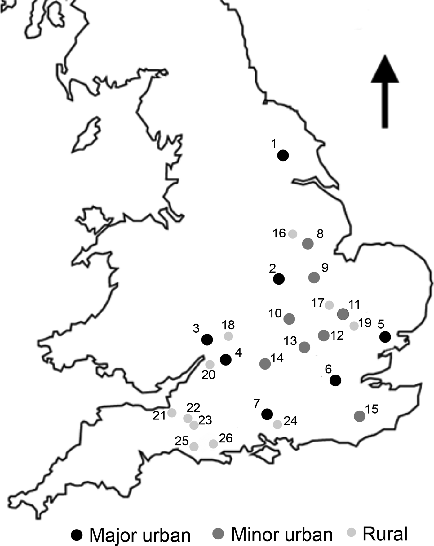
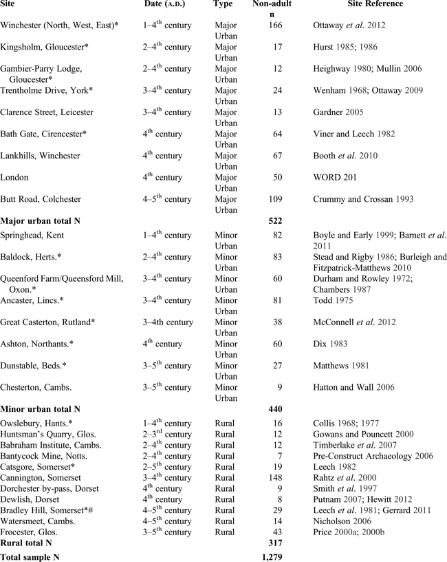
In the most comprehensive overview to date, 26 first- to fifth-century sites were included. The sample comprises 1,279 individuals (0–17 years), with 953 non-adult skeletons recorded by the author and osteological data from a further 326 gathered from (un)published reports (Table 1, fig. 1). Three sites yielded a limited number of first-century burials (Winchester (north, west and east), Springhead, Owslebury). Sources include the Museum of London Wellcome Osteological Database (WORD),Footnote 31 published site reports and grey literature reports from the commercial sector. Poundbury Camp was deliberately excluded from analysis, since the site is not representative of non-adult health in major urban settlements as demonstrated in detail by Rohnbogner and Lewis.Footnote 32 Sites were divided into major urban (coloniae, civitas capitals), minor urban (small towns, roadside settlements) and rural categories (villae, farmsteads, villages), following settlement classifications discussed in the literature.Footnote 33 Settlement classification in Roman Britain is complex and the terms major urban, minor urban and rural seek to acknowledge and describe the economic and administrative status of walled and unenclosed settlements, enabling comparisons in health across three distinct living environments. Age categories correspond with developmental stages and were used to minimise bias and allow cross-site comparison: perinate (<42 weeks gestation), 0.0–1.0 years, 1.1–2.5 years, 2.6–6.5 years, 6.6–10.5 years, 10.6–14.5 years and 14.6–17.0 years. The sample recorded by the author was aged using tooth crown and root formation stages,Footnote 34 long-bone length regression formulaeFootnote 35 and skeletal fusion and maturation.Footnote 36 A variety of pathological conditions described below were taken into consideration to explore ill-health in Romano-British children.
Map of sites with non-adult remains included in survey.

INDIVIDUAL SITE ASSEMBLAGE DETAILS

* site samples recorded by the author to attain primary data.
# non-adult burials were recovered from within the settlement boundaries associated with a fourth-century building (Leech et al. Reference Leech, Besly and Everton1981), rather than from the fifth-century cemetery (Gerrard Reference Gerrard2011).
READING THE SIGNS: NON-ADULT PALAEOPATHOLOGY
A range of skeletal lesions may develop during childhood, as a result of blood-borne diseases, non-specific stress and infection, metabolic disease and trauma. The presence of pathological conditions, together with the age of the affected individual, allows observations on the prevalence of ill-health within a population. In turn, social, behavioural and biological factors can be explored, including child-rearing practices, living environment and maternal health.
BLOOD-BORNE DISEASES
Pitting and porosity on the outer table of the skull (porotic hyperostosis) and orbital roof (cribra orbitalia) indicate blood-borne disorders. Non-adults display lesions more frequently than adults due to a reduced capacity of sustaining higher red blood cell production.Footnote 37 The palaeopathological literature has ascribed cribra orbitalia and porotic hyperostosis to iron deficiency anaemia, arising from excessive blood loss, insufficient dietary iron intake, intestinal parasites or diarrhoea.Footnote 38 Lesions also stem from vitamin B12/B9 deficiency in megaloblastic anaemias which may co-occur with iron deficiency.Footnote 39 The informative value of cribra orbitalia and porotic hyperostosis is considerable, including general childhood health, maternal health and feeding practices. A largely plant-based diet will leave the mother deficient in vitamin B12 and iron. In turn, the breastfed infant is nursed on milk deficient in vitamins and minerals. Prolonged breastfeeding without adequate supplementation in the growing child will eventually lead to deficiency. Lastly, haemolytic anaemias, such as thalassaemia, are recognisable in the osteological record.Footnote 40
STRESS AND INFECTION
Enamel hypoplasia
Enamel hypoplasia are linear bands or pits in tooth crown enamel, a retrospective record of early childhood stress.Footnote 41 Defects can affect all teeth and are most often recorded on the crowns of the anterior permanent dentition, formed until the age of four.Footnote 42 Generally, enamel hypoplasia are a non-specific indicator of poor health and stressors may include trauma, mal- or undernutrition, fever and infection.Footnote 43 Deciduous canines may be affected by unilateral pitted enamel defects, arising from weakened bone structure coupled with minor trauma from normal motor development during infancy. Their aetiology is complex and probably not linked with a systemic cause.Footnote 44
Non-specific infection
Endocranial lesions are found on the inner table of the skull.Footnote 45 Lesions may occur secondary to infection, trauma, tumours or meningitis and have been observed in cases of tuberculosis and vitamin deficiencies.Footnote 46 Sub-periosteal new bone formation is recognisable as raised, porous patches of new bone.Footnote 47 This type of lesion is an inflammatory response to infection, trauma, circulatory disorders, joint disease, haematological disease, skeletal dysplasia and metabolic or neoplastic disease.Footnote 48
Tuberculosis
Tuberculosis is a disease of malnutrition, poverty and overcrowding. It is a chronic infectious disease of the lungs, skin, lymph nodes, intestines and, in rare cases, bones and joints.Footnote 49 The bacterial genus specific to humans, Mycobacterium tuberculosis, is spread via airborne droplets, sputum or human waste and can be transmitted from mother to foetus.Footnote 50 Ingesting infected animal products will spread the animal equivalent bacillus, Mycobacterium bovis, from bovines to humans.Footnote 51
METABOLIC DISEASE
Vitamin D deficiency
The prohormone vitamin D is absorbed via the intestine or formed by the skin's dermal cells in response to ultraviolet light and is vital to the formation and maintenance of healthy bone structure.Footnote 52 In children, especially infants, chronic vitamin D deficiency has its most marked impact, leading to rickets and osteomalacia. Rachitic children exhibit unmineralised bone that is porous in appearance and, when mechanical forces are applied, is prone to characteristic bending deformities. Apart from exposure to sunlight and associated cultural practices, rickets also informs on calcium deficiency, female health and patterns in transitional feeding.Footnote 53
Scurvy
The clinical condition of scurvy results from chronic vitamin C deficiency. Non-adult skeletons, especially those of infants and young children are more likely to exhibit scorbutic lesions due to rapid growth.Footnote 54 Skeletal growth is slowed down and sub-periosteal haematomas occur at weakened walls of small blood vessels, yielding characteristic porous patches of new bone, particularly in the skull.Footnote 55 Apart from direct evidence for a lack of fresh fruits and vegetables, scurvy in archaeological populations is a vehicle for exploring resource stress, social hierarchies, ecology and behaviour.Footnote 56
TRAUMA
Dislocation, alteration to the shape of the bone, partial and complete breaks are distinct skeletal injuries observable in the osteological record.Footnote 57 Childhood fractures are a vital tool for providing information on occupational stress and exposure to trauma. Paediatric bone may yield distinctive fractures that can quickly lose visibility in the palaeopathological record due to fast remodelling.Footnote 58
The following methods were used by the author. Crude prevalence of enamel hypoplasia was recorded macroscopically.Footnote 59 Infections of a non-specific nature were recorded using sub-periosteal new bone formation and endocranial lesions.Footnote 60 Tuberculosis was diagnosed in the presence of lytic lesions in the spine with visceral rib lesions and widespread new bone formation.Footnote 61 Cribra orbitalia and porotic hyperostosis were scored according to Stuart-Macadam.Footnote 62 A diagnosis of thalassaemia was based on the presence of costal osteomas or ‘rib-within-a-rib’,Footnote 63 osteopenia and generalised cortical thickening, cribra orbitalia and porotic hyperostosis.Footnote 64 Vitamin D deficiency was identified following the criteria of Ortner and Mays, Ortner, and Brickley and Ives.Footnote 65 Vitamin C deficiency was scored according to Brickley and IvesFootnote 66 and the ‘Ortner criteria’ for lesions on the skull and metaphyseal ends of long bones.Footnote 67
As expected, working with secondary palaeopathological data presents limitations, as recording methods and reporting standards may differ. The Chi-squared 3 × 2 non-parametric statistic (X2) was used to test for differences between groups. False positives were avoided by setting the confidence interval at 99.5 per cent (p = 0.005) and using the test sparingly only when percentages indicated great differences between groups.
RESULTS
AGE-AT-DEATH
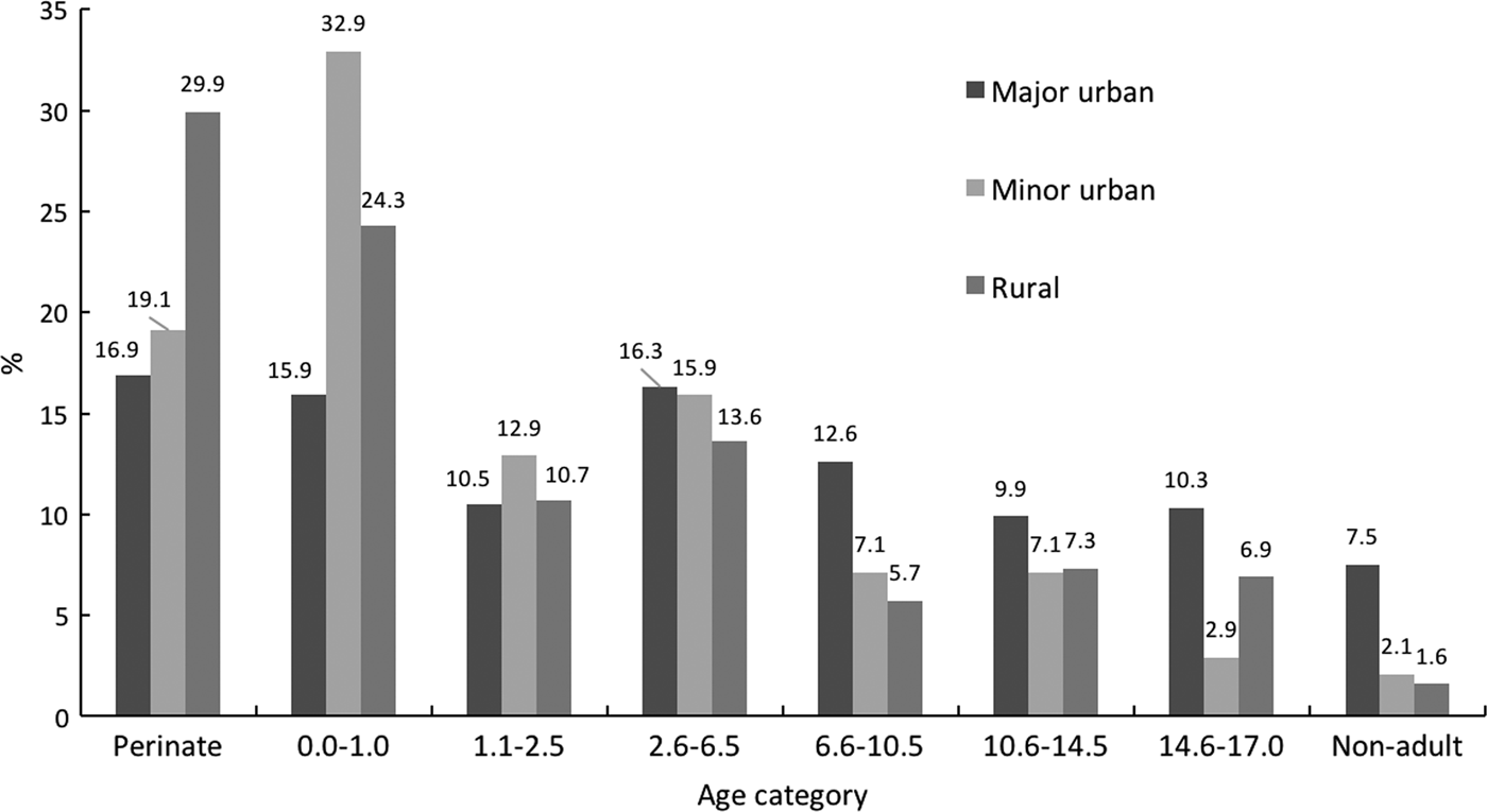
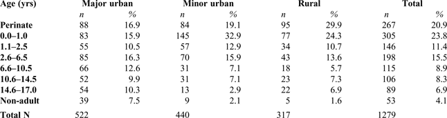
There were significantly fewer perinates (16.9 per cent, n = 88; X2 = 21.71, d.f. = 2, p < 0.001) and infants (15.9 per cent, n = 83; X2 = 38.25, d.f. = 2, p < 0.001) reported from major urban sites. Most striking were the high numbers of rural perinates (29.9 per cent, n = 95) and infants from minor urban sites (32.9 per cent, n = 145). In the 6.6–10.5 year old group, significantly more individuals come from major urban cemeteries than elsewhere (12.6 per cent, n = 66; X2 = 14.98, d.f. = 2, p < 0.001). The 14.6–17.0 year olds were significantly more frequent in major urban cemeteries (10.3 per cent, n = 54; X2 = 20.25, d.f. = 2, p < 0.001) compared with minor urban sites (2.9 per cent, n = 13) (Table 2; fig. 2).
Percentage distribution of ages-at-death.

AGES-AT-DEATH OF NON-ADULTS FROM MAJOR URBAN, MINOR URBAN AND RURAL SETTLEMENTS OF ROMAN ENGLAND

% of site total
THE PATHOLOGY
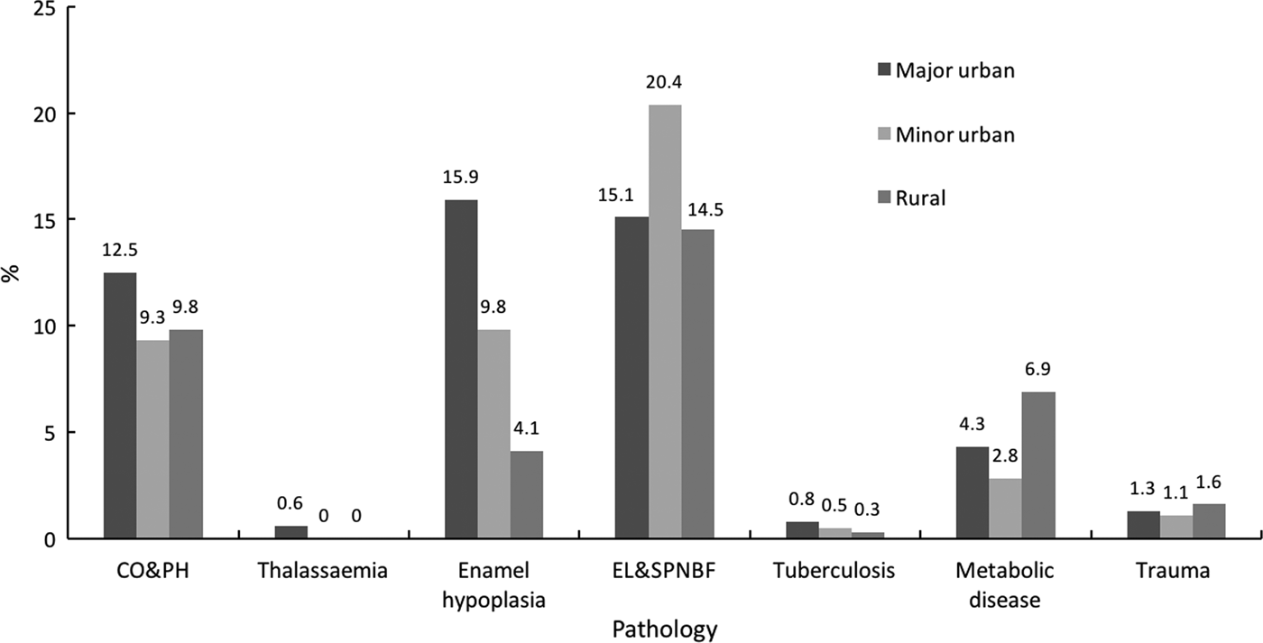
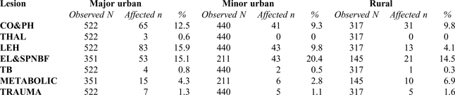
The distribution of lesions across the settlement types was not uniform (Table 3). Elevated rates of cribra orbitalia and porotic hyperostosis, thalassaemia, enamel hypoplasia and tuberculosis were found in the major urban cohort (fig. 3). No additional cases of probable thalassaemia were reported, other than those already discussed in the literature.Footnote 68 Infants were excluded from analysis for infectious and metabolic diseases, as their bones are increasingly reactive and porous, mimicking lesions of pathological origin.Footnote 69 Deficiency diseases of vitamin D and C in children aged 1.1 years and older were most frequent in the rural sample at 6.9 per cent (n = 10) (fig. 3).
Overview of percentage distributions of pathological lesions (CO: cribra orbitalia, PH: porotic hyperostosis, EL: endocranial lesions, SPNBF: sub-periosteal new bone formation).

NUMBER AND PERCENT OVERVIEW OF NON-ADULT PATHOLOGY BY LESION

% affected of observed by age group and site type; CO: cribra orbitalia, PH: porotic hyperostosis, THAL: thalassaemia, LEH: linear enamel hypoplasia, EL: endocranial lesions, SPNBF: sub-periosteal new bone formation, TB: tuberculosis.
HAEMATOPOIETIC LESIONS
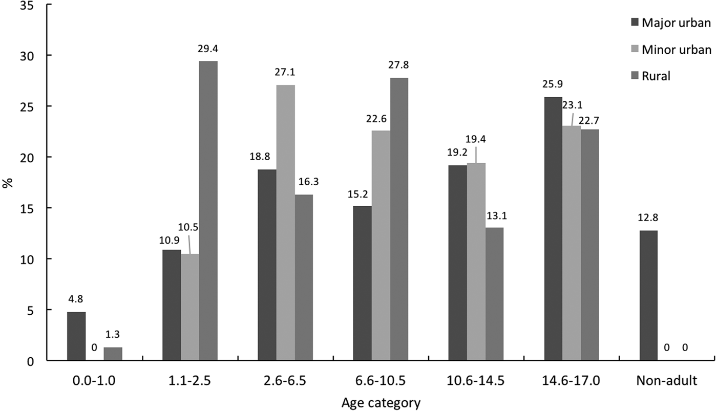
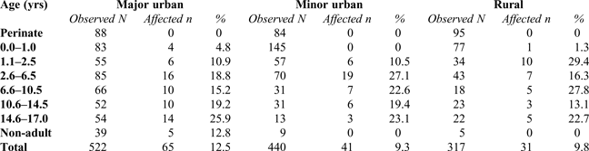
High rates of cribra orbitalia and porotic hyperostosis were observed in the rural children aged 1.1–2.5 (29.4 per cent, n = 10) and 6.6–10.5 years old (27.8 per cent, n = 5). The rates reported from minor urban sites were elevated in 2.6–6.5 year olds (27.1 per cent, n = 19), where rates from major urban and rural sites were similar at 18.8 per cent (n = 16) and 16.3 per cent (n = 7) respectively (Table 4; fig. 4).
Percentage distribution of haematopoietic lesions by age group.

NUMBER AND PERCENT OF HAEMATOPOIETIC SKELETAL LESIONS (CRIBRA ORBITALIA AND POROTIC HYPEROSTOSIS)

% affected of observed by age group and site type
STRESS AND INFECTION
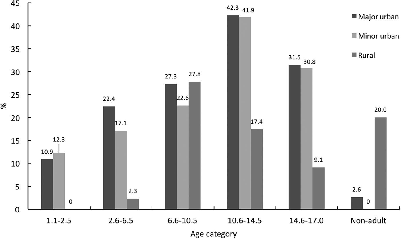
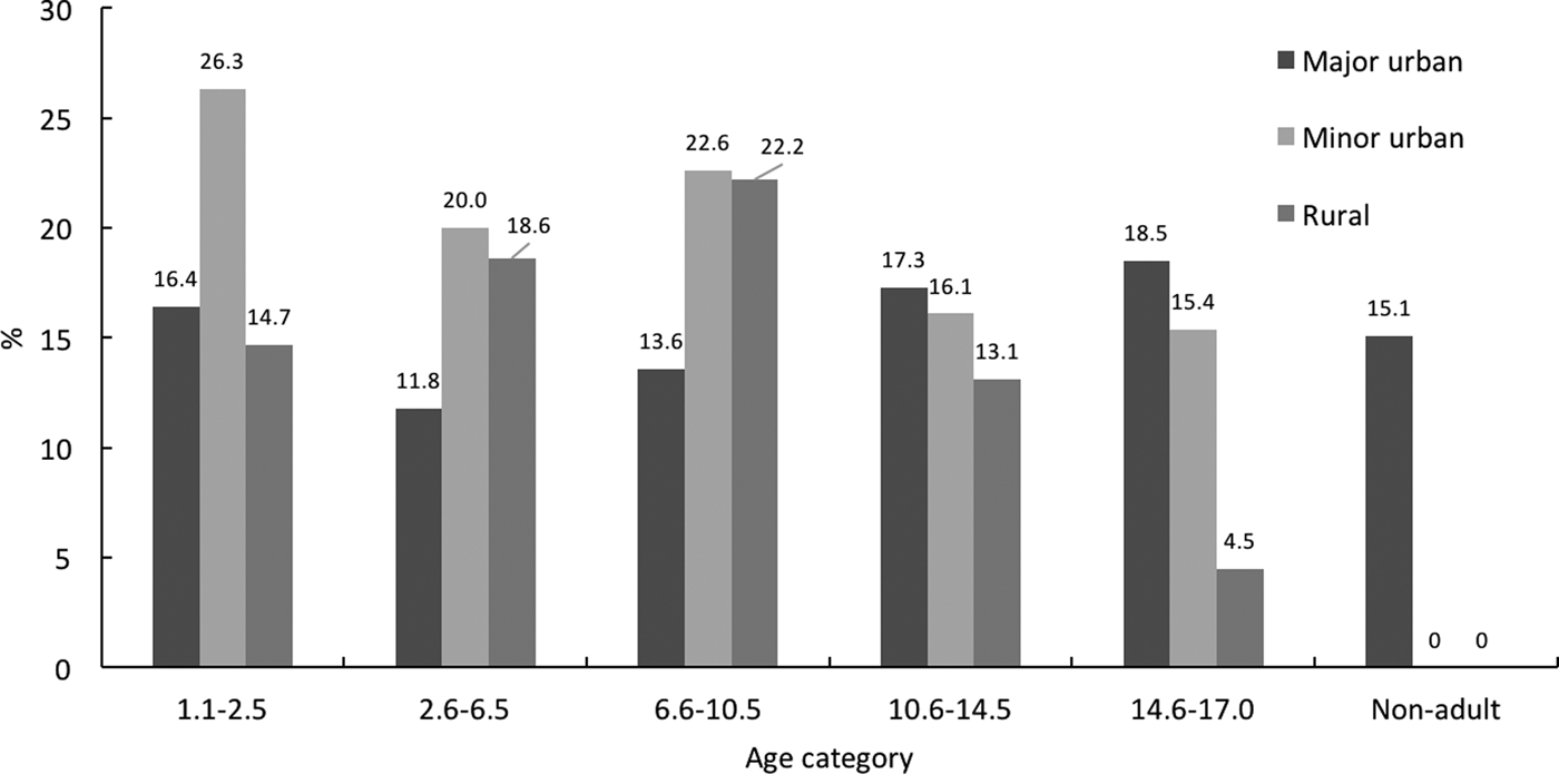
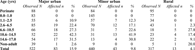
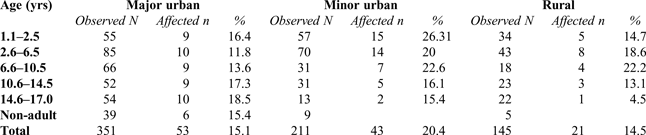
Enamel defects were significantly more frequent in major urban sites (15.9 per cent, n = 83; X2 = 29.43, d.f. = 2, p < 0.001). Enamel hypoplasia in deciduous canines were reported in urban non-adults up to 2.5 years old. In 6.6–10.5 year olds, rates are similar in the major urban (27.3 per cent, n = 18) and rural sites (27.8 per cent, n = 5). From 10.6 years and older, the rural enamel hypoplasia rate is lower than in the urban sites, although not statistically significant (Table 5; fig. 5). Endocranial lesions and sub-periosteal new bone formation occurred at similar rates in major urban (15.1 per cent, n = 53) and rural contexts (14.5 per cent, n = 21), and were most prevalent in the minor urban sample (20.4 per cent, n = 43). Children from minor urban contexts showed the highest rate amongst the 1.1–2.5 year olds (26.3 per cent, n = 15). In 2.6–6.5 and 6.6–10.5 year olds, lesions occurred at similar rates in the minor urban and rural sites at rates of 20.0 per cent (n = 14)/18.6 per cent (n = 8) and 22.6 per cent (n = 7)/22.2 per cent (n = 4). In 10.6–14.5 and 14.6–17.0 year olds, major urban rates were highest. In 14.6–17.0 year olds, the rural rate was low at 4.5 per cent (n = 1), although the result was not statistically significant (Table 6; fig. 6).
Percentage distribution of enamel hypoplasia by age group.

Percentage distribution of sub-periosteal new bone formation and endocranial lesions by age group.

NUMBER AND PERCENT OF CRUDE PREVALENCE RATES OF ENAMEL HYPOPLASIA

% affected of observed by age group and site type
NUMBER AND PERCENT OF LESIONS INDICATIVE OF INFLAMMATION/INFECTION (SUB-PERIOSTEAL NEW BONE FORMATION, ENDOCRANIAL LESIONS)

% affected of observed by age group and site type
TUBERCULOSIS
Individuals with lesions that are either strongly suggestive of, or possible tuberculosis are few (n = 9). Suspected and probable cases were reported from all three site types, with a bias towards major urban settlements (0.8 per cent, n = 4).Footnote 70 A further two probable cases were identified at minor urban Ancaster and Ashton and one possible case at rural Cannington.Footnote 71
METABOLIC DISEASE
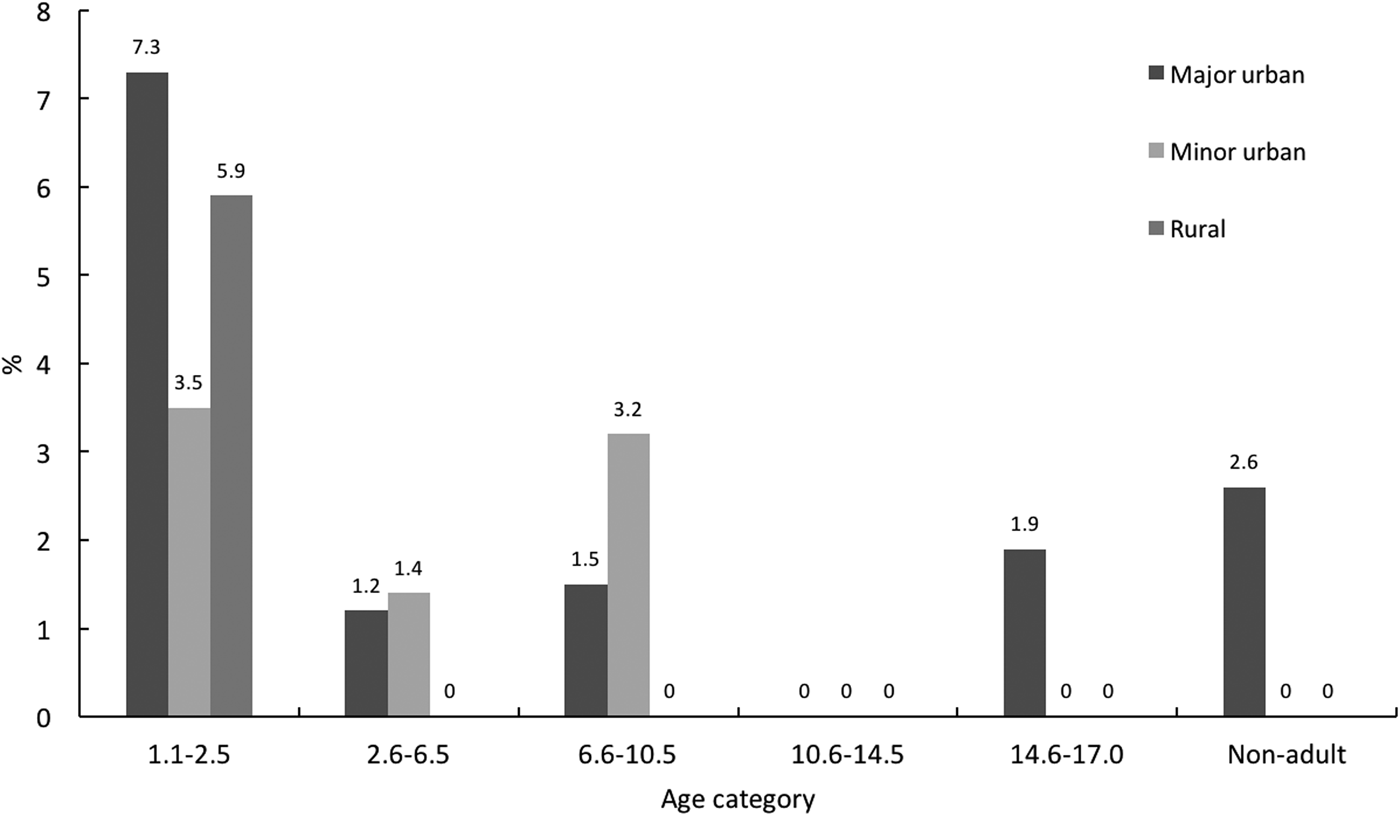
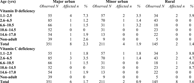
Vitamin D deficiency was reported in 14 individuals (2.0 per cent) of all ages, except amongst 10.6–14.5 year olds. Rickets was most frequent in the 1.1–2.5 year age group (5.5 per cent, n = 8), where it was most prevalent in major urban sites (7.3 per cent, n = 4) and lowest in minor urban settlements (3.5 per cent, n = 2). In the rural sample, vitamin D deficiency was only reported in 1.1–2.5 year olds at 5.9 per cent (n = 2). An adolescent female from Lankhills, Winchester, was the oldest individual with vitamin D deficiency. Lesions were healed implying that rickets occurred earlier in childhood (Table 7; fig. 7).Footnote 72
Percentage distribution of vitamin D deficiency by age group.

NUMBER AND PERCENT OF VITAMIN D AND C DEFICIENCIES

% affected of observed by age group and site type
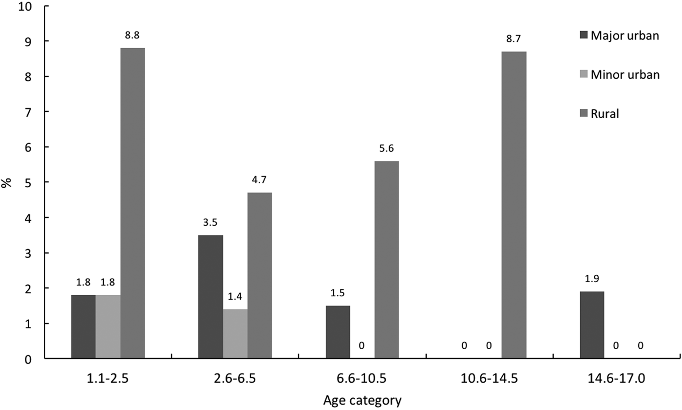
A total of 16 individuals (2.3 per cent) across all ages were reported as vitamin C deficient. Prevalence was highest in 1.1–2.5 year olds (3.4 per cent, n = 5), where 8.8 per cent (n = 3) of rural individuals were affected and only 1.8 per cent (n = 1) of non-adults from major urban and minor urban settlements. The distribution was not significant (X2 = 3.84, d.f. = 2). In the minor urban sample, scurvy was absent from the age of 2.6–6.5 years. The rural cohort exhibited higher prevalence rates across all age groups. The oldest affected individual was a 14.6–17.0 year old from Bath Gate, Cirencester, with healed lesions (Table 7; fig. 8).
Percentage distribution of vitamin C deficiency by age group.

TRAUMA
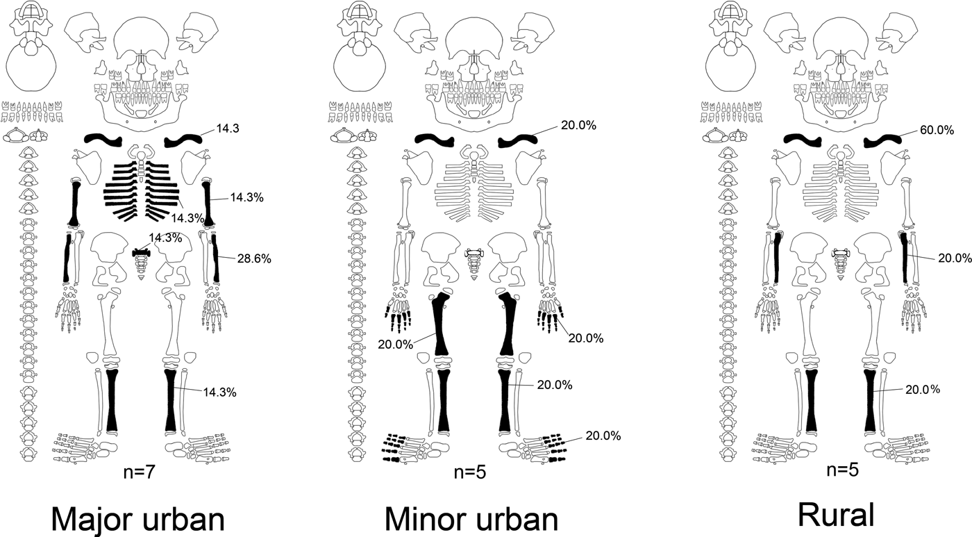
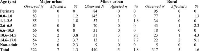
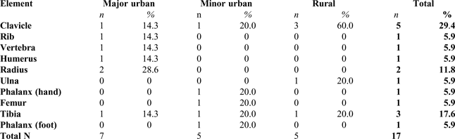
Trauma affected 17 individuals from all three settlement types, the highest rate at 1.6 per cent (n = 5) in the rural sample. The majority of trauma was identified in children aged 10.6 years and older (64.7 per cent, n = 11). Higher fracture rates were seen in 10.6–14.5 year olds from minor urban sites. Rural 14.6–17.0 year olds exhibited the highest fracture rate at 9.1 per cent (n = 2) (Table 8). The left side (n = 9) was more often affected than the right (n = 5). Clavicular fractures were most frequent (29.4 per cent, n = 5), followed by tibial (17.6 per cent, n = 3) and radial fractures (11.8 per cent, n = 2), the latter most common in the major urban sample (n = 2, 28.6 per cent).Footnote 73 Minor urban non-adults exhibited similar fracture rates of the clavicle, long bones of the lower limbs and hand/foot phalanges (fig. 9). Clavicular fractures were most frequent in rural non-adults (60.0 per cent, n = 3) (Table 9).
Distribution of fracture locations by settlement type. Template adapted from Roksandic (Reference Roksandic2003, fig. 2).

NUMBER AND PERCENT OF TRAUMA

% affected of observed by age group and site type
FRACTURE SITES BY SKELETAL ELEMENT

% of total fractures by site
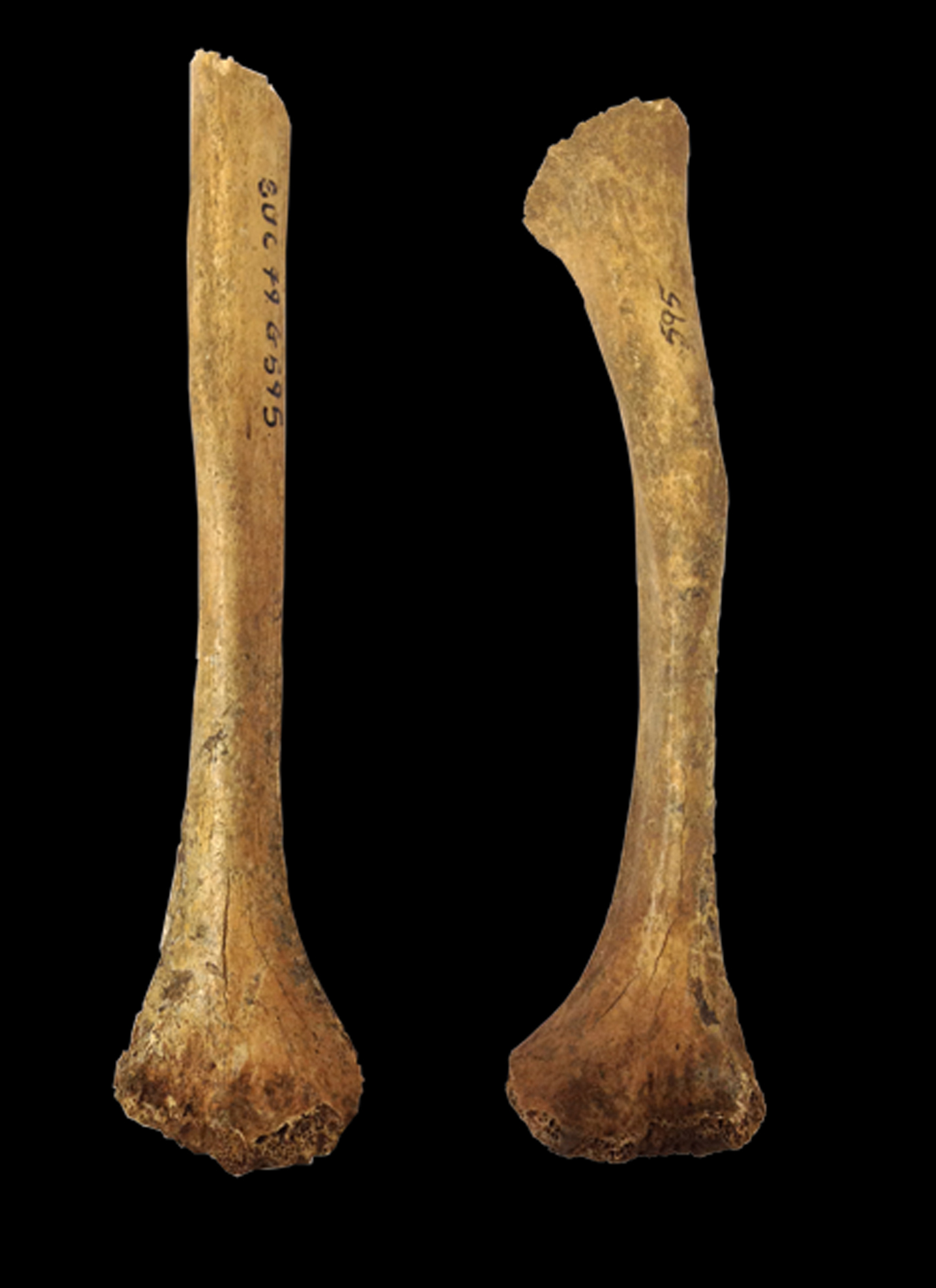
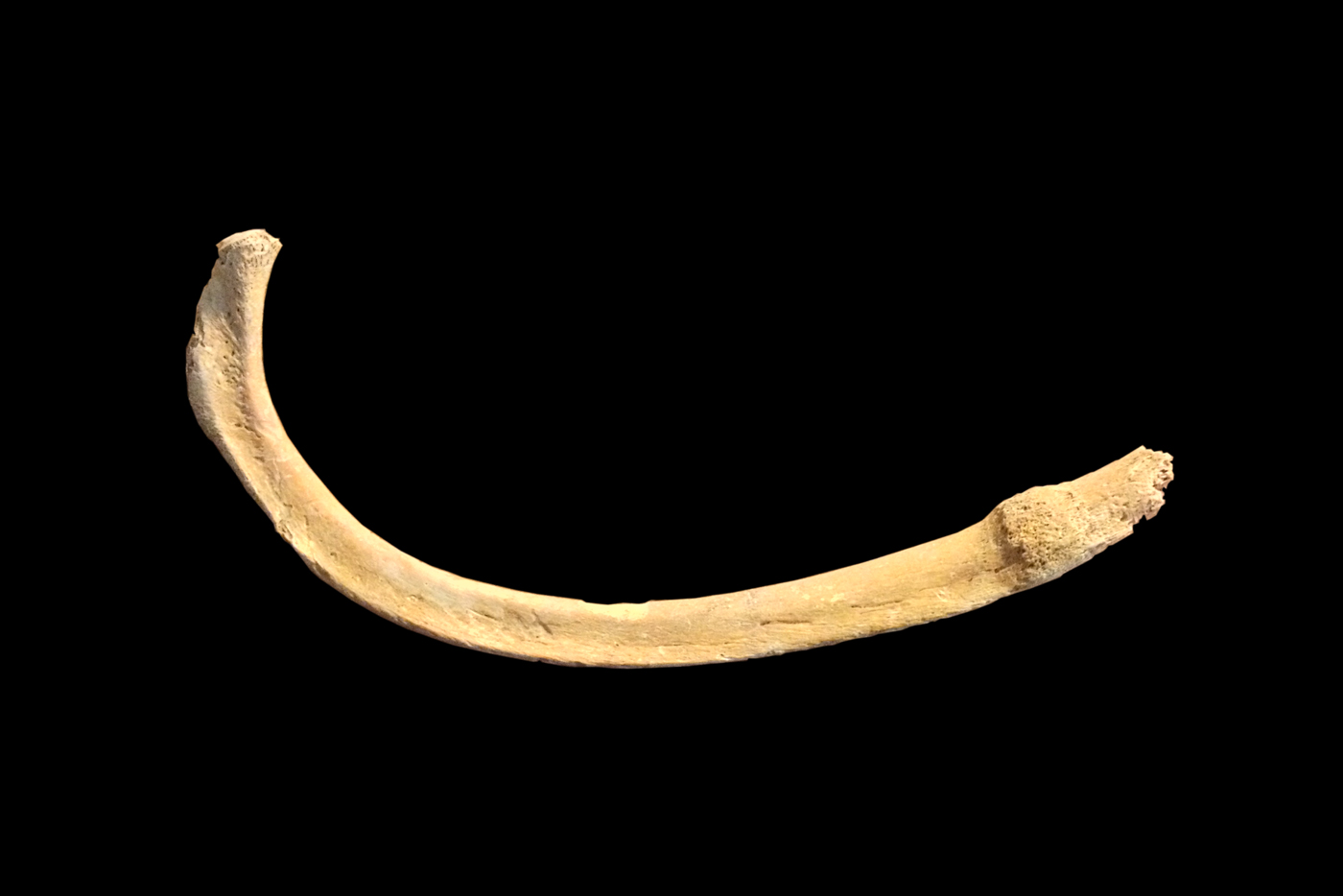
Falls onto the shoulder, outstretched arm or hand may cause fractures to the clavicle, humerus, radius or ulna (figs 10 and 11).Footnote 74 Tibial and femoral fractures require considerable force either through violent injury, a direct blow to the element or a strong twisting force.Footnote 75 The youngest individual with trauma was a Winchester infant with a healing rib fracture (fig. 12). The fracture may have arisen due to direct trauma,Footnote 76 complications in labour,Footnote 77 be a stress fracture from repeated coughing or vomiting,Footnote 78 or even be caused by accidental, over-zealous swaddling and infant handling.Footnote 79
Digital radiograph of right clavicle with healed fracture (top) at the mid-shaft. Bottom is left clavicle for comparison. From Queensford Farm/Mill, skeleton 51/232. (With permission from Oxfordshire Museums Service)

Right: left humerus with fracture at the mid-shaft, resulting in prominent angulation and changes to the proximal and distal epiphyseal ends. Left: right humerus for comparison. From Butt Road, Colchester, skeleton 595. (With permission from Colchester and Ipswich Museums)

Infant left rib with fracture callus. From Winchester (Victoria Road East cemetery), skeleton 444. (With permission from Hampshire Cultural Trust)

DISCUSSION
This study is the largest scale analysis of non-adult health in Roman Britain to date, specifically focusing on morbidity and mortality of 0–17 year olds from urban and rural contexts. Achieving as broad an overview as possible, the study relies on data compiled through primary analysis by the author and on relevant data drawn from (un)published reports and databases (secondary data). This approach presented challenges. The secondary data are flawed due to inconsistent methods and variation in recording. Hence, the study provides a broad-brush approach, yielding observations of initial trends that can be explored in more detail using the primary data alone.Footnote 80 Osteological analysis is reliant on individuals that have died and been buried at a particular siteFootnote 81 and can never be truly representative of all individuals that have lived and died in major urban, minor urban or rural settlements. The chronology of the sample means that the majority of sites date to the later phases of Roman Britain when inhumation was the more common funerary rite. Limitations are also introduced by excavation and preservation bias, dictating the spatial and geographical spread of the sample. The North is poorly represented with only one major urban site, Trentholme Drive, York. The West Country is represented by a cluster of rural sites in Dorset and Somerset. Instead, the focus is primarily on the central and, to a lesser extent, southern areas of Roman Britain (fig. 1). The rural sample is comparatively small, a product of fewer commercial investigations of rural sites, a focus on urban archaeology and the possibility of a bias towards cremation rites on rural sites. In the adolescents aged 14.6–17.0 years, we have to consider the possibility of an artificial peak due to different ageing techniques followed in reports and the subsequent inclusion of older individuals. Some interesting trends in demography, stress and metabolic disease between the urban and rural groups have become apparent. Age-at-death and the overall distribution of enamel hypoplasia reached statistical significance between urban and rural groups at 99.5 per cent confidence while a peak in vitamin C deficiency is apparent in the rural children.
INFANT MORTALITY: REALITY OR MIRAGE?
High levels of perinatal burials in the rural sample and high representation of infants in minor urban cemeteries were witnessed. Neonatal deaths are caused by endogenous (i.e. biological and genetic) factors and reflect problems inherent at birth, which are most commonly seen in rural groups today.Footnote 82 Scott and Duncan suggested that environmental factors, including infection, nutrition, poisoning and accidents, are decisive in post-neonatal mortality.Footnote 83 These would reflect different stresses experienced by mothers and babies in urban and rural environments in Roman Britain. Previous studies have shown that urbanisation and increased industrialisation in past populations triggered elevated post-neonatal mortality.Footnote 84 The overcrowding, pollution and infection seen in major urban environments would have presented greater risks for post-neonatal mortality.Footnote 85 The results for perinatal and infant mortality suggest that these dangers did not exist in Romano-British urban sites. This seems unlikely and differences are probably due to differential infant burial rites and recovery.Footnote 86 Some rural sites have high proportions of neonate burials, often recovered from within the settlement boundaries, including Owslebury, Hampshire (43.8 per cent of total non-adult burials), Catsgore, Somerset (52.6 per cent), and Frocester (81.4 per cent) and Huntsman's Quarry (83.3 per cent) in Gloucestershire. The large percentage of perinates reported at Frocester is even more remarkable for the total absence of older infants, suggesting this may be a designated perinatal burial site. Some major urban cemeteries had low numbers of perinates, particularly the London burial grounds (4.0 per cent) and Butt Road, Colchester (2.8 per cent), with no perinates at Trentholme Drive, York, and the Gloucester cemeteries. Low representation of infants in managed cemeteries is not exclusive to Roman Britain.Footnote 87 Distinct rites for those dying within the first year of life may have been a contemporary ritual practised across pockets of the Empire. Pliny the Elder describes children as lacking a soul until the age of teething at around six months old,Footnote 88 possibly accounting for differential treatment in death.Footnote 89 Funerary practice may have dictated the interment of babies and infants within the settlement boundaries, in clusters in a dedicated area of the cemetery, or at a separate site altogether, perhaps even in ‘infant corners’ that are yet to be excavated, ultimately impacting on the urban and rural rates of infant mortality observed in this study.Footnote 90
THE WEANLING'S DILEMMA
Pathological lesions in children up to 2.5 years old attest to inadequate strategies in supplementary feeding. Insufficient calcium in urban weanlings may have been a problem, as supported by the highest rate of rickets in 1.1–2.5 year olds from major urban sites (7.3 per cent). Calcium deficiency prompting rickets is usually seen after the age of two years old following inadequate weaning strategies.Footnote 91 However, as we cannot differentiate between rickets caused by either calcium or vitamin D deficiency, we have to consider the effect of clothing, childcare practices and living environment.Footnote 92 Mothers in major urban towns may have kept their children indoors to recover and rest. Subsequently, vitamin D deficiency ensued, possibly exacerbated by low calcium levels in the diet, prolonged breastfeeding and gastrointestinal maladies that limited calcium absorption and ultimately caused rachitic lesions.Footnote 93
A slight increase in mortality is observed in 1.1–2.5 year olds from minor urban sites, which may correspond with the peak in infection observed in this age group and attest inadequate weaning strategies.Footnote 94 Rural weanlings demonstrate elevated levels of scurvy, cribra orbitalia and porotic hyperostosis when compared to their urban counterparts. Suitable foods may have been scarce. Vitamin C deficiency may have been a product of cereal-based supplementary foods, cooking/boiling of fresh produce and nursing by a deficient mother or wet nurse.Footnote 95 Anaemia may have been prompted by infection, high pathogen load and parasites introduced by a diet low in iron and unhygienic feeding practices.Footnote 96
IMPLICATIONS OF CHILDHOOD TUBERCULOSIS
Generally, evidence for infection was present in all three site types. Reporting of tuberculosis was low overall, but reinforces notions of a crowded and unsanitary urban environment.Footnote 97 Low-quality air may have been a problem in both town and country, as demonstrated by skeletal lesions indicative of respiratory infections such as bronchitis.Footnote 98 Smoke off the hearths and poor ventilation would have caused respiratory disorders in rural dwellings, while close contact with livestock would have caused transmission of T. bovis to humans.Footnote 99 The effects of childhood tuberculosis in Roman Britain may have been felt widely. Blindness, deafness and mental retardation follow when the initial infection spreads to the meninges of young children under four years old.Footnote 100
ADOLESCENCE AND WORKING LIVES
Proportionately higher rates of 6.6–10.5 and 14.6–17.0 year olds were observed in major urban settlements. Their lower numbers in non-urban cemeteries indicate that older children may have migrated to major urban settlements. Infections of non-specific origin were on the rise in rural children until the age of 6.6–10.5 years, after which they declined from 22.2 per cent to 4.5 per cent in 14.6–17.0 year olds. An inverse pattern was apparent in the major urban cohort, with infections among the lowest until 6.6–10.5 years, followed by an increase to 18.5 per cent in 14.6–17.0 year olds. This opposite relationship further supports the hypothesis of the migration of older children into the towns. Migration between rural and urban populations has been suggested previously in regional studies from Dorset and London.Footnote 101 The urban environment exposed incoming children to new pathogens and demanding physical labour, eventually raising mortality.Footnote 102 Although there is limited documentary evidence, the existence of child (forced) labour and a young start to the working life is feasible.Footnote 103 Children from as young as five years old would have been tasked with household chores, with working life commencing in the early teens or even pre-teens.Footnote 104
The majority of the rural population would have performed strenuous agricultural labour.Footnote 105 Workers needed adequate nutrition for energy expenditure in this physically demanding job, while also being exposed to accidents and infectious diseases via livestock. Fracture locations in the rural cohort may bear witness to working with traction animals. Spinal degeneration in a 16-year-old female from Cannington further demonstrates the extent of intense physical activity.Footnote 106 Although enamel hypoplasia affected fewer individuals in the rural cohort, rates soared in the 6.6–10.5 year age group to the highest in the sample (27.8 per cent). Rural children who were strong enough to survive adverse conditions in early childhood faced an event aged 6.6–10.5 years old that caused elevated mortality. Non-specific infection, cribra orbitalia and porotic hyperostosis were all higher in the rural 6.6–10.5 year olds, compared to their major urban peers. We may suggest that the higher incidence of ill-health in this age group marks the biological signature of a social age transition whereby children commenced their working life. This tallies with what is known from historical sources regarding the shifting social status of children from the age of around seven years.Footnote 107
Apart from agricultural labour, industrial activity such as quarrying may have taken place on some rural sites, including Cannington, Somerset,Footnote 108 possibly accounting for the rise in mortality in 14.6–17.0 year olds from rural cohorts (6.9 per cent). Equally, the major urban towns were constantly under construction, with some buildings unsafe.Footnote 109 Indeed, five of the seven (71.4 per cent) fractures sustained by non-adults from major urban sites may stem from falls. Additionally, the densely populated poor districts and close proximity of living and working space would have exposed adolescent inhabitants to health risks in large towns.Footnote 110
DISEASES OF DEPRIVATION?
Unexpectedly, rickets and scurvy were present in rural children. Since rickets was only found in 1.1–2.5 year olds, it is linked to calcium deficiency in young rural children or childcare practices. Perhaps these included shielding the poorly child from the sun. Scurvy was found in all but the oldest age group in the rural sample. Vitamin C levels in the body are affected by reduced nutritional intake, increased requirements, malabsorption or genetic causes.Footnote 111 Especially in young children, fussy eaters, religious dietary practices, low socio-economic background, neglect, infection, inflammation, anaemia, gastrointestinal diseases, and infections and deficiencies in the pregnant and breastfeeding mother have to be considered.Footnote 112 Scorbutic lesions not only indicate absence of certain foods, but also more widespread under- or malnutrition secondary to food insecurity, preferential feeding and shortcomings in subsistence economy.Footnote 113 Rural children suffered with haematopoietic stress, demonstrated by high rates of cribra orbitalia and porotic hyperostosis in 1.1–2.5 and 6.6–10.5 year olds. Children would have sustained chronic health insults in rural environments, with nutritional stress, infections, pathogens and parasites affecting both children and their mothers.Footnote 114
The literature on dietary variation in Roman Britain indicates that the consumption of animal and plant foods was not uniform across the social strata. Meat was especially less accessible to lower-status individuals.Footnote 115 A cereal-based diet introduces excessive amounts of phytates which inhibit intestinal absorption of iron.Footnote 116 The haematopoietic and metabolic deficiencies observed in the rural children may therefore attest to lower status. Vitamin C deficiency is prevented by ingesting small amounts of fruits and vegetables, such as a cup of leek or pear each day.Footnote 117 Social change and status differences dictate the foods people have access to, affecting those at the bottom of the social ladder most profoundly.Footnote 118 Populations experience a negative impact on health during rapid economic change or modernisation.Footnote 119 For Roman Britain, or England more specifically, malnutrition in the rural children may be a result of landownership, affecting food security and distribution.Footnote 120 The Roman conquest was followed by a rapid population increase and extensive reorganisation of the rural landscape.Footnote 121 The rural population was forced to adapt, supply the urban population and army and provide produce for trade and taxes.Footnote 122 Agricultural production then further increased towards the end of Roman rule.Footnote 123 During the fourth century, taxation changed from money into kind, tenants were legally tied to the estate and land tenancy became hereditary.Footnote 124 The rural population was under increasing strain, while landowners were relieved of having to provide food and accommodation to their (bonded) workforce.Footnote 125 Patronage systems may be seen from the fourth and fifth centuries, in some respects similar to later feudalism in medieval Europe.Footnote 126 Pressure mounted due to events on the Continent, including famine in the Rhineland.Footnote 127 Britain had become one of the main exporters of crops to the Rhine army, which further increased demand for produce while manpower remained the same.Footnote 128 Additionally, the Church as a powerful new landowner has to be considered, negotiating land distribution, tenancy and taxation.Footnote 129 Ultimately the rural population felt the strain, as the part of society from which resources are extracted is most affected by political instability, economic change and food shortages.
CONCLUSION
This study has presented palaeopathological and age-at-death data for 1,279 non-adults from 26 Romano-British sites, providing the most comprehensive survey of children from urban and rural Roman Britain to date. The data allow us to deliberately contrast ill-health between town and country, albeit with a bias towards later Roman phases and sites from central and southern England. This provides promising new insights into Romano-British life, from the possibility of distinct burial rites for the death of the youngest, to the movement of children from rural to urban areas and the start of their working lives. The study also highlights the adversity faced by some children, the impact on their health and, most importantly, some distinct differences in the stressors between urban and rural environments. Rural settlements posed threats to the well-being of children, certainly at a more pressing scale than expected, and with important implications for our current understanding of everyday lived realities in rural Roman Britain. To date, the vast majority of Roman Britons living in the countryside have remained archaeologically silent, even more so the women and children, similar to the ‘voiceless poor’Footnote 130 in developing and developed countries today. The findings presented in this article not only suggest a certain kind of lifestyle for the rural population, but also urge us to reconsider current ideas of living and working in Roman Britain and how we source our understanding of daily life for those outside the urban centres. Social stratification of Romano-British society may be to blame and should remain on the research agenda to gauge its full effect on population health and well-being.
ACKNOWLEDGEMENTS
I am particularly grateful to Drs Mary Lewis and Hella Eckardt for their interest, suggestions and comments. This research would not have been possible without access to skeletal collections and I am indebted to Robert Kruszynski, Natural History Museum London; Keith Fitzpatrick-Matthews, North Hertfordshire District Council Museums Service; Dr Simon Mays, English Heritage/Historic England; Christiane Jeuckens and David Moon, Oxfordshire Museums Service; Dr Jo Buckberry, Biological Anthropology Research Centre at the University of Bradford; Steve Minnitt, Somerset Heritage Centre; David Allen and Helen Rees, Hampshire Cultural Trust; Sarah Wilson, Peterborough Museum; Lorraine Cornwell, Rutland County Museum; Dr Alison Brooks and Heather Dawson, Corinium Museum; Dr Paul Sealey and Jessica Dowdell, Colchester and Ipswich Museums; Timothy Vickers, Luton Culture. fig. 9 was adapted and reproduced with the kind permission of Dr Mirjana Roksandic. I want to thank David Moon at Oxfordshire Museums Service, Jessica Dowdell at Colchester and Ipswich Museum, and Helen Rees at Hampshire Cultural Trust, for granting permission for photography and publication of figs 10, 11 and 12. This research was funded by an AHRC PhD studentship.