Introduction
Cognitive dysfunction is common in early stages of Parkinson’s disease (PD), affecting up to half of newly diagnosed individuals at disease onset (Janvin, Aarsland, Larsen, & Hugdahl, Reference Janvin, Aarsland, Larsen and Hugdahl2003), many of whom are at risk of developing dementia as the disease progresses (Aarsland, Andersen, Larsen, Lolk, & Kragh-Sorensen, Reference Aarsland, Andersen, Larsen, Lolk and Kragh-Sorensen2003; Williams-Gray et al., Reference Williams-Gray, Evans, Goris, Foltynie, Ban, Robbins and Barker2009). Cognitive impairment in PD is related to functional impairment in activities of daily living and lower quality of life (Klepac, Trkulja, Relja, & Babic, Reference Klepac, Trkulja, Relja and Babic2008), with earlier occurrence of cognitive impairment associated with more rapid decline (Janvin, Larsen, Aarsland, & Hugdahl, Reference Janvin, Larsen, Aarsland and Hugdahl2006; Uc et al., Reference Uc, McDermott, Marder, Anderson, Litvan and Como2009). Cognitive deficits in PD are heterogeneous, manifesting most reliably as executive dysfunction (Dirnberger & Jahanshahi, Reference Dirnberger and Jahanshahi2013; Foltynie, Brayne, Robbins, & Barker, Reference Foltynie, Brayne, Robbins and Barker2004; Miller, Neargarder, Risi, & Cronin-Golomb, Reference Miller, Neargarder, Risi and Cronin-Golomb2013), impaired learning and memory (Bronnick, Alves, Aarsland, Tysnes, & Larsen, Reference Bronnick, Alves, Aarsland, Tysnes and Larsen2011; Lewis, Cools, et al., Reference Lewis, Cools, Robbins, Dove, Barker and Owen2003), and decreased psychomotor processing speed (Uc et al., Reference Uc, Rizzo, Anderson, Qian, Rodnitzky and Dawson2005). The pathophysiology of cognitive impairment in PD is complex, and is thought to involve cortical networks distinct from the circuitry subserving classic motor symptoms of PD (Bosboom, Stoffers, & Wolters, Reference Bosboom, Stoffers and Wolters2004; Huang et al., Reference Huang, Mattis, Tang, Perrine, Carbon and Eidelberg2007). The pathogenesis and progression of cognitive impairment in PD is still not clearly understood (Barone et al., Reference Barone, Aarsland, Burn, Emre, Kulisevsky and Weintraub2011). Elucidating the networks involved at the earliest stages of cognitive impairment could inform non-pharmacological treatment.
Cognition arises from dynamic interactions of distributed brain regions operating as large-scale networks (Bressler & Menon, Reference Bressler and Menon2010). Recent large-scale network analyses exploring cognitive function reveal three core neurocognitive networks that are disrupted across many neuropsychiatric disorders (Menon, Reference Menon2011): the salience network (SN), the default-mode network (DMN), and the central executive network (CEN) (Greicius, Krasnow, Reiss, & Menon, Reference Greicius, Krasnow, Reiss and Menon2003; Menon & Uddin, Reference Menon and Uddin2010; Seeley et al., Reference Seeley, Menon, Schatzberg, Keller, Glover, Kenna and Greicius2007). Typically, in healthy individuals, the SN and CEN increase in activation during cognitive tasks requiring attention to external stimuli (Dosenbach et al., Reference Dosenbach, Visscher, Palmer, Miezin, Wenger, Kang and Petersen2006), whereas DMN activity is suppressed (Greicius et al., Reference Greicius, Krasnow, Reiss and Menon2003; Raichle et al., Reference Raichle, MacLeod, Snyder, Powers, Gusnard and Shulman2001). The SN is thought to be responsible for detecting and filtering information necessary to maintain goal-directed behavior by shifting attention between external and internal processes (Menon, Reference Menon2011; Seeley et al., Reference Seeley, Menon, Schatzberg, Keller, Glover, Kenna and Greicius2007), mechanistically observed as SN-mediated switching in activation between the CEN and DMN (Menon, Reference Menon2011; Seeley et al., Reference Seeley, Menon, Schatzberg, Keller, Glover, Kenna and Greicius2007). These patterns are observable during cognitive tasks as well as the resting state (Sridharan, Levitin, & Menon, Reference Sridharan, Levitin and Menon2008).
In PD, the hallmark features of striatal dysfunction and concomitantly altered corticostriatal-thalamo-cortical neurocircuitry (Kish, Shannak, & Hornykiewicz, Reference Kish, Shannak and Hornykiewicz1988; Ravina et al., Reference Ravina, Marek, Eberly, Oakes, Kurlan, Ascherio and Shoulson2012) suggest that distributed cortical networks are affected by disease progression (Monchi, Petrides, Mejia-Constain, & Strafella, Reference Monchi, Petrides, Mejia-Constain and Strafella2006; Moustafa, Krishna, Eissa, & Hewedi, Reference Moustafa, Krishna, Eissa and Hewedi2013). Specifically, the SN is affected by striatal disruption as striatal neurons are highly interconnected with neurons in the insular cortex (Chikama, McFarland, Amaral, & Haber, Reference Chikama, McFarland, Amaral and Haber1997; Fudge, Breitbart, Danish, & Pannoni, Reference Fudge, Breitbart, Danish and Pannoni2005), a key node of the SN. Striatal dysfunction and the parallel loss of D2 signaling in the insula (Christopher, Marras, et al., Reference Christopher, Marras, Duff-Canning, Koshimori, Chen, Boileau and Strafella2014) are thought to disrupt SN activity, impairing its function in switching between other brain networks, including the CEN and DMN (Menon & Uddin, Reference Menon and Uddin2010). Our previous work has shown that functional coupling between the striatum and SN decreases as disease severity increases (Putcha, Ross, Cronin-Golomb, Janes, & Stern, Reference Putcha, Ross, Cronin-Golomb, Janes and Stern2015), emphasizing that the communication between the striatum and SN becomes dysfunctional as PD progresses.
Although interactions among all three core neurocognitive networks are relevant for efficient cognition, functional coupling between the SN and the DMN in particular is critically important for performing tasks requiring cognitive control, or switching attention between externally and internally salient stimuli (Fransson & Marrelec, Reference Fransson and Marrelec2008; Menon & Uddin, Reference Menon and Uddin2010; Sridharan et al., Reference Sridharan, Levitin and Menon2008). In response to externally salient events requiring a high level of cognitive effort, the SN is activated (Seeley et al., Reference Seeley, Menon, Schatzberg, Keller, Glover, Kenna and Greicius2007) and the DMN is suppressed (Buckner, Andrews-Hanna, & Schacter, Reference Buckner, Andrews-Hanna and Schacter2008), whereas during internally focused attention, such as self-monitoring or memory retrieval, the DMN is activated (Spreng, Mar, & Kim, Reference Spreng, Mar and Kim2009). This pattern results in anti-correlated activity between the SN and DMN during episodes of successful cognitive effort. Several studies have demonstrated that the SN has a causal influence on activity within the DMN across a range of cognitive tasks (Chiong et al., Reference Chiong, Wilson, D’Esposito, Kayser, Grossman, Poorzand and Rankin2013; Jilka et al., Reference Jilka, Scott, Ham, Pickering, Bonnelle, Braga and Sharp2014; Sridharan et al., Reference Sridharan, Levitin and Menon2008), suggesting that SN dysfunction impacts the modulation of DMN activity. We postulate that SN dysfunction due to PD-related striatal disruptions could also impact the interaction between the SN and DMN, leading to cognitive impairment.
In addition to striatal connections to the SN and DMN, the striatum is also connected with cortical areas that comprise the CEN through reciprocal circuitry with the dorsolateral prefrontal cortex and posterior parietal cortex (Middleton & Strick, Reference Middleton and Strick2000; Postuma & Dagher, Reference Postuma and Dagher2006), which display abnormal activations in PD during cognitively demanding tasks (Lewis, Dove, Robbins, Barker, & Owen, Reference Lewis, Dove, Robbins, Barker and Owen2003; Schendan, Tinaz, Maher, & Stern, Reference Schendan, Tinaz, Maher and Stern2013; Tinaz, Lauro, Hallett, & Horovitz, Reference Tinaz, Lauro, Hallett and Horovitz2015; Tinaz, Schendan, & Stern, Reference Tinaz, Schendan and Stern2008). Our previous work has demonstrated diminished functional coupling between the SN and CEN as well as aberrant positive functional coupling between the CEN and DMN in individuals with PD compared to age-matched control participants (Putcha et al., Reference Putcha, Ross, Cronin-Golomb, Janes and Stern2015), suggesting that neurocognitive network interactions become dysfunctional in early stages of PD.
A unifying framework presenting large-scale network connectivity of neurocognitive networks allows a systematic examination of cognitive dysfunction (Bressler & Menon, Reference Bressler and Menon2010). Here, we investigate the association between cognitive performance and functional coupling across three neurocognitive networks in non-demented individuals with PD compared to age- and education-matched healthy control participants (MC). We hypothesized that we would observe cognitive deficits in PD compared to MC participants. We also hypothesized that better cognitive performance would be associated with anti-correlated functional coupling between the SN and DMN and positive functional coupling between the SN and CEN, consistent with the patterns observed in young neurologically normal adults (Fox, Snyder, et al., Reference Fox, Laird, Fox, Fox, Uecker, Crank and Lancaster2005; Sridharan et al., Reference Sridharan, Levitin and Menon2008). Identifying associations between cognitive changes in PD and functional communication between the core neurocognitive networks has important implications for extending our understanding of the neural mechanisms of cognitive impairment before the onset of dementia.
Methods
Participants
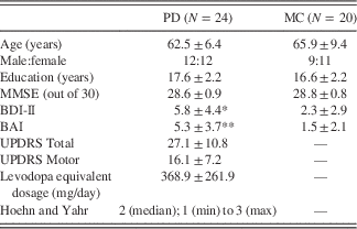
The cohort studied here are the same individuals reported in Putcha et al., Reference Putcha, Ross, Cronin-Golomb, Janes and Stern2015. Twenty-four individuals diagnosed with PD (12 female, mean age 62.5 years, 2 left-handed) and 20 age- and education-matched control participants (MC; 11 female, mean age 65.9 years, 2 left-handed) participants (Table 1). All participants provided informed consent in a manner approved by the Institutional Review Board of Boston University and the Partners Human Research Committee. All participants were screened for other neurological and psychiatric illness. Please refer to Putcha et al. (Reference Putcha, Ross, Cronin-Golomb, Janes and Stern2015) and the Supplementary Materials for further detail.
Participant characteristics

Note. Values presented in the table are means ± standard deviations, unless otherwise noted.
* Group differences at a significance level of p<.05, **p<.005.
MMSE=Mini-Mental State Examination; UPDRS=Unified Parkinson’s Disease Rating Scale; BDI-II=Beck Depression Inventory, 2nd Edition; BAI=Beck Anxiety Inventory.
Neuropsychological Assessment
An abbreviated three-domain neuropsychological assessment consisting of one test per domain (executive function, psychomotor speed/fine motor dexterity, and verbal memory) was used to assess cognitive abilities. Each of these cognitive tasks required the individual to focus on external demands and suppress internal self-referential thoughts, thereby exhibiting cognitive control of attentional resources. To compare performance across domains, performance scores from each test were converted to Z-scores, normalized on the basis of the mean and standard deviation of the age- and education-matched control participants (MC). Independent samples t-tests were conducted on these Z-scores to determine group differences on cognitive test performance. Effect sizes were also calculated and reported based on established methods (Lakens, Reference Lakens2013). Standard multiple linear regression was conducted to investigate whether functional connectivity between the core neurocognitive networks (SN, DMN, CEN) related to cognitive performance. Functional coupling measures and group membership, as well as the interaction of functional coupling and group membership were entered as predictors of cognitive performance across neuropsychological tests. We also conducted hierarchical linear regression entering age and education as demographic variables in Step 1, and our variables of interest in Step 2, to determine if the demographic variables were significantly affecting our findings. Subsequent mediational analyses using Sobel’s test of mediational significance was conducted to further assess significant interaction effects. Within-group analysis was conducted using Pearson’s correlations, between cognitive performance and the functional coupling of neurocognitive networks. Bonferroni correction was applied to address the multiple correlations across three cognitive tests, so p-values less than.016 are considered significant (.05 divided by 3).
Executive functioning (set-shifting) was assessed with the Trail Making Test, consisting of two subtests, Parts A and B (Tombaugh, Reference Tombaugh2004). Trails A is a test of simple attention and psychomotor speed, in which participants connect numbered circles in ascending order (1–2–3, etc.). Trails B is a measure of combined visual search, psychomotor speed, cognitive flexibility, and the ability to shift and maintain response set. Participants sequentially alternate between alphanumeric sequences (1–A–2–B, etc.). Test–retest reliability varies with age range and population studied (Dikmen, Heaton, Grant, & Temkin, Reference Dikmen, Heaton, Grant and Temkin1999; Strauss, Sherman, & Spreen, Reference Strauss, Sherman and Spreen2006), but is considered adequate for Part A (0.79) and Part B (0.89). Time to completion on Trails B was used as our primary measure of executive function as it represents a holistic measure of both lower-order executive function (sustained attention) and higher-order set-switching abilities (Kortte, Horner, & Windham, Reference Kortte, Horner and Windham2002). As a secondary step to isolate only the set-switching ability, the time to complete Trials A was subtracted from the time to complete Trails B (“Trails B minus A”) to isolate only the higher-order set-switching executive function.
Psychomotor speed/fine motor dexterity was assessed with the Purdue Pegboard Test (Tiffin & Asher, Reference Tiffin and Asher1948). This test evaluates complex, visually guided coordinated movements in a timed manner. This test has high test–retest reliability (0.81–0.89; Buddenberg & Davis, Reference Buddenberg and Davis2000) and construct validity (0.78), as it correlates highly in normal adults with finger tapping and other manual dexterity tasks (Doyen & Carlier, Reference Doyen and Carlier2002). The performance score indicates the average number of pegs successfully placed within a 30-s trial period. The left and right hands were evaluated in separate trials, and their scores were then averaged to create a composite across hands of psychomotor speed (Purdue Pegboard).
Verbal memory was assessed with the Rey Auditory Verbal Learning Test – 30 min Delay Recall condition (RAVLT Recall) (Rey, Reference Rey1964). Participants recalled as many words as possible from a previously learned list after a 30-min delay. This test has adequate test–retest reliability overall (0.60–0.70), with the delayed recall score among the more reliable subscores (Mitrushina, Satz, Chervinsky, & D’Elia, Reference Mitrushina, Satz, Chervinsky and D’Elia1991).
Neuroimaging Procedure
Each scanning session included 20 min of structural imaging sequences followed by resting state data acquisition lasting 6 min and 35 s, during which the participants were asked to remain still and maintain eyes-open fixation on a projected image of a white cross on a black background. Please refer to Putcha et al., Reference Putcha, Ross, Cronin-Golomb, Janes and Stern2015, as well as the Supplementary Materials for further details.
Motion Correction and ICA-Based Denoising
From our initial data set of 26 PD and 24 MC participants, we identified and excluded 2 individuals with PD and 4 matched control individuals who demonstrated excess motion (greater than 2 mm absolute displacement) from our analysis, using the Artifact Detection toolkit made for SPM8 software. Further details on these procedures can be found in our Supplemental Materials.
Inter-network Functional Coupling
The default mode (DMN), central executive (CEN), and salience networks (SN) were defined using a previously published set of templates from the BrainMap Database (Fox, Laird, et al., Reference Fox, Laird, Fox, Fox, Uecker, Crank and Lancaster2005; Laird et al., Reference Laird, Fox, Eickhoff, Turner, Ray, McKay and Fox2011; Laird, Lancaster, & Fox, Reference Laird, Lancaster and Fox2005). The DMN comprises medial prefrontal cortex, posterior cingulate/precuneus cortices, and bilateral posterior parietal cortex. The CEN is divided into left- and right-hemisphere localized CEN; the left-hemisphere localized network (L-CEN) comprises left-lateralized dorsolateral prefrontal cortex and inferior frontal gyrus, as well as posterior parietal cortex, while the right-hemisphere localized network (R-CEN) comprises right-lateralized dorsolateral prefrontal cortex and bilateral posterior parietal cortices. The SN comprises bilateral anterior insula/frontal opercula and the dorsal anterior cingulate gyrus. FSL’s dual regression approach was used to calculate the orthogonalized subject specific timecourses and spatial maps for each network of interest within each of our individual subject’s data (Beckmann, Mackay, Filippini, & Smith, Reference Beckmann, Mackay, Filippini and Smith2009; Cole et al., Reference Cole, Beckmann, Long, Matthews, Durcan and Beaver2010; Filippini et al., Reference Filippini, MacIntosh, Hough, Goodwin, Frisoni, Smith and Mackay2009; Janes, Farmer, Frederick, Nickerson, & Lukas, Reference Janes, Farmer, Frederick, Nickerson and Lukas2014). Subject-specific timecourses were extracted from the SN, DMN, R-CEN, and L-CEN. Correlation coefficients (Pearson’s r) were computed between the SN and DMN, the SN and CEN (both right and left hemisphere), and between the CEN (both right and left hemisphere) and DMN to determine the extent of inter-network functional coupling at the group level, and subsequently Fisher-z-transformed for use in further regression and correlational analyses. For further detail on this analysis, please refer to our previous publication on these findings (Putcha et al., Reference Putcha, Ross, Cronin-Golomb, Janes and Stern2015). To investigate if PD disease duration, disease severity, or dopamine replacement medication were related to these inter-network functional coupling measures, correlation coefficients (r) were computed between disease duration (number of years), disease severity (Hoehn and Yahr stage), levodopa equivalent dosage, and the functional coupling values (z) described above.
Volumetric Analysis
To ensure that volumetric differences between MC and PD participants were not impacting our results, the following analyses were conducted. MP-RAGE images were processed using FreeSurfer (version 5.3.0) (http://surfer.nmr.mgh.harvard.edu). Standard preprocessing of structural volumes produced reconstructions that were used to determine if there were any areas of cortical thinning or subcortical atrophy in the PD group compared to MC using the QDEC utility. We also compared striatal volume between MC and PD groups to determine if there were any volumetric differences.
Results
No Group Differences in Age, Education, Male:Female Ratio, Overall Mental Status, Cortical Thinning, or Brain Volume
As previously reported in this participant sample (Putcha et al., Reference Putcha, Ross, Cronin-Golomb, Janes and Stern2015), MC and PD participants were matched on age (p>.2), education (p>.1), male:female ratio (Χ 2=.11; p>0.7), and MMSE scores (p>.5; range 25.7 to 29.7). There were no between-group differences in whole brain cortical thinning or subcortical atrophy (p>.05, Bonferroni corrected), and no group differences in striatal volume after controlling for intracranial volume (p>.4).
Executive Functioning
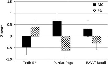
Set-shifting ability was assessed using the Trail Making Test consisting of two subtests (Parts A and B). We found that individuals with PD were impaired in set-shifting ability compared to MC individuals on Trails B (t=3.14; p=.003; 95% CI [−9.7, −0.32], Hedges’s g s=0.95; Figure 1) and Trails B minus A (t=2.5; p=.016; 95% CI [−8.67, −0.16], Hedges’s g s=0.76). We found no group differences in Trails A performance, the measure of simple attention (p>.1). Note that higher numbers on Trails indicate longer time to completion (worse performance).
Cognitive performance. PD participants performed significantly worse than MC participants across multiple cognitive domains, including executive function (Trail Making Test Part B; t=−3.14; p=.003), psychomotor processing speed (Purdue Pegboard; t=5.3; p<.001), and delayed verbal recall (RAVLT 30 min Delay Recall; t=2.05; p=.04). *Negative Trails B scores indicate better (faster) performance on this test.

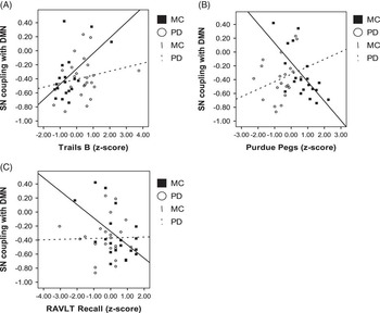
Functional coupling between the SN and DMN (beta=0.42; p=.04), but not group membership (beta=0.35; p=.10), explained a significant amount of variance in Trails B performance. There was no significant interaction between group membership and functional coupling between SN and DMN (beta=−0.18; p=0.5) in predicting Trails B performance. Within-group correlational analysis suggests that performance on this task was significantly related to functional coupling between the SN and DMN in MC participants only (Trails B; r=0.53, p=.02; Figure 2A), and we did not observe this association in the PD group alone (r=0.20=5; p=.26; Figure 2A). We found no other significant association between set-shifting ability and functional coupling between any of the other neurocognitive networks.
SN coupling with DMN relates to cognition in MC but not PD. Functional coupling between the Salience Network (SN) and the Default Mode Network (DMN) is related inversely to better cognitive performance in MC across all three cognitive domains: (A) Executive function (Trail Making Test- Part B; r=0.53; p=.02), (B) Psychomotor Processing Speed (Purdue Pegboard; r=−0.68; p=.001), and (C) Verbal Memory Recall (RAVLT 30-min Delay; r=−0.52; p=.02). This association suggests that anti-correlations between the SN and DMN are related to better cognitive performance in MC. There are no significant associations between cognitive performance and functional coupling between these networks in PD. Each data point represents an individual participant.

This analysis was repeated predicting performance on Trails B minus A, the composite measure designed to isolate set-shifting from simple attention and visual scanning. We found that functional coupling between the SN and DMN (beta=0.45; p=.03), but not group membership (beta=0.19; p=.4), explained a significant amount of variance in Trails B minus A performance. There was no significant interaction between group performance and functional coupling between SN and DMN (beta=−0.3; p=.24) in predicting Trails B minus A performance. Within-group correlation analysis revealed a significant correlation between SN-DMN functional coupling and the Trails B minus A performance in the MC group (r=0.60; p=.007), but not the PD group (r=0.11; p=.63).
This analysis was repeated predicting performance on Trails A, the measure of simple attention. We found that neither functional coupling between the SN and DMN (beta=0.38; p=.07), nor group membership (beta=0.34; p=.3), predicted Trails A performance. The interaction between group performance and functional coupling between SN and DMN did not predict Trails A performance (beta=−0.05; p=.8). Within-group correlational analysis did not reveal a significant association between SN-DMN functional coupling and Trails A performance in the MC group (r=0.37; p=.11) or the PD group (r=0.35; p=.10).
Covarying age and education into Step 1 of a hierarchical linear regression did not change these results and by themselves, these demographic variables did not predict performance on the Trail Making Test, F(2,39)=1.2, p=.31. There were no other significant main effects of functional connectivity between any of the other neurocognitive network pairs in predicting Trail Making Test performance, although main effect of group was observed across these other models in predicting performance on this test emphasizing group differences on cognitive test performance.
Psychomotor Speed
Psychomotor speed/fine motor dexterity was assessed with the Purdue Pegboard Test. We found that PD participants were impaired on this task compared to MC participants (t=5.32; p<.001; 95% CI [−7.53 1.75]; Hedges’s g s =1.60; Figure 1).
Functional coupling between the SN and DMN (beta=−0.52; p=.001), but not group membership (beta=−0.21; p=.2), explained Purdue Pegboard performance. We also found a significant interaction between group membership and functional coupling between SN and DMN (beta=0.69; p=.002). However, the Sobel test of mediational significance suggests that group membership does not mediate this relationship (Sobel t=1.18; p=.23). Within-group correlational analysis within the MC group alone revealed a significant association between Purdue Pegboard performance and coupling between the SN and DMN (r=−0.68; p=.001; Figure 2B), and no association in the PD group alone (r=0.27; p=.23; Figure 2B).
Age and education in Step 1 of a hierarchical linear regression did predict performance on Purdue Pegboard Test (F(2,39)=4.1; p=.02). Over and above this effect, we still observed a significant main effect of group membership (beta=−0.35; p=.03), a significant main effect of functional coupling between the SN and DMN (beta=−0.45; p=.005), and a significant interaction between group and functional coupling (beta=0.56; p=.01). There were no other significant main effects of functional connectivity between any of the other neurocognitive network pairs (SN-CEN, CEN-DMN) in predicting Purdue Pegboard performance, although a main effect of group was observed across these other models in predicting performance on the Purdue Pegboard Test emphasizing group differences on cognitive test performance.
Verbal Memory Retrieval
Verbal memory retrieval was assessed using the RAVLT 30-min Delay Recall condition. The PD group showed poorer performance than the MC group (t=2.21; p=.04; 95% CI [−7.97 1.21], Hedges’s g s =0.62; Figure 1).
Functional coupling between the SN and DMN (beta=−0.48; p=.024), but not group membership (beta=−0.04; p=.8), explained a significant amount of variance in RAVLT Delayed Recall performance. The interaction between group membership and SN-DMN functional coupling did not significantly predict performance on the RAVLT delayed recall (beta=0.47; p=.08). Within-group correlational analysis examining the MC group separately revealed a significant association between performance on RAVLT Delayed Recall and functional coupling between the SN and DMN (r=−0.52; p=.02; Figure 2C), and no such association within the PD group (r=0.03; p=.9; Figure 2C).
Age and education in Step 1 of a hierarchical linear regression did not change these results and by themselves, these demographic variables did not explain a significant amount of variance in predicting cognitive performance on the RAVLT delayed recall condition (F(2,39)=6.5; p=.5). There were no other significant main effects of functional connectivity between any of the other neurocognitive network pairs in predicting RAVLT verbal delayed recall performance (SN-CEN, p>.5; CEN-DMN, p>.2), although main effect of group was observed across models in predicting performance on this test.
Associations with Disease and Levodopa Equivalent Dosage
No measures of functional coupling were related to disease duration, disease severity (Hoehn and Yahr stage), or levodopa equivalent dosage (LED). No cognitive measures were related to LED. Psychomotor speed (Purdue Pegboard) was the only cognitive measure score related to disease duration (r=−0.45; p=.04), such that longer disease duration was related to worse performance.
Between-Groups Inter-network Functional Coupling
Across all subjects, L-CEN was significantly and negatively correlated with the DMN (r=−0.45; p=.002) and positively correlated with the SN (r=0.66; p<.001). As previously reported in this participant sample (Putcha et al., Reference Putcha, Ross, Cronin-Golomb, Janes and Stern2015), inter-network functional coupling was significantly different between groups, such that SN coupling with R-CEN was diminished in PD compared to MC (t=2.45; p=.019; 95% CI [−7.63 0.43], Hedges’s g s =0.69), and R-CEN coupling with DMN was aberrantly positive in PD compared to the expected anti-correlation between these networks observed in the MC group (t=−2.06; p=.04, 95% CI [−8.08 −0.001], Hedges’s g s =0.59).
Surprisingly, here we did not observe differences in functional coupling between the SN and DMN in the PD group compared to the MC group (p>.6), and there were no significant correlations between cognitive performance and functional coupling between any of the three neurocognitive networks within the PD group alone. We did not find any evidence that functional coupling between any of the other neurocognitive networks predicts task performance in any of the regression models conducted.
Discussion
There were two main findings in this study. First, we found evidence of cognitive dysfunction across domains of executive functioning, psychomotor speed, and verbal memory delayed recall in a group of non-demented individuals with PD compared to age- and education-matched control participants, replicating what has been previously reported (Barone et al., Reference Barone, Aarsland, Burn, Emre, Kulisevsky and Weintraub2011; Kudlicka, Clare, & Hindle, Reference Kudlicka, Clare and Hindle2011). Our novel finding is that functional coupling between the SN and the DMN predicts performance in all three cognitive domains regardless of group membership. Within-group analysis suggest this finding may be more robust in the control participants, as the significant correlations between SN-DMN functional coupling and performance on each of these measures were observed in the MC participants, but not in PD alone. The results suggest that functional coupling between the SN and DMN is important for successful cognition across domains. We postulate that PD-related striatal disruption may lead to abnormal network dynamics, exacerbating cognitive dysfunction as the disease progresses.
Cognitive impairment in early stages of PD is present in many individuals, but is often under-recognized using routine screening methods (Mamikonyan et al., Reference Mamikonyan, Moberg, Siderowf, Duda, Have, Hurtig and Weintraub2009). In the present study, we found evidence of cognitive deficits across tests of executive functioning, psychomotor speed, and verbal memory recall in PD individuals with MMSE scores that were comparable to those of age- and education-matched control participants. Successful cognitive performance on these tasks requires switching between externally and internally salient stimuli, resulting in anti-correlated network coupling between the SN and the DMN. Our findings demonstrate that successful performance on tasks requiring a high level of cognitive control is related to anti-correlated intrinsic functional coupling between the SN and DMN.
Large-scale functional networks exert coordinated effects across distributed cortical areas during many different cognitive functions (Bressler & Menon, Reference Bressler and Menon2010). In particular, the three core neurocognitive networks examined here (SN, DMN, CEN) are thought to interact dynamically to influence cognitive task performance (Fox, Snyder, Vincent, & Raichle, Reference Fox, Snyder, Vincent and Raichle2007; Seeley et al., Reference Seeley, Menon, Schatzberg, Keller, Glover, Kenna and Greicius2007; Sridharan et al., Reference Sridharan, Levitin and Menon2008). The SN plays the critical role of responding to external events that are behaviorally salient, and switching between activation of the DMN and CEN (Menon & Uddin, Reference Menon and Uddin2010). The SN becomes dysfunctional in PD as its key nodes, the anterior insula and dorsal anterior cingulate cortex, become direct targets of PD-related striatal disruption (Christopher, Koshimori, Lang, Criaud, & Strafella, Reference Christopher, Koshimori, Lang, Criaud and Strafella2014; Christopher, Marras, et al., Reference Christopher, Marras, Duff-Canning, Koshimori, Chen, Boileau and Strafella2014). This suggests that striatal dysfunction directly impacts the functional integrity of the insula, and, therefore, the SN, in individuals with PD.
Interactions between the SN and the DMN are crucial in the control of attention required for demanding cognitive tasks. In healthy young adults, greater anti-correlation between the SN and DMN has been associated with more efficient cognitive control (Kelly, Uddin, Biswal, Castellanos, & Milham, Reference Kelly, Uddin, Biswal, Castellanos and Milham2008). Failures of DMN deactivation have been associated with reduced sustained attention (Weissman, Roberts, Visscher, & Woldorff, Reference Weissman, Roberts, Visscher and Woldorff2006), and are observed across many neurological diseases (Leech & Sharp, Reference Leech and Sharp2014). In PD, it is proposed that as the SN becomes dysfunctional it is no longer able to suppress DMN activity effectively (Bonnelle et al., Reference Bonnelle, Ham, Leech, Kinnunen, Mehta, Greenwood and Sharp2012; Jilka et al., Reference Jilka, Scott, Ham, Pickering, Bonnelle, Braga and Sharp2014). Decreased intra-network DMN connectivity in PD at rest was associated with decreased processing speed and increased time on tasks requiring a high level of cognitive control (Disbrow et al., Reference Disbrow, Carmichael, He, Lanni, Dressler, Zhang and Sigvardt2014), suggesting that DMN integrity is critical for efficient cognition. Compared with cognitively intact individuals, individuals with PD who have mild cognitive impairment also demonstrated dopaminergic deficits within the SN and DMN that have been associated with disrupted memory retrieval (Christopher et al., Reference Christopher, Duff-Canning, Koshimori, Segura, Boileau, Chen and Strafella2015).
Surprisingly, we did not observe group differences in functional coupling between the SN and DMN. This may be explained by the fact that we examined functional coupling between timecourses across entire networks, rather than focusing on each of the individual nodes comprising each network. Previous work has established decreased functional connectivity within just the posterior nodes of the DMN (medial temporal lobes and inferior parietal cortices) but not within the anterior node (medial prefrontal cortex) (Tessitore et al., Reference Tessitore, Esposito, Vitale, Santangelo, Amboni, Russo and Tedeschi2012). Furthermore, the medial prefrontal cortex has demonstrated comparable task-related deactivations compared with control participants, while the posterior cingulate cortex showed less deactivation during task than expected (van Eimeren, Monchi, Ballanger, & Strafella, Reference van Eimeren, Monchi, Ballanger and Strafella2009). These findings suggest that the posterior DMN nodes in particular may be dysfunctional. Investigations into the functional coupling between the SN and these posterior hubs of the DMN may reveal the dysfunctional coupling we hypothesized. Future longitudinal follow-up investigations would also help to elucidate the progression of network-level cortical changes over time.
One of the primary findings of this study was that functional coupling between the SN and the DMN predicts cognitive performance across multiple cognitive domains. In contrast, we did not observe an association between cognitive performance and SN-CEN interactions, as hypothesized. In our previous study (Putcha et al., Reference Putcha, Ross, Cronin-Golomb, Janes and Stern2015), we reported on alterations in intrinsic SN-CEN and CEN-DMN functional coupling between MC and PD. In this follow up study, we examined associations between functional coupling and cognitive performance, and did not find a link between cognition and SN-CEN coupling, but we did with SN-DMN connectivity. This finding is in line with recent work emphasizing the role of the DMN over the CEN in highly demanding cognitive tasks (Disbrow et al., Reference Disbrow, Carmichael, He, Lanni, Dressler, Zhang and Sigvardt2014; Duan et al., Reference Duan, Liao, Liang, Qiu, Gao, Liu and Chen2012). In contrast with the DMN, nodes within the CEN are not a major site of dopamine deficiency in PD (Christopher et al., Reference Christopher, Duff-Canning, Koshimori, Segura, Boileau, Chen and Strafella2015) and the CEN does not demonstrate intra-network connectivity changes compared to healthy control participants in early stages of PD (Disbrow et al., Reference Disbrow, Carmichael, He, Lanni, Dressler, Zhang and Sigvardt2014). Taken together, this evidence suggests that although the CEN is an important network for efficient cognition (Menon, Reference Menon2011), coupling between the SN and DMN is of primary importance in supporting cognitive performance in both healthy aging and PD.
It is worth noting that PD participants in this study were evaluated during peak levels of dopaminergic medications, which limits our ability to address questions related to the effects of dopamine on neurocognitive network interactions in the absence of medication effects. These results should be regarded as conservative. In addition, dopaminergic medications used to treat Parkinson’s disease have been shown to affect striatal activity, but not cortical activity (Martinu, Degroot, Madjar, Strafella, & Monchi, Reference Martinu, Degroot, Madjar, Strafella and Monchi2012). Although our measure of levodopa equivalent dosage (LED) did not correlate significantly with any measure of functional coupling or with cognitive performance, we cannot completely rule out the possibility that dopamine medication is related in some way to the association between functional network coupling and cognitive performance in our PD group. There is some evidence that dopaminergic therapy diminishes DMN integrity (Krajcovicova, Mikl, Marecek, & Rektorova, Reference Krajcovicova, Mikl, Marecek and Rektorova2012). However, examining the network connectivity of individuals with PD on their typical medication regimens has great utility as most patients experience cognitive dysfunction and disrupted activities in daily living even during peak medication states (Narayanan, Rodnitzky, & Uc, Reference Narayanan, Rodnitzky and Uc2013). It is also worth noting that dopamine replacement therapy does not ameliorate cognitive disturbances specific to attentional set-shifting and other aspects of executive functioning (Lewis, Slabosz, Robbins, Barker, & Owen, Reference Lewis, Slabosz, Robbins, Barker and Owen2005; Poletti & Bonuccelli, Reference Poletti and Bonuccelli2013), which are subserved by the neurocognitive networks examined in our present study. We also acknowledge the possibility that the low doses of anxiolytic and antidepressant medications could also have an effect on functional network disruption, although it is unlikely as only three PD participants were receiving these medications and our results did not change when excluding them.
We provide evidence in this study of functional coupling between SN and the DMN relating to cognitive task performance. This finding may be more robust in our healthy older adults, as this association was observed strongly in the MC group alone but not in the PD group. We postulate that, as these PD participants progress in disease severity, dysfunctional neurocognitive network interaction specific to the SN and DMN may underlie the progression of cognitive deficits ultimately leading to dementia. This work supports previous investigations highlighting the SN and DMN as networks critical for supporting high-level cognitive functioning, which has clinical implications for non-pharmacological treatment. With converging evidence suggesting that these networks become vulnerable in early stages of PD progression, the SN and DMN represent potential targets of non-invasive treatment approaches, such as repetitive transcranial magnetic stimulation, which may serve to improve quality of life and extend independence in daily living.
Acknowledgments
The authors declare no competing financial interests. This work was supported by the National Institute of Health R01 NS067128. Scanning was carried out at the Athinoula A. Martinos Center for Biomedical Imaging at the Massachusetts General Hospital, which receives funding from P41EB015896, a Biotechnology Resource Grant supported by the National Institute of Biomedical Imaging and Bioengineering (NIBIB), National Institutes of Health. This work also involved the use of instrumentation supported by the NIH Shared Instrumentation Grant Program and/or High-End Instrumentation Grant Program; specifically, grant number(s) S10RR022976 and S10RR019933. We also acknowledge salary funding for ACJ from grant number K01DA029645. Drs. Janes and Stern share co-last authorship.
Supplementary Material
Supplementary materials can be found online. Please visit journals.cambridge.org/jid_INS.