Case report
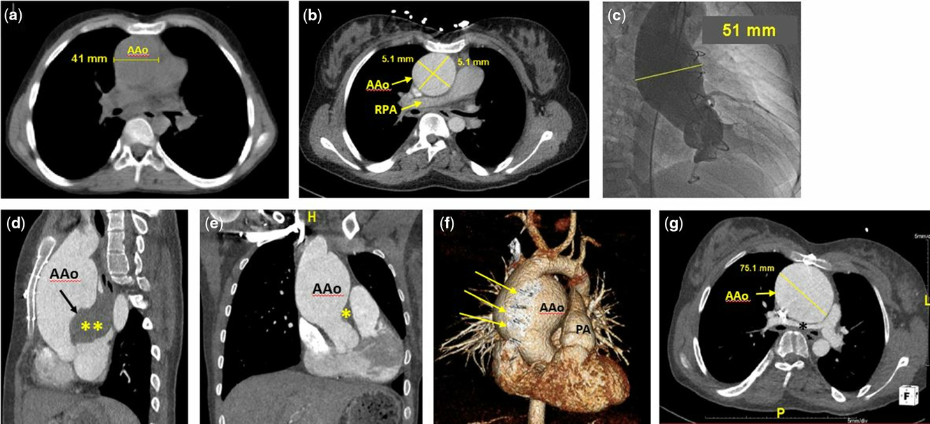
This case reports a 17-year-old Amish female status post-orthotopic heart transplantation at 6 weeks of age for end stage heart failure. Genetic testing revealed a homozygous gene mutation (3330+2T>G) in the MYBPC3 gene, which is associated with lethal, infantile hypertrophic cardiomyopathy. The patient is a non-smoker with no family history of connective tissue disease. She developed severe diffuse coronary artery vasculopathy with anginal symptoms and underwent a second orthotopic heart transplant at 6 years of age with a structurally normal allograft. She had a cardiac CT when she was 8 years old for concerns of lymphoproliferative disorder that revealed an ascending aortic diameter of approximately 4.4 cm. This dimension was stable over time by serial echocardiographic measurements. Four months after her 16th birthday, the patient was admitted to the hospital for community-acquired pneumonia. Her fever resolved with intravenous antibiotics, but her cough persisted and she was re-admitted a month later for concerns of a chronic infection. Bronchoscopy revealed 50% narrowing of the upper trachea in the AP dimension, 30% narrowing of the right main stem bronchus, and 60% narrowing of the left main stem bronchus. A chest CT demonstrated tree incidental aneurysmal dilatation of the ascending aorta measuring 5.1 cm × 5.1 cm (Fig 1a and b).
CT chest and aortic angiogram imaging demonstrating progression of ascending aortic dilatation. ( a ) CT chest without contrast axial view of ascending aorta with maximal diameter of 44 mm (April, 2008). ( b ) CT chest with contrast axial view of ascending aorta with maximal diameter of 51 mm × 51 mm (March, 2015). ( c ) Aortic root angiogram showing maximal aortic diameter of 51 mm. ( d ) CT chest with contrast sagittal view of ascending aortic aneurysm with posterior rupture (stars) through intimal flap (black arrow) compressing the left atrium and very close approximation to the sternum. ( e ) CT chest with contrast coronal view of ascending aortic aneurysm with rupture (asterisks) and compression of left atrium. ( f ) Volume rendered image of ascending aortic aneurysm with apposition to sternal wires (arrows). ( g ) CT chest with contrast axial view showing maximal diameter of ascending aorta of approximately 75 mm with compression of the right pulmonary artery (black asterisk). AAo = ascending aorta; PA = pulmonary artery; RPA = right pulmonary artery.
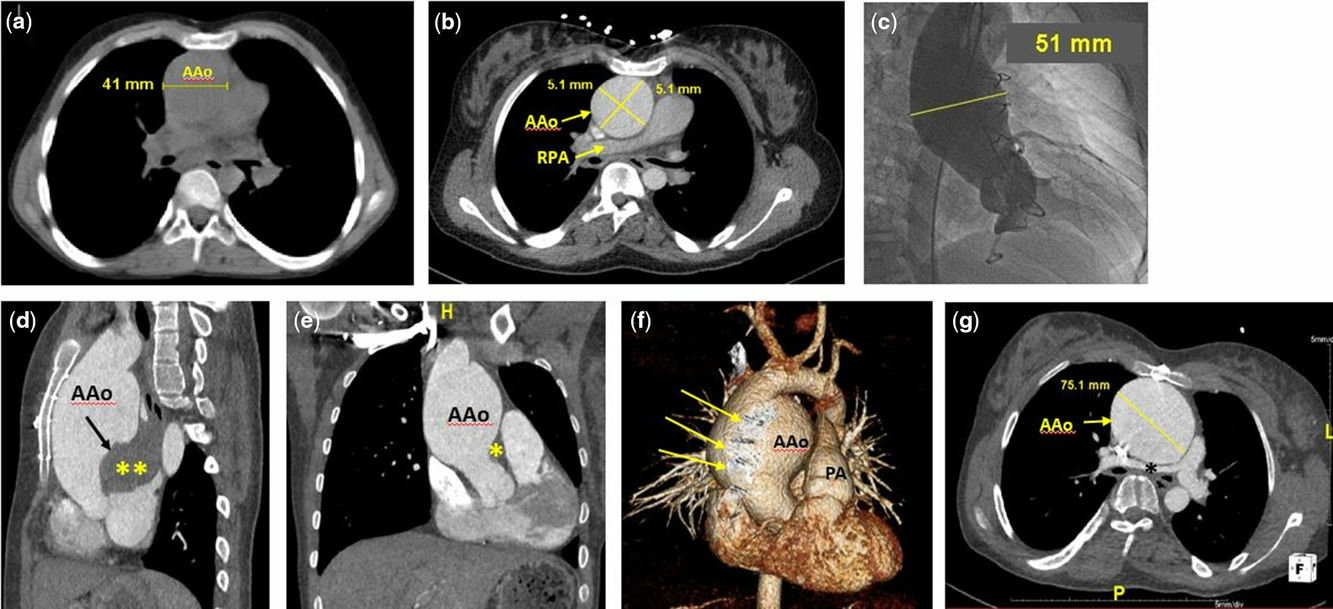
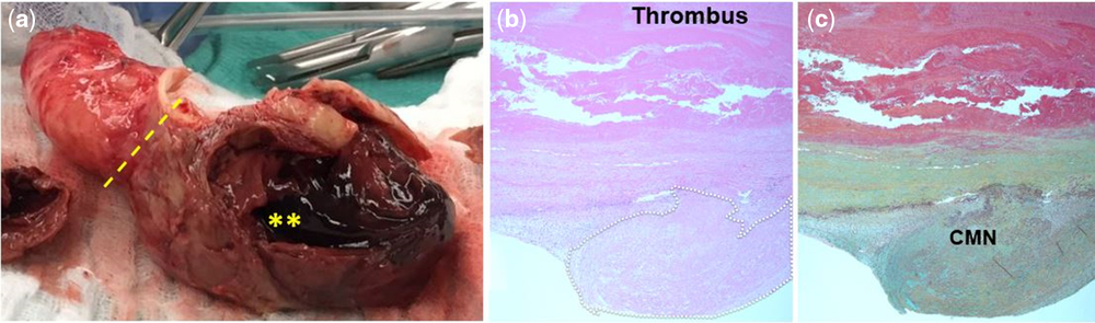
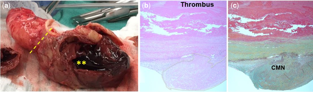
In the absence of known connective tissue disease or family history of dissection, and in light of the slow progression of her aortic dilatation, the patient did not meet criteria for surgical intervention based on the 2010 ACCF/AHA guidelines. She was started on enalapril 2.5 mg by mouth twice daily for afterload reduction to help attenuate further dilatation, but this was discontinued after a few months for concerns that it was contributing to her dry cough and intermittent wheezing, which did not resolve after evaluation by Pulmonology and initiation bronchodilator therapy. An aortic root angiogram was performed during her surveillance cardiac catheterisation and showed a stable ascending aortic dimension of 5.1 cm (Fig 1c). However, a routine follow-up echocardiogram a few months later revealed a further increase in her ascending aortic dimension to 6 cm with an abrupt calibre change at the transverse aorta to 2.6 cm. Cardiac CT confirmed a type A-contained rupture that dissected along the posterior aspect of the proximal ascending aorta with a maximum diameter of 7 cm (Fig 1d–g). There was compression of the left atrium and the right pulmonary artery by the aneurysmally dilated ascending aorta (Fig 1g). She denied any chest pain, shortness of breath, diaphoresis, pallor, pre-syncope, or syncope since her recent cardiac catheterisation. She underwent immediate repair of the ascending aorta and hemiarch with a 28-mm Dacron tube graft. Chest exploration revealed a posterior rupture of her native aorta, with compression of the roof of the left atrium (Fig 2a). The aneurysm began approximately 1 cm above the anastomosis between native and donor aorta and the dissection did not cross suture line. There was no donor–recipient aortic mismatch noted in the operative reports for both of her transplantations. Post-operatively, she required an intravenous esmolol infusion for hypertension, but was quickly transitioned back to oral enalapril at her pre-operative dose. Her cough and wheezing did not recur post-operatively. Surgical pathology showed moderate cystic medial degeneration, healed dissection, and pseudoaneurysm with thrombus formation (Fig 2b).
Surgical specimen and histology. ( a ) Excised portion of severely dilated ascending aorta with ruptured type A dissection. Note the marked change in calibre past the area of dissection (dotted line) and visible thrombus (asterisk). ( b ) PAS stain through full thickness area of dissection showing thrombus and loss of aortic wall integrity (outlined area). ( c ) Movat Pentachrome of the same full thickness area of dissection. Area of CMN demonstrates complete disorganisation of the aortic wall collagen fibres and loss of wall integrity. CMN = cystic medial necrosis; PAS = Periodic acid-Schiff.
Discussion
Aortic dissection causes significant morbidity and mortality in adults, predominantly affecting men greater than 65 years of age. Reference Larson and Edwards1 Risk factors for AD in adults have been widely studied and guidelines for treatment are well established. This is not the case in the paediatric population (i.e. age < 21 years), where the incidence of AD is low, ranging from 0.67 to 3.5% of all dissections. Reference Fikar and Fikar2 As with adults, bicuspid aortic valve, connective tissue disease, and pre-existing aortic aneurysm are all predisposing factors, Reference Fikar and Koch3 but CHD also plays a large role. In fact, in one of the largest retrospective reviews of AD in children and young adults (i.e. age < 30 years) to date, CHD was associated with AD 38% of the time. Reference Shamszad, Barnes and Morris4 Complicating matters further is the fact that several forms of CHD are associated with moderate ascending aortic dilatation without a high incidence of dissection. Reference Stulak, Dearani and Burkhart5 Therefore, with such different predisposing factors, it becomes unclear if adult treatment guidelines can be appropriately applied to children.
Aortic complications following orthotopic heart transplantation have also been documented. In adults, the incidence is estimated between 1 and 2%. Reference Vigano, Rinaldi and D’Armini6 In children, the occurrence is significantly lower and is often associated with infection, aortic augmentation as part of staged Fontan palliation, or known connective tissue disease. Reference Lang, Frazier and Collins7 In a review of 453 adults who underwent heart transplantation, Vigano et al found 9 (2%) who developed aortic complications, which they grouped into 4 categories: primary acute aortic rupture, bacterial and mycotic pseudoaneurysms of the ascending aorta, aneurysm and dissection of the native aorta, and dissection in heterotopic transplant. The aetiology of AD in the early post-operative period was related to systemic hypertension, size mismatch between the native and donor aorta, and technical errors in creating the anastomosis while true aneurysm and dissection of the native aorta was associated with atherosclerosis and immunosuppression-induced hypertension and diabetes.
In the absence of known bicuspid valve or connective tissue disease, the most recent ACCF/AHA guidelines recommend surgical intervention for thoracic aortic aneurysms exceeding 5.5 cm or growing at a rate of >0.5 cm annually. Reference Hiratzka, Bakris and Beckman8 Our patient had slowly progressive ascending aortic dilatation for years which did not meet treatment guidelines by either criteria. Her transplanted aortic valve was trileaflet with no regurgitation, and she did not possess any other known predisposing risk factors for AD, such as family history, hypertension, or tobacco use. From a transplant perspective, she had no episodes of rejection, was never treated for a mediastinal infection, and does not have diabetes or hypertension, which are known side effects of long-term immunosuppression. Moreover, review of her operative records does not indicate that there was significant donor–recipient aortic mismatch. She was appropriately treated with an angiotensin-converting enzyme inhibitor, though this was discontinued due to concerns that her cough and wheezing were a medication side effect. Her ascending aortic diameter remained stable for several months despite stopping the enalapril, as was confirmed by aortic root angiogram, but a surveillance echocardiogram revealed a completely asymptomatic type A dissection 6 months after the catheterisation. In retrospect, the dry cough may have been due to compression of her bilateral main stem bronchi by the aortic aneurysm as opposed to a medication side effect.
Also noteworthy in this case is the patient’s homozygous MYBPC3 (3330+2T>G) gene mutation, which is known to cause lethal infantile hypertrophic cardiomyopathy. Reference Zahka, Kalidas and Simpson9 It is possible that this mutation may also affect aortic integrity, but since death always occurs within the first year of life, this consequence may not have sufficient time to manifest. Alternatively, the patient may have a second homozygous recessive mutation or even de novo mutation for aortopathy, which is quite plausible given the high incidence of recessive disorders in the Amish population. However, the negative family history speaks against this when taking into consideration the large size of the patient’s family.
Post-anastomotic aortic dilatation is common in children after orthotopic heart transplantation, and cases of aortic aneurysm and dissection have been reported in both children and adults. However, our patient did not have any of the known risk factors for dissection in either the general or heart transplant population and developed this potentially life-threatening complication despite appropriate surveillance. This case suggests that children with known aortic dilatation status post-orthotopic heart transplant may be at increased risk of dissection and may require surgical intervention sooner than is recommended by the current guidelines.
Financial Support
This research received no specific grant from any funding agency, commercial, or not-for-profit sectors.
Conflicts of Interest
None.
Ethical Standards
N/A.