Introduction
As the type species (Toriyama and Tomaru, Reference Toriyama, Tomaru, Murphy, Fauquet and Bishop1995) of the genus Tenuivirus, Rice stripe virus (RSV) is transmitted by Laodelphax striatellus (the small brown planthopper); the virus contains negative-sense, single-stranded RNA with several of its genes being expressed by an ambisense strategy (Zhu et al., Reference Zhu, Hayakawa, Toriyama and Takahashi1991, Reference Zhu, Hayakawa and Toriyama1992; Takahashi et al., Reference Takahashi, Toriyama, Hamamatsu and Ishihama1993; Hamamatsu and Ishihama, Reference Hamamatsu and Ishihama1994; Toriyama et al., Reference Toriyama, Takahashi, Sano, Shimizu and Ishihama1994). Since 1999, Rice stripe disease has reached epidemic proportions over a large area of rice-planting regions in China, resulting in tremendous damage to rice production in local areas, and causing widespread concern (Wei, Reference Wei2003). In 2007, by investigating Rice stripe disease in Jiangsu, Henan, Yunnan, Shandong, Anhui, Hubei and Zhejiang Provinces, the field incidence of this disease was generally 5–10%, up to over 20% in seriously afflicted areas, with ‘bald fields’ occurring in some places, so this disease has become the main rice disease at the local level. At present, the lack of disease-resistant strains is a major contributing factor to this extensive disease outbreak; therefore, exploring the molecular mechanism of interaction between RSV and its host is of great importance to solving this problem.
In this study, real-time reverse transcriptase–polymerase chain reaction (RT-PCR) and high performance liquid chromatography (HPLC) have been used. Thus, the expression of the auxin synthetase gene YUCAA1 mRNA and the changes of endogenous auxin content in rice (Oryza sativa subsp. japonica) plants and rice suspension cells infected by RSV were analysed to determine the effect of auxin on the interaction between RSV and rice. Furthermore, the change of RSV replication in infected rice plants was quantitatively determined following the treatment of infected plants by eliminating endogenous auxin and providing artificially synthesized auxin externally, and will provide the essential experimental basis for further research into auxin signal conduction during the interaction between RSV and rice.
Material and methods
Plant material and virus source
Oryza sativa L. subsp. japonica cv. Wuyujing 3 was adopted as the experimental plant. The virus was collected from a rice field in Hongze, Jiangsu Province, showing typical RSV leaf blight disease. After RT-PCR identification, the virus was transferred by viruliferous Laodelphax striatellus and then stored in rice plants (Taizhong 1). The diseased plants were used for RSV purification, in accordance with the method of Toriyama (Reference Toriyama1982).
Reagents and devices
The RNA extraction kit, Trizol Reagent, was produced by Invitrogen Corporation (California, USA); the TaKaRa EXScriptTM RT-PCR Kit was purchased from TaKaRa (Otsu, Japan); the fluorescent ratio PCR device, MiniOpticonTM System was manufactured by Bio-Rad Laboratories, Inc. (California, USA); the Agilent 1100 HPLC system was the product of Agilent Technologies, Inc. (Palo Alto, California, USA).
Virus inoculation and sample treatment
We inoculated young rice at the two-leaf stage with viruliferous Laodelphax striatellus, then placed them into Murashige and Skoog (MS) liquid culture, and cultured at them at 28°C under alternating conditions of 14 h light and 10 h darkness. Samples were collected at 0, 4, 8, 12 and 16 days after inoculation. Each of the collected samples was placed in liquid nitrogen, and ground into powder. Some samples were subjected to RSV determination by RT-PCR, and others were stored at −80°C for later use. YUCAA1 mRNA expression and endogenous auxin content of the RSV-positive samples were measured, with healthy plants at corresponding growth phases as the blank control for every batch of samples.
Whole rice plants infected during the initial period were treated by one of the following three methods: (1) soaked with 30 μmol/l indole acetic acid (IAA) for 16 h; (2) soaked with KPSC buffer solution (10 μmol/l potassium phosphate, pH 6.0, 2% sucrose, 50 μmol/l chloramphenicol) for 16 h, replacing the buffer solution once every 2 h (processing in KPSC buffer solution can eliminate the endogenous auxin in plants; Jain et al., Reference Jain, Kaur, Tyagi and Khurana2006a); and (3) diseased plants were first treated by KPSC buffer solution then transferred to 30 μmol/l IAA solution for 16 h. Samples of plants from each of the three treatments were collected at fixed times each day and stored at −80°C for further study.
Rice suspended cell culture and RSV infection
For suspended cell culture of Wuyujing 3 rice and RSV infection, the method of Yang et al. (Reference Yang, Wang, Wang, Yie and Tien1996) was followed. Samples were collected at 0, 16, 32, 48 and 64 h (five batches) after inoculation, with healthy suspended cells of rice at the corresponding growth phases as blank controls for every batch of samples.
Primer design
PerlPrimer software was used for real-time RT-PCR specific primer design (Marshall, Reference Marshall2004). In order to ensure specificity of designed primers, the selected primers were analysed comparatively using BLAST software and the GenBank database. The RSV CP gene primer was homologous with all known RSV CP genes, but not homologous with gene sequences of the rice genome. The primer of the rice auxin synthetase gene was not homologous with other rice gene sequences.
The specific primer for the RSV CP gene was:
5′ end: 5′-RTTGACAGACATACCAGCCAG-3′ (R=A/G)
3′ end: 5′-CATCATTCACTCCTTCCAAATAACY-3′ (Y=C/T).
The specific primer of rice auxin synthetase gene YUCAA1 was
5′ end: 5′-TCATCGGACGCCCTCAACGTCGC-3′
3′ end: 5′-GGCAGAGCAAGATTATCAGTC-3′.
The rice eukaryotic elongation factor 1-alpha gene (eEF-1α) was used as the internal reference gene (Jain et al., Reference Jain, Nijhawan, Tyagi and Khurana2006b); primers were:
5′ end: 5′-TTTCACTCTTGGTGTGAAGCAGAT-3′
3′ end: 5′-GACTTCCTTCACGATTTCATCGTAA-3′.
Total RNA extraction and real-time RT-PCR detection
Extraction included taking a 50 mg plant sample, grinding the sample while still frozen at the temperature of liquid nitrogen, then extracting total RNA in 1 ml Trizol, according to the instructions provided by the manufacturer.
Using the ExscriptTM RT Reagent Kit (TaKaRa) to conduct the reverse transcription reaction, the 20 μl reverse transcription reaction system contained the following: total RNA, 1 μg; 3′-end primer (10 pmol/l), 1 μl; 5×ExscriptTM buffer, 4 μl; ExscriptTM RT Enzyme Mix I, 1 μl; finally made up to 20 μl with RNase-free distilled water (dH2O). The reaction conditions were as follows: 42°C for 15 min and 85°C for 5 s. We took 1 μl of the cDNA solution as a template, and added the following reagents: 2×Premix Ex TaqTM, 25 μl; 1 μl 3′-end primer; 1 μl 5′-end primer (10 pmol/l); and 22 μl dH2O.
Real-time PCR was conducted in 50 μl volume and 48 wells of the MiniOpticonTM System. The procedure included 10 s of denaturation at 95°C, followed by 45 cycles of denaturation (95°C for 6 s) and annealing (62°C for 20 s). Then, the specificity of the amplification products was identified by both 1.5% agarose gel electrophoresis and the dissociation curve of each gene.
We diluted the cDNA solution of each gene by 40, 41, 42, 43 and 44 with EASY Dilution and took 1 μl of each as a template to conduct real-time PCR sensitivity detection and produce a standard curve. According to the reference gene, all samples were normalized (corrected using the initial RNA amount), and we then determined the relative expression in different samples of each objective gene. This was repeated three times for each sample, and biological detections were repeated at least twice.
HPLC measurement of endogenous auxin
Endogenous auxin in different samples was extracted according to Kelen et al. (Reference Kelen, Cubuk Demiralay, Sen and Ozkan2004), using the Agilent 1100 HPLC system. A SupelcosilTM LC-18 analytical column (5.0 μm, 4.6 mm×250.0 mm) was used as the chromatographic column, methanol:water:acetic acid (45.0:54.2:0.8, v/v) as mobile phase; the flow rate was 0.5 ml/min, and UV detection wavelength was 254.0 nm. We followed the external standard method, fixed quantity, with the HPLC reagent, Fluka, as a nominal sample. The recovery rate was measured using mixed nominal sample during the above-mentioned purification and separation process, and the sample measurement was corrected according to the recovery rate.
Results
Primer specificity validation and real-time RT-PCR accuracy assay of auxin synthetase gene YUCAA1 and RSV CP gene
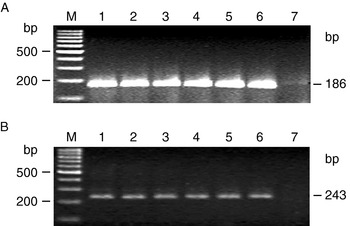
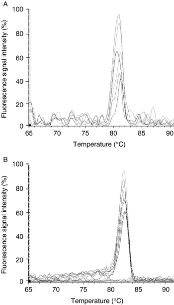
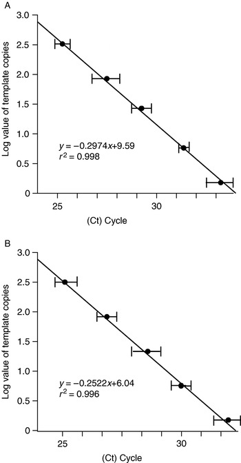
Rice auxin synthetase gene YUCAA1 and RSV CP gene were amplified by using real-time RT-PCR, and a single band of each was obtained, with sizes 186 and 243 bp, respectively (Fig. 1A, B), which corresponded to the anticipated design, and their melting curves each had a single peak (Fig. 2A, B). Using a series of fourfold dilutions of cDNA as templates, the standard curves of YUCAA1 and CP gene were drawn, their r 2 values were 0.998 and 0.996, respectively (Fig. 3A, B). Both were more than 0.995, demonstrating good linear coefficient and high accuracy; this is suitable for relative quantitative analysis of objective genes in different samples. This indicated that these primers were suitable for the SYBR Green I method-based real-time RT-PCR detection.
Detection of (A) the rice auxin synthase gene YUCAA1 and (B) RSV CP gene by real-time RT-PCR. Lanes: M, 100 bp ladder; 1–6, PCR products of YUCAA1 (A) and CP gene (B); 7, blank control.

Melting curves (replicates) of rice auxin synthase gene YUCAA1 (A) and RSV CP gene (B).

Standard curves of rice auxin synthase gene YUCAA1 (A) and RSV CP gene (B). Values in the figures represent the means±SE of three replicates.

Effect of RSV infection on auxin synthetase gene expression
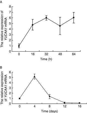
Comparing the YUCAA1 expression of different samples with the blank control, RSV infection significantly up-regulated YUCAA1 mRNA expression in suspended cells of rice. After 16 h of RSV infection, expression was increased to 4.76 times that of healthy cells, and 5.98 times after 32 h of infection. But after 48 h of infection, the amount expressed was slightly decreased to 4.57 times. After 64 h of infection, the expression amount was increased again to 6.03 times (Fig. 4A).
Influences on YUCAA1 mRNA expression in rice suspension cells (A) and rice plants (B) infected with RSV. Values in the figures represent the means±SE of three experimental replicates.

After RSV infection of rice plants, up-regulation of rice YUCAA1 mRNA expression occurred only between days 4 and 8, with 5.16 and 1.38 times the expression of healthy plants, respectively. From days 12 to 16 after infection, the amounts YUCAA1 mRNA expression in diseased plants were 0.20 and 0.17 times that of healthy rice, respectively (Fig. 4B). By day 16 after infection, the typical stripes of RSV leaf blight disease were visible.
Effect of RSV infection on rice endogenous auxin
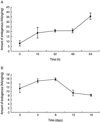
Figure 5A indicates that endogenous auxin content was significantly increased in RSV-infected suspended cells of rice compared with non-infected suspended cells. After 16–48 h of infection, the endogenous auxin content level in cells was high – 2.34–2.59 times that of healthy cells. After 64 h of virus infection, the endogenous auxin content level had increased further −4.43 times higher than that of healthy cells.
Influences on endogenous IAA in rice suspension cells (A) and rice plants (B) infected by RSV. Values in the figures represent the means±SE of three experimental replicates.

In rice plants, at days 4 and 8 after RSV infection, the endogenous auxin showed a tendency to increase, being 1.31 and 1.38 times that of healthy rice plants, respectively. At day 12 after RSV infection, the endogenous auxin content in diseased plants started to decrease, being 0.81 times that of healthy rice at the same growth phase. At day 16 after virus infection, the signs of rice stripe disease appeared, and the endogenous auxin content of infected plants was 0.73 times that of healthy rice at the same growth phase (Fig. 5B).
Changes in RSV replication in plants under different treatments
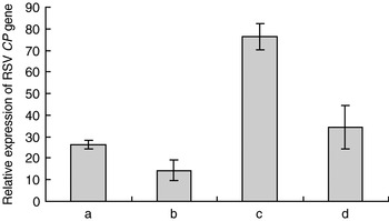
To determine the influence of RSV replication on endogenous auxin change in plants, the expression of the RSV CP gene was studied. A comparative analysis was conducted between diseased rice plants treated with the KPSC buffer solution and IAA solution, and non-treated diseased rice plants. The results indicated that the expression of RSV CP increased approximately three times in rice plants after they were treated with KPSC buffer to deplete the endogenous auxins, and decreased by 45% after IAA treatment. The expression of the RSV CP gene decreased by 55% after IAA treatment was given after KPSC buffer treatment (Fig. 6).
Influences on RSV replication in diseased rice plants treated with KPSC buffer and IAA. a, Diseased rice plants, untreated; b, diseased rice plants treated with IAA; c, diseased rice plants treated with KPSC buffer; d, diseased rice plants treated with IAA after treatment with KPSC buffer. Values in the figure represent the means±SE of three experimental replicates.

Discussion
The host plants showed abnormal growth and development after they were infected by the plant virus, including shortening, leaf discoloration, chlorosis and piebaldism, yellowing, absence of apical dominance, and even death. During the interaction between plant pathogens and host, various factors are involved in the genesis of these phenotypes, such as the salicylic acid signal conduction pathway (Yalpani et al., Reference Yalpani, Silverman, Wilson, Kleier and Raskin1991; Shirasu et al., Reference Shirasu, Nakajima, Rajasekhar, Dixon and Lamb1997; Bartsch et al., Reference Bartsch, Gobbato and Bednarek2006) and ethylene signal conduction pathway (Lorenzo et al., Reference Lorenzo, Piqueras, Sanchez-Serrano and Solano2003). An interaction between pathogen and host has also been reported to affect auxin signal conduction. For example, the interaction between Tobacco mosaic virus (TMV) replicase gene and PAP1, an auxin regulating factor, in TMV-infected Arabidopsis thaliana may interfere with the normal location of PAP1, resulting in its functional loss, causing the disturbance of normal auxin signal conduction and specific disease signs (Padmanabhan et al., Reference Padmanabhan, Goregaoker, Golem, Shiferaw and Culver2005). At the same time, the auxin signalling pathway is involved in natural immunity reactions in plants, and bacterial flagellin may induce miRNA393 to give negative regulation of auxin receptor genes TIR1 and AFBs in Arabidopsis thaliana, therefore inhibiting auxin signal conduction and resulting in disease resistance (Navarro et al., Reference Navarro, Dunoyer and Jay2006).
The overexpression of OsWAKY31 may significantly increase the resistance of rice to Magnaporthe grisea infection, by eliminating its sensitivity to the growth caused by externally synthesized auxin (Zhang et al., Reference Zhang, Peng and Guo2008). These studies showed that the auxin signal conduction pathway plays an important role in pathogenesis of plant pathogens, the microRNA-mediated immunological mechanism of host plants, and disease resistance of hosts.
The present research team carried out a study on transcriptomics of the interaction between RSV and rice, by utilizing the Affymetix gene chip of the entire rice genome. We found that 12 related genes of the auxin signal conduction pathway (Aux/IAAs, SAURs, GH3s and ARFs) indicated a change of expression in the transcription spectrum of RSV-infected rice, with nine genes being auxin early reaction genes (5 Aux/IAAs genes, 3 SAURs genes and 1 GH3s gene) (unpublished). In the auxin signal conduction pathway, auxin mainly controls the growth and development of plants through regulating the expression of early reaction genes (Abel and Theologis, Reference Abel and Theologis1996; Jain et al., Reference Jain, Kaur, Tyagi and Khurana2006a, Reference Jain, Kaur, Garg, Thakur, Tyagi and Khuranac, Reference Jain, Tyagi and Khuranad). To validate whether RSV infection causes a change of endogenous auxin content in rice, resulting in a change of expression of these early reaction genes, the present authors chose suspended cells of rice and rice plants as the studied material, and comparatively analysed the change of expression of the auxin synthetase gene, YUCAA1, and endogenous auxin content during RSV infection, replication and onset of the disease process. Compared with healthy suspended cells of rice, expression of the rice auxin synthesis gene YUCAA1 increased in RSV-infected rice cells within 0–64 hours, resulting in increased endogenous auxin content in suspended cells of rice. However, at the plant level, rice YUCAA1 expression increased in diseased plants at the initial phase of RSV infection, and endogenous auxin content also increased. But, at later phases of RSV infection, especially after signs of disease occurred (at 16 days after inoculation), YUCAA1 expression significantly decreased in diseased plants, and the endogenous auxin content was significantly lower than that in healthy rice.
Thus RSV infection may result in a change of endogenous auxin synthesis in rice. At the initial phase of infection, endogenous auxin synthesis significantly increased, but after typical signs occurred, the endogenous auxin synthesis was inhibited in diseased plants. These results are consistent with the past results of this research group. In microarray analysis during the transcriptomics study on the interaction between RSV and rice, the selected material comprised diseased plants at 7 days after the onset of symptoms. The results analysis of the study indicated that the endogenous auxin content in diseased plants at 7 days after disease onset was lower than that in healthy rice at the corresponding phase. The decrease in endogenous auxin content may cause decreased expression of some early reaction genes in the auxin signal conduction pathway (Jain et al., Reference Jain, Kaur, Tyagi and Khurana2006a, Reference Jain, Kaur, Garg, Thakur, Tyagi and Khuranac, Reference Jain, Tyagi and Khuranad). In microarray analysis, the expression of OsIAA6, OsIAA9, OsIAA31 and GH3-5 decreased, possibly caused by decreased auxin content in the diseased plant, which was consistent with the conclusion of a study using synthetic auxin treatment by Jain et al. (Reference Jain, Kaur, Tyagi and Khurana2006a, Reference Jain, Kaur, Garg, Thakur, Tyagi and Khuranac).
From this, we conclude that the different phases after RSV infection can be the main factors causing the difference of auxin at both the cell and plant level, and the auxin signal conduction pathway is also involved in the interaction between RSV and rice, which is a dynamic process. It is generally considered that the initial phase of infection is the most intense phase of interaction between pathogens and host, and also the most complicated phase involving all kinds of signal conduction. The process of RSV infection of rice has a long latent period, showing signs in 10–30 days. From the perspective of the interaction between plant pathogens and host, using diseased plants as study material of interaction between RSV and host, the initial phase of interaction between RSV and rice is often neglected, because the interaction between RSV and host is at a late phase when the signs occur. In addition, during the culture of rice, the study results on the interaction between RSV and rice vary greatly due to the influence of many abiotic environmental factors, such as temperature, light and humidity. Therefore, the RSV-infected rice suspended cell system provides a beneficial platform for studying the initial phase of the interaction between RSV and host, RSV infection replication and virus assemblage, etc.
This study also indicated that elimination of endogenous auxin in diseased plants could significantly improve RSV replication, but treatment with artificially synthesized auxin could make RSV content in diseased plants decrease rapidly. It is presumed that the elimination of endogenous auxin and the external application of artificially synthesized auxin may influence the growth and development of the host plant, thus indirectly influencing RSV replication, because raw materials, energy and environment must be provided by the host cells during the synthesis of virus components and virion assemblage of RSV as an obligate parasite. This also indicates the inter-influence and interconnection between the auxin signal conduction pathway and RSV infection process of rice, and a cross-talk molecule might exist between them. Certainly, these studies are not far enough advanced to thoroughly reveal the auxin signal conduction pathway of interaction between RSV and rice. It is necessary to detect and validate functionally downstream genes in the auxin signal conduction pathway, such as the auxin receptor gene, auxin early reaction genes and transcription inhibition factors, and so on, based on the present results of interactions between RSV and rice.
Acknowledgements
This research was funded by the Major Project of Chinese National Programs for Fundamental Research and Development (No. 2006CB100203), Doctoral Fund of the Ministry of Education of China (No. 20040389002; No. 20050389006) and National Natural Science Foundation of China (No. 30671357).