Various coagulase negative staphylococci (CNS) are the most commonly isoloated bacteria in subclinical mastitis of sheep and goats (Haenlein, Reference Haenlein2002; Leitner et al. Reference Leitner, Chaffer, Caraso, Ezra, Kababea, Winkler, Glickman and Saran2003, Reference Leitner, Chaffer, Shamay, Shapiro, Merin, Ezra, Saran and Silanikove2004a, Reference Leitner, Merin and Silanikoveb; Hall & Rycroft, Reference Hall and Rycroft2007). The bacteria are found on the udder and adjacent skin and are therefore difficult to control during lactation. Moreover, diagnosing infected glands is difficult because most infections are not recognized visually and only milk bacterial testing at a laboratory accompanied by elevation in somatic cell count (SCC) can confirm the animal's condition. Previously it was shown that milk from glands infected by CNS had significantly higher SCC and poorer milk quality compared with milk from uninfected glands. Infected glands also had reduced milk yield (Poutrel et al. Reference Poutrel, de Cremoux, Ducelliez and Verneau1997; Haenlein, Reference Haenlein2002; Leitner et al. Reference Leitner, Chaffer, Caraso, Ezra, Kababea, Winkler, Glickman and Saran2003). Among the CNS, Staphylococcus haemolyticus is a frequently isolated pathogen in dairy cows and small ruminants (Las Heras et al. Reference Las Heras, Dominguez, Lopez, Paya, Pena, Mazzucchelli, Garcia and Fernandez-Garayzabal2000; Chaffer et al. Reference Chaffer, Leitner, Zamir, Winkler, Glickman, Ziv and Saran2003). However, it always appears sporadically and not as a major pathogen for the herd. Staph. haemolyticus is a Gram-positive, coagulase-negative, coccus that appears as single 'grape-like' clusters and causes moderate to strong haemolysis on blood agar (Balows et al. Reference Balows, Hausler, Herrmann, Isenberg and Shadomy1991).
Recently, in a dairy goat herd of ~250 milking animals supplying milk for yogurt and cheese, milk coagulation appeared to be disturbed, resulting in low quality products. Individual milk samples from all the milking goats were tested using the California mastitis test (CMT) and bacteriological culture. A total of 25·6% (46/180 goats) had elevated SCC and growth of atypical highly mucoid colonies with CMT >2, however, mostly from goats with no clinical signs of mastitis. Drying-off and antibiotic treatment of all infected and uninfected goats resulted in very low curing at the next parturition, forcing the herdsman to control the outbreak by routine monitoring and culling of infected does. Individual testing and culling for two lactations resulted in clearing the herd of the bacteria. The present study describes the identification steps attempted to identify the bacteria and to compare it with other CNS including Staph. haemolyticus. Characterizing and identifying the bacterium which caused the outbreak was important since most of the goat milk in Israel is transported to large dairies and therefore it was important to alert the dairy sector that such a pathogen exists.
Materials and Methods
Animals
The dairy goat farm consisted of ~250 goats of the Shami breed in lactations 1–6 (with 30% yearly replacement) and their milk was used for the owners' yogurt and cheese production. The goats were maintained in 4-m2 closed sheds, with an additional 4 m2 of open space for each animal. The average individual milk yield in the flock was ~403 L/year, with 4·3% fat and 3·9% protein. The goats were machine-milked daily at 6·00 and 17·00. Milk samples from every udder-half after cleaning and disinfecting the teats for determination of SCC were preserved with Broad Spectrum Microtabs® II (D & F Control Systems, Inc., Dublin CA, USA) and sent to the Central Laboratory of the Israeli Cattle Breeders Association, Caesarea, and analysed using the Fossomatic 360 (Foss Electric, Hillerød, Denmark) which was calibrated with goat milk. In addition, CMT (KERBL, Buchbach, Germany) was also performed as an animal-side fast test. Every sample was tested also for the presence of bacteria. Owing to the large number of animals infected (25·6%; 46/180 goats) with atypical highly mucoid colonies accompanied by elevated SCC (>1·5×106 cells/ml) most of which were goats with no clinical signs of mastitis, all the lactating goats were dried off and treated with cephalosporin COBACTAN®DC (150 mg active Cefquinome; Intervet, The Netherlands) as prescribed by the manufacturer, a full tube per half-udder at drying-off. Three days postpartum a sample from each udder-half was cultured. The results revealed low curing as well as new infections with the same bacterial species. Therefore, a programme was instituted of monitoring all the animals 3 d post partum (CMT and bacterial culture) followed by fortnightly CMT testing, culture of all does with a CMT >2, and culling of the infected goats. This programme was practised for two consecutive years.
Bacteriological examinations
Bacteriological analysis was performed according to accepted standards of the National Mastitis Council (Oliver et al. Reference Oliver, Gonzalez, Hogan, Jayarao and Owens2004). An aliquot (0·01 ml) from every milk sample was spread onto blood-agar plates (Bacto-Agar; Difco Laboratory, Becton, Dickinson & Co., France) containing 5% washed sheep red blood cells and onto MacConkey plates. All plates were incubated at 37°C and examined for growth after 18 h and 42 h. Colonies suspected to be staphylococci were tested for coagulase (tube test) (Anilab, Rehovot, Israel). Strain identification was carried out with the API STAPH-IDENT 32 Staph Kit (BioMerieux S.A., Marcy-l'Etoile, France). When the percentage of micrococci-like bacteria that matched the test strain exceeded 90%, the strain identification was regarded as specific.
16S ribosomal DNA analysis
Representative isolates of the atypical CNS bacteria (ET14L; ET193L; ET321R) as well as previously identified Staph. haemolyticus (KS487; NH246; NH249) and Staph. simulans (KS177; KS4012) strains were submitted to 16S rDNA analysis. Briefly, chromosomal DNA was extracted from each isolate by using the Alkali-lysis technique, as described by Hartas et al. (Reference Hartas, Hibble and Sriprakash1998). Bacteria-domain-targeted PCR primers were used to amplify a conserved region of bacterial DNA coding for 16S rDNA (1392 bp), as described previously (Amann et al. Reference Amann, Ludwig and Schleifer1995). The following primers, purchased from Sigma Genosys (Israel) were used: 11F (5′-GTTTGATCMTGGCTCAG-3′) and 1392R, (5′-ACGGGCGGTGTGTAC-3′). PCR was performed in a Biometra/Tgradient thermocycler. Reactions were carried out in a final volume of 40 μl, with 0·5 pmol of each primer and 20 μl of PCR reaction master mix (Fermentas, Ontario, Canada). PCR conditions were: 95°C for 4 min followed by 36 cycles of 94°C for 30 s, 58°C for 30 s and 72°C for 1·5 min. The final cycle was followed by extension at 72°C for 10 min. PCR products were visualized on 1% agarose gel and following a purification step were sent for sequence determination (Hy Laboratories, Rehovot, Israel). Sequence identity was determined with the BLAST program (Altschul et al. Reference Altschul, Madden, Schaffer, Zhang, Zhang, Miller and Lipman1997).
Susceptibility test
Susceptibility testing was performed on the eight bacterial strains using the disk diffusion technique on Mueler-Hinton agar (DIFCO) (Bauer et al. Reference Bauer, Kirby, Sherris and Turck1966). Commercially available disks: penicillin G (10 units/disk), neomycin (10 units/disk), erythromycin (15 units/disk), cephalotin (10 units/disk), trimethoprin-sulphamethoxazol (SXT) (1·25–23·75 μg/disk) and cloxacillin (CX) (1 μg/disk) were applied as recommended, and the plates were incubated at 37°C for 24 h. The interpretation of susceptibility or resistance was done as recommended (Barnes-Pallesen et al. Reference Barnes-Pallesen, Blackmer, Britten, Bushnell, Van Damme and Wellcome1987).
Results
At the first testing (year I) 46/180 milking goats were found to be infected with atypical highly mucoid colonies accompanied by elevated cell count (>1·5×106) mostly from goats with no clinical signs of mastitis. To minimize spread of the infection to the whole herd, and possibly to other herds, it was decided to treat all the milking goats, infected and uninfected, with intramammary antibiotic dry-off treatment. Animals were treated with COBACTAN®DC after a susceptibility test to a range of antimicrobials which found the bacteria susceptible to cephalotin. At the next parturition (year II), of the positive goats treated, 78·3% (36/46) remained infected with the said bacteria. New infections with the same bacteria were found in 22/134 goats that were negative at dry-off and treated, as well as in 10/44 young does that entered the herd, resulting in 30·4% (68/224) of the milking goats infected with the bacteria. Therefore, a policy of monitoring all the animals 3 d post partum (CMT and culture) followed by bi-weekly monitoring, and culling of all the infected goats was used to clear the bacteria from the herd in two years (year III).
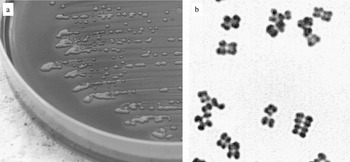
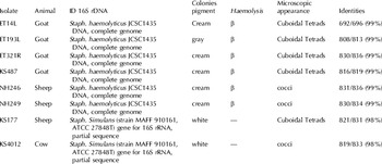
Several isolates of the atypical CNS bacteria (ET14L; ET193L; ET321R) were chosen for further microbiological and biochemical characterization and were compared with several other staphylococci from our collection, namely Staph. haemolyticus (KS487; NH246; NH249) and Staph. simulans (KS177; KS4012) that were isolated from cow, sheep or goats (Table 1). Species identification performed with the API STAPH-IDENT 32 kit showed >99·4% identity for all strains with T ranging between 0·49 and 0·84. The three atypical CNS were found to be Staph. haemolyticus (99·9% with T of 0·84). 16S rDNA sequencing confirmed the biochemical tests and assured that the three strains were indeed Staph. haemolyticus (99% identity). Table 1 summarizes the genetic and biochemical characterization of the strains and describes several phenotypic traits. All strains showed mucoid, smooth, opaque colonies, after incubation for 24 h at 37°C on blood agar. Continuous incubation at room temperature up to 48 h did not change the appearances of any of the isolates except the three atypical Staph. haemolyticus strains. These bacteria, which grew slowly during the first 24 h incubation and did not initially appear mucoid, continued to grow and became highly mucoid after 48 h (Fig. 1a). All the isolates except for the two Staph. simulans showed β haemolysis on blood agar plates.
(a) Mucoid colonies of atypical Staph. haemolyticus after 24-h incubation at 37°C and an additional 24 h at room temperature. (b) Microscopic cuboidal tetrad clusters of atypical Staph. haemolyticus. Gram staining (×1000).

Genetic and phenotypic characterization of the atypical CNS isolates and some reference CNS strains, Staphylococcus haemolyticus (ET14L; ET193L; ET321R), Staph. haemolyticus (KS487; NH246; NH249), Staph. simulans (KS177; KS4012) derived from different animals

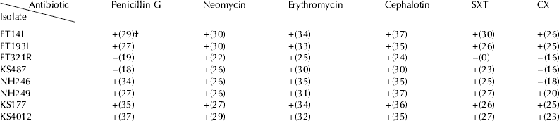
Staph. haemolyticus KS487 was found to be resistant to penicillin G, Staph. haemolyticus ET321R was resistant to SXT and four bacteria were resistant to CX. All strains were sensitive to neomycin, erythromycin and cephalotin (Table 2).
Susceptibility test using the disk diffusion technique on Mueler-Hinton agar of the atypical Staphylococcus haemolyticus (ET14L; ET193L; ET321R), Staph. haemolyticus (KS487; NH246; NH249) and Staph. simulans (KS177; KS4012)
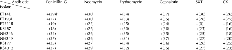
† Figures in parenthesis denote the inhibition diameter in mm, and the +/− stand for sensitive (+) or resistance (−) according to the manufacturer's instructions
Microscopically, most of the isolates tested, including the three atypical Staph. haemolyticus strains, showed unique cuboidal tetrad clusters, reminiscent of those of the genus Sarcina (Fig. 1b).
Discussion
The incidence of subclinical mastitis in dairy animals ranges from 15% to >50% (Wilson et al. Reference Wilson, Gonzalez and Das1997; Leitner et al. Reference Leitner, Chaffer, Caraso, Ezra, Kababea, Winkler, Glickman and Saran2003; Janosi & Baltay, Reference Janosi and Baltay2004; Leitner et al. Reference Leitner, Chaffer, Shamay, Shapiro, Merin, Ezra, Saran and Silanikove2004a, Reference Leitner, Merin and Silanikoveb, Reference Leitner, Merin, Silanikove, Ezra, Chaffer, Gollop, Winkler, Glickman and Saranc; Pitkala et al. Reference Pitkala, Haveri, Pyorala, Myllys and Honkanen-Buzalski2004). The major types of bacteria involved in subclinical mastitis are various CNS that are found on the udder and adjacent skin (Haenlein, Reference Haenlein2002; Bergonier & Berthelot, Reference Bergonier and Berthelot2003; Bergonier et al. Reference Bergonier, De Cremoux, Rupp, Lagriffoul and Berthelot2003; Leitner et al. Reference Leitner, Chaffer, Zamir, Mor, Glickman, Winkler, Weisblit and Saran2000, Reference Leitner, Chaffer, Caraso, Ezra, Kababea, Winkler, Glickman and Saran2003, Reference Leitner, Chaffer, Shamay, Shapiro, Merin, Ezra, Saran and Silanikove2004a, Reference Leitner, Merin and Silanikoveb, Reference Leitner, Merin, Silanikove, Ezra, Chaffer, Gollop, Winkler, Glickman and Saranc). Staph. haemolyticus is not considered a major udder pathogen in dairy cattle or small ruminants. In the present case, the outbreak was caused by an atypical Staph. haemolyticus strain which caused increased migration of leucocytes to the gland and low milk quality for cheese production. In most instances it did not cause signs of clinical infection and was noticed only after coagulation abnormalities appeared. The elevation of SCC seen with other CNS infections, which was associated with loss of curd yield and deterioration in its quality (Leitner et al. Reference Leitner, Chaffer, Shamay, Shapiro, Merin, Ezra, Saran and Silanikove2004a, Reference Leitner, Merin and Silanikoveb) was most pronounced in this case. Subclinical infections in dairy animals represent a problem because identification is not obvious and increase in the SCC in infected glands is modest [about (300–500)×103 cells/ml]. Therefore subclinicaly infected animals tend to be milked into the bulk milk tank where their substandard milk mixes with the milk from uninfected glands.
At the herd level, antibiotic treatment and drying-off of all the animals failed, although the bacteria were sensitive to the drug in vitro. Only routine bacterial testing and culling of all the positive goats eradicated the bacteria from the herd. The failure to cure infected udders from the bacteria with the COBACTAN®DC was not expected; however, it should be noted that not all bacteria causing udder infection sensitive to a specific antibiotic at the laboratory testing are successful when treating animals at the farm. Further study is needed if antibiotic treatment for this specific bacterium is to be the choice.
The outbreak caused by atypical CNS underlines the need for accurate biochemical and genetic methods for ultimate identification of the pathogen, since the current attitude to CNS in general is as a 'minor pathogen'. The involvement of Staph. haemolyiticus in this case suggests that it might be equipped with virulence factors adapted for the specific host. Understanding the pathogenicity of this strain together with development of DNA-based techniques for rapid identification of the pathogen might facilitate early detection and control of this pathogen.