Introduction
Congestive heart failure (CHF) is a common consequence of most diseases of the heart that affects about 0.4% of the population aged 45 years or over (Davies et al., Reference Davies2001; Vos and Begg, Reference Vos and Begg2007). CHF is a leading cause of hospitalization, morbidity and mortality in Western societies and its presence undermines the physical and mental well-being of patients (Hobbs et al., Reference Hobbs, Kenkre, Roalfe, Davis, Hare and Davies2002; Koenig, Reference Koenig2006). Recent evidence from observational studies suggests that CHF may also compromise intellectual function by affecting attention, memory, visuospatial skills and reasoning (Almeida and Flicker, Reference Almeida and Flicker2001; Almeida and Tamai, Reference Almeida and Tamai2001b; Beer et al., Reference Beer2009; Sauve et al., Reference Sauve, Lewis, Blankenbiller, Rickabaugh and Pressler2009), although it is not entirely clear if these deficits are better explained by co-existing medical conditions or if they are progressive in nature.
Coronary artery disease accounts for nearly two-thirds of all cases of CHF in adults (Gheorghiade and Bonow, Reference Gheorghiade and Bonow1998) and, not surprisingly, these conditions share numerous risk factors such as hypertension, diabetes, obesity, smoking and sedentary lifestyle (Kalogeropoulos et al., Reference Kalogeropoulos2009). Coronary artery disease and associated risk factors have been associated with depression and cognitive impairment (Almeida et al., Reference Almeida, Hulse, Lawrence and Flicker2002; Reference Almeida2007; Reference Almeida, Calver, Jamrozik, Hankey and Flicker2009; Bruce et al., Reference Bruce2003; Almeida and Pfaff, Reference Almeida and Pfaff2005; Whooley, Reference Whooley2006; Lautenschlager et al., Reference Lautenschlager2008), thus highlighting the possibility of confounding when investigating mental health outcomes of people with CHF (vascular diseases and risk factors have been associated with brain changes that result in depression and cognitive impairment) (Almeida et al., Reference Almeida2007; Moorhouse and Rockwood, Reference Moorhouse and Rockwood2008). The CHF Italian Study Group attempted to address this issue by comparing the cognitive performance of patients with CHF with that of people with other cardiovascular diseases (Trojano et al., Reference Trojano, Antonelli Incalzi, Acanfora, Picone, Mecocci and Rengo2003). They found that older adults with CHF had lower scores than controls on tasks measuring attention, verbal fluency and memory, and that the presence of significant cognitive impairment was associated with the severity of CHF (as measured by the New York Heart Association Class), and the presence of hypertension and depressive symptoms (Trojano et al., Reference Trojano, Antonelli Incalzi, Acanfora, Picone, Mecocci and Rengo2003). However, their control group was highly heterogeneous (it included people with hypertension, rheumatic fever, ischemic heart disease, diseases of pulmonary circulation and of the pericardium and endocardium) and had a different distribution of risk factors compared with CHF participants, so that doubt remains about the specific contribution of CHF to the cognitive deficits that they observed.
The origin of the cognitive deficits associated with CHF is also unclear. Left ventricular ejection fraction (Zuccala et al., Reference Zuccala, Cattel, Manes-Gravina, Di Niro, Cocchi and Bernabei1997; Almeida and Tamai, Reference Almeida and Tamai2001b), cerebral blood flow (Almeida and Tamai, Reference Almeida and Tamai2001a; Alves et al., Reference Alves2005), biochemical imbalances (Gunstad et al., Reference Gunstad2006), and regional brain atrophy (Woo et al., Reference Woo, Macey, Fonarow, Hamilton and Harper2003; Beer et al., Reference Beer2009) have all been linked to the cognitive impairment of patients with CHF. In addition, preliminary evidence suggests that appropriate treatment can ameliorate attention and memory (Almeida and Tamai, Reference Almeida and Tamai2001a; Tanne et al., Reference Tanne2005), although data from cardiac transplant patients indicate that some deficits may be irreversible (Schall et al., Reference Schall, Petrucci, Brozena, Cavarocchi and Jessup1989). Longitudinal studies could also offer valuable information about the possible causal link between CHF and cognitive impairment, as one would expect these deficits to worsen with ongoing exposure to the hemodynamic and metabolic changes associated with the disease. Unfortunately, there are virtually no longitudinal data on this topic, with the only available follow-up survey showing that the cognitive performance of patients with CHF either remains stable or improves modestly over a follow-up period of 12 months (Stanek et al., Reference Stanek2009).
The question of whether CHF causes cognitive decline is important because current approaches to the management of CHF require the active participation of the patients, who are expected to adhere to a complex medication regime, as well as monitoring their symptoms, weight, water and salt intake (Riegel et al., Reference Riegel2009). The concern, in this case, is that patients may have trouble complying with such expectations if CHF causes cognitive impairment, and this would lead to suboptimal treatment, more frequent health complications and greater use of health services (Riegel et al., Reference Riegel, Vaughan Dickson, Goldberg and Deatrick2007). Moreover, a better understanding of the association between CHF and cognitive decline may enhance our appreciation of the mechanisms that ultimately lead to the development of dementia in later life.
We designed this study to determine if adults with CHF associated with left ventricular dysfunction show evidence of cognitive decline over a period of two years compared with adults with normal left ventricular function with and without a clinical history of coronary artery disease. We hypothesized that participants with CHF would show evidence of cognitive deficits and decline compared with controls free from overt coronary artery disease, but not compared with controls with symptomatic coronary artery disease.
Methods
Study design
The Heart-Mind Study was a two-year longitudinal investigation of the cognitive performance of adults with CHF, coronary heart disease and controls. We collected endpoints of interest between May 2006 and March 2010 in Perth, Western Australia.
Participants
Participants were community volunteers who responded to advertisements about the study in the local media, or who were referred to the Heart-Mind Study by their primary care physician or hospital consultant. Referring hospital services included Royal Perth Hospital, Fremantle Hospital, and Sir Charles Gairdner Hospital. The Ethics Committees of the Royal Perth, Fremantle and Sir Charles Gairdner Hospitals approved the study protocol and all participants offered written informed consent. This investigation was conducted according to the principles expressed in the Declaration of Helsinki.
We excluded from participation people with a prior clinical history of stroke or cardiac arrest, hearing or visual impairment of sufficient severity to interfere with assessment procedures, Mini-Mental State Examination (MMSE) total score lower than 24 (Crum et al., Reference Crum, Anthony, Bassett and Folstein1993), or who showed evidence of clinically significant depressive or anxiety symptoms (Hospital Anxiety and Depression Scale sub-scores greater than 10) (Zigmond and Snaith, Reference Zigmond and Snaith1983). The latter were excluded because clinically significant anxiety and depressive symptoms can compromise performance on cognitive tests. We also excluded volunteers younger than 45 years of age, and those who were not fluent in written/spoken English or who reported a recent history of myocardial infarction (30 days or less).
Participants with CHF showed evidence of left ventricular dysfunction (ejection fraction – EF – lower than 0.4) and clinical symptoms consistent with the diagnosis of CHF for at least six months, such as exertion dyspnea, orthopnea, fatigue and weakness, dry cough, dizzy spells or palpitations, or physical signs of systemic edema (CHF group). We excluded from the study adults with New York Heart Association Functional Classification (NYHA) class IV (Miller-Davis et al., Reference Miller-Davis, Marden and Leidy2006) at the time of assessment because of the significant severity of illness and high short-term mortality (Castel et al., Reference Castel2010). Hence, these participants had treated and stable CHF.
Controls with coronary heart disease (CAD) displayed clinical and biochemical evidence of past myocardial infarction (i.e. elevated CK, CKMB, troponin T or troponin I), normal left ventricular function (EF > 0.6) and no clinical symptoms of CHF (CAD group). Controls without overt CAD denied symptoms or history suggestive of angina, myocardial infarction and CHF, and had normal left ventricular function (EF > 0.6) (control group). We did not collect systematic data on the use of medications from our participants.
Outcomes
The Cambridge Cognitive Examination of the Elderly Revised (CAMCOG) was the primary outcome measure of this study (Williams et al., Reference Williams, Huppert, Matthews and Nickson2003). The CAMCOG is a brief neuropsychological battery that assesses various aspects of cognitive functioning, including orientation, language, memory, attention and concentration, praxis, perception, calculation, and executive functions. The assessment produces a total summary score that can range from 0 to 105, with higher scores indicating better performance. The CAMCOG has robust psychometric properties and its scores are sensitive to change over time (Cullum et al., Reference Cullum2000; Williams et al., Reference Williams, Huppert, Matthews and Nickson2003).
Other cognitive outcomes of interest included immediate, and short and long-delayed recall as measured by the California Verbal Learning Test (CVLT) (Delis et al., Reference Delis, Freeland, Kramer and Kaplan1988; Woods et al., Reference Woods, Delis, Scott, Kramer and Holdnack2006), and the Digit Copy and Coding subtests of the Wechsler Adult Intelligence Scale (Wechsler, Reference Wechsler1997).
Other measures
We measured quality of life with the short form health survey (SF36) (Ware et al., Reference Ware1998), and symptoms of depression and anxiety with the Hospital Anxiety and Depression Scale (HADS) (Zigmond and Snaith, Reference Zigmond and Snaith1983). The SF36 produces summary scores for physical (PCS) and mental health (MCS) that range from 0 to 100 (the standardized mean for the population is 50 with standard deviation of 10), whereas the HADS produces depression and anxiety subscores that range from 0 to 21 each. All participants provided information about their age, gender, marital status and schooling (highest degree achieved). We measured their pre-morbid intelligence quotient (IQ) with the Cambridge Contextual Reading Test (Beardsall and Huppert, Reference Beardsall and Huppert1994), and calculated the left ventricular EF based on the results of an echocardiogram and the following formula: EF = (final diastolic volume – final systolic volume) / final diastolic volume. Perth Echo Services completed all echocardiograph assessments, and the coefficient of variation for the estimation of EF was lower than 5%. We also used the 6-Minute Walk Test as a safe and non-invasive index of the functional capacity of participants (Bittner, Reference Bittner1997).
Procedures for the collection of outcomes and other measures
Participants recorded demographic information at the initial assessment, which also included testing with the MMSE (as detailed above) (Crum et al., Reference Crum, Anthony, Bassett and Folstein1993) and cardiovascular history, including symptoms of CHF, angina, myocardial infarction, stroke and rating with the NYHA class (Miller-Davis et al., Reference Miller-Davis, Marden and Leidy2006). Consenting and potentially eligible participants were then referred for an echocardiographic examination to determine their left ventricular EF which we used, together with clinical history and review of medical notes, to allocate participants to the CHF, CAD and control groups.
Participants returned for the 120-minute face-to-face collection of outcomes within two weeks of completion of the echocardiogram. In addition to this baseline assessment, information about the outcomes of interest was collected again after 12 and 24 months.
Statistical methods
The data were managed and analyzed with Stata version 11.1 (StataCorp, College Station, TX). We used descriptive statistics to summarize baseline data, cross-tabulation to determine their distribution according to group membership (Pearson's chi-square statistic: χ2), and analysis of variance (one way analysis of variance or Kruskal-Wallis test) followed by Student's t-tests (mean and respective standard deviation – SD) or Mann-Whitney tests (median and the inter-quartile range – IQR) to compare the distribution of numerical variables for participants with CHF with controls.
We then used multilevel regression models to investigate the outcomes of interest according to group allocation (Stata command xtmixed and xtlogit). This enabled us to use all available data from participants (including those for whom outcome data were missing) and to take into account the intra-person correlation generated from the repeated measures of the same participant. Initially, we investigated the effect of the interaction between time of assessment and outcomes, but excluded the interaction effects because they made no obvious statistical contribution to the final models (p > 0.5). The basic models were adjusted for intra-individual changes over time and the baseline measure of each outcome (for example, by taking into account the cognitive score of participants at baseline). Age, gender, IQ, HADS-D and HADS-A were then added to a fully adjusted model, with the resulting coefficients indicating the mean difference and respective 95% confidence interval of the mean difference (95% CI) between the groups compared with non-CAD controls over 24 months. Alpha was set at 5% and all statistical tests reported are two-tailed.
Results
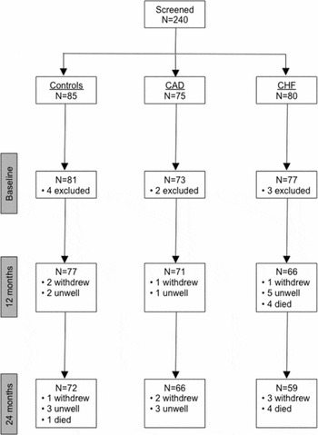
We screened 240 people, of whom nine did not fulfill criteria to join the study. Of those included, 77 had CHF NYHA class I to III, 73 had a documented clinical history of CAD but no CHF, and 81 did not have CHF and showed no evidence of clinically relevant coronary artery disease (controls). Figure 1 shows the flow of participants during the follow-up period of 24 months.
Flow of participants during the two years of follow-up according to group membership: controls with normal ventricular function and no clinical history of coronary heart disease, adults with clinical history of coronary artery disease (CAD), and adults with congestive heart failure (CHF). Assessments took place at baseline and again after 12 and 24 months.

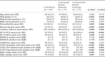
At the baseline assessment, participants in the three study groups were well balanced for age (range: 45 to 86 years), marital status and educational achievement. However, there were more men with CHF compared with CAD and controls, and the premorbid IQ of people with CHF was, on average, 2 points lower than the IQ of controls and participants with CAD. As expected, people with CHF had lower EF and walked a shorter distance in the 6-minute walk test than controls and adults in the CAD group (Table 1).
Demographic, clinical and cognitive characteristics of controls without coronary heart disease and participants with documented clinical history of coronary heart disease or congestive heart failure at the baseline assessment

p-value1: p-value from statistics comparing people with congestive heart failure with controls.
p-value2: p-value from statistics comparing people with congestive heart failure with coronary heart disease.
SD = standard deviation of the mean; IQR = inter-quartile range of scores.
SF36 PCS = short form health survey of physical health; SF36 MCS = short form health survey of mental health; HADS-A = Hospital Anxiety and Depression Scale – Anxiety subscale; HADS-D = Hospital Anxiety and Depression Scale – Depression subscale; CAMCOG = Cambridge Cognitive Examination of the Elderly Revised; CVLT = California Verbal Learning Test.
Participants with CHF had lower PCS and MCS ratings than controls, but there was no significant difference in the total scores of the HADS-D and HADS-A. Compared with people in the CAD group, CHF participants had lower PCS, higher HADS-A, but similar MCS and HADS-D. Lastly, participants with CHF had lower cognitive scores in all measures of cognitive function compared with controls, but CAMCOG, digit coding and digit copying scores were not significantly different from people in the CAD group.
Main results for cognitive outcomes
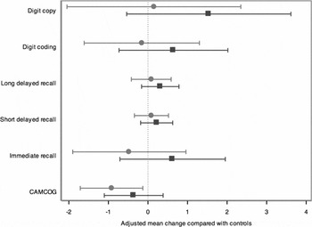
Figure 2 shows the adjusted changes of all cognitive outcomes relative to controls for CHF and CAD participants during the follow up period. The average CAMCOG score of controls during follow up was 95.1 (95% CI = 94.0, 96.3) and the scores of participants with CHF and CAD declined 3.1 (95% CI = −4.7, −1.5; p < 0.001) and 1.4 points (95% CI = −3.0, 0.2; p = 0.095) compared with controls (adjusted for intra-individual variability). We then adjusted the model for the CAMCOG score of participants at baseline, as well as for age, gender, IQ, and HADS-D and HADS-A scores: the CAMCOG scores of participants with CHF were, on average, 0.9 points (95% CI = −1.7, −0.1; p = 0.022) lower than the scores of controls. Compared with controls, the adjusted scores of participants with CHF did not change significantly over two years for immediate recall (adjusted mean difference = −0.5, 9% CI = −1.9, 1.0; p = 0.519), short delayed recall (adjusted mean difference = 0.1, 95% CI = −0.3, 0.5; p = 0.688), long delayed recall (adjusted mean difference = 0.1, 95% CI = −0.4, 0.6; p = 0.747), digit coding (adjusted mean difference = −0.2, 95% CI = −1.6, 1.3; p = 0.833) and digit copying (adjusted mean difference = 0.1, 95% CI = −3.3, 0.1). Of note, all participants with CAD and normal controls continued to have EF > 0.6, whereas all participants with CHF continued to have EF < 0.4 by the end of the follow-up period of two years.
Mean change in cognitive scores over two years of adults with congestive heart failure (circle) and coronary artery disease (squares) relative to controls (vertical dotted line – all values to the left of this line represent worsening relative to controls). The whiskers represent the 95% confidence interval of the mean difference. All values were adjusted for age, gender, intelligence quotient, Hospital Anxiety and Depression Scale – Depression subscale (HADS-D), and Hospital Anxiety and Depression Scale – Anxiety subscale (HADS-A), and respective cognitive scores at baseline.

We also completed a series of planned analyses comparing the cognitive performance of participants with CHF and CAD over two years. There were no between-group differences on the CAMCOG (adjusted mean difference = −0.7, 95% CI = −1.4, 0.1; p = 0.083), CVLT immediate recall (adjusted mean difference = −1.2, 95% CI = −2.6, 0.1; p = 0.068), CVLT short delayed recall (adjusted mean difference = −0.6, 95% CI = −0.5, 0.3; p = 0.482), CVLT long delayed recall (adjusted mean difference = −0.3, 95% CI = −0.7, 0.2; p = 0.256), digit coding (adjusted mean difference = −0.6, 95% CI = −1.9, 0.7; p = 0.334), and digit copying (adjusted mean difference = −1.4, 95% CI = −3.5, 0.7; p = 0.193).
Finally, we performed supplementary analyses to clarify if the two-year between-group difference in CAMCOG scores could be explained by poor functional capacity and self-perceived health, as measured by the 6-minute walk test and PCS scores. Inclusion of the results of the 6-minute walk test in the model weakened the association between CHF and cognitive decline compared with controls (adjusted mean difference = −0.7, 95% CI = −1.6, 0.1; p = 0.073). The same occurred after the inclusion of PCS scores in the model (adjusted mean difference = −0.8, 95% CI = −1.6, 0.1; p = 0.068). Left ventricular function, as measured by EF, did not fully account for the decline in CAMCOG scores of participants with CHF compared with controls (adjusted mean difference = −2.0, 95% CI = −4.0, −0.0; p = 0.050).
Results for mood and quality of life outcomes
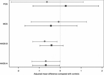
Participants in all study groups had low scores on the HADS-A and HADS-D, although people in the CAD (adjusted mean difference = 0.7, 95% CI = 0.1, 1.3; p = 0.039) and CHF groups (adjusted mean difference = 0.7, 95% CI = 0.0, 1.4; p = 0.048) experienced a slight increase in the severity of anxiety symptoms during follow-up compared with controls. Similarly, the severity of depressive symptoms increased amongst participants in the CHF (adjusted mean difference = 0.7, 95% CI = 0.1, 1.3; p = 0.014) but not in the CAD group (adjusted mean difference = 0.4, 95% CI = −0.1, 1.0; p = 0.144). Figure 3 summarizes these findings.
Mean change in Hospital Anxiety and Depression Scale – Anxiety subscale (HADS-A), Hospital Anxiety and Depression Scale – Depression subscale (HADS-D), Mental (MCS) and Physical Health (PCS) summary measures of the SF-36 over two years of adults with congestive heart failure (circle) and coronary artery disease (squares) relative to controls (vertical dotted line – all values to the left of this line represent worsening relative to controls). The whiskers represent the 95% confidence interval of the mean difference. All values were adjusted for age, gender and respective baseline scores.

We also calculated the population average odds of clinically significant anxiety and depression during follow-up in the CAD and CHF groups relative to controls, defined as a score of 11 or greater on the HADS-A and HADS-D, respectively. The average proportion of controls, CAD and CHF participants with anxiety was 1.3%, 4.4% and 6.2%. Likewise, the average proportion of controls, CAD and CHF participants with depression during follow-up was 0.9%, 2.9% and 5.2%, respectively. These analyses were adjusted for age, gender and PCS scores.
The MCS scores of participants with CAD and CHF did not change significantly compared with the scores of controls (adjusted mean MCS difference = −0.4, 95% CI = −1.7, 0.8; p = 0.488 for CAD and −0.1, 95% CI = −1.4, 1.3; p = 0.923 for CHF). The analysis of PCS outcomes yielded similar findings (adjusted mean PCS difference = 0.3, 95% CI = −1.3, 1.8; p = 0.738 for CAD and −0.6, 95% CI = −2.3, 1.1; p = 0.481 for CHF compared with controls).
Discussion
The results of this study confirmed that adults with CHF show evidence of cognitive decline over two years compared with controls with normal left ventricular function and no history of coronary artery disease. We also found that people with stable CHF experience a relative decline in general cognitive function over two years compared with cardiologically healthy controls, but not compared with people with past clinical history of coronary artery disease. Finally, we observed a slight excess of clinically significant anxiety and depressive symptoms amongst people with CHF compared with cardiologically healthy controls, but no obvious difference between people with CHF and controls with coronary artery disease.
The Heart-Mind Study recruited well-characterized samples of adults with CHF, coronary artery disease and cardiologically healthy controls who were closely followed for two years. Loss to follow-up was minimal and, in the case of CHF, was mostly due to the death of participants (10.4%). Importantly, our approach to the analysis of the data enabled us minimize the potential bias due to survival, as it permitted the inclusion of all available data into the models (even for people who were later lost to follow-up). This is important because loss to follow-up is more frequent amongst participants with worse health, and such a loss could bias the results of the study towards the null (and patients with CHF are generally less well than those with coronary heart disease or controls free of cardiovascular problems). In other words, the approach that we used to analyze our data decreases the probability that the observed lack of difference between the groups over time was due to survivorship bias. This is also the first study to monitor change in cognitive function amongst patients with CHF over an extended period of time, and to take into account the non-specific effects of concomitant cardiovascular illness. We acknowledge, however, that our study sample was formed by a group of motivated volunteers, and this may restrict somewhat the external validity of our findings. In addition, our CHF sample included only people with systolic dysfunction, so that it remains unclear if these results can be extended to patients with CHF due to non-systolic cardiac illness. Our study groups were not perfectly balanced for gender, premorbid IQ and HADS scores, although differences between groups were minimal and our analyses have taken into account the possibility of confounding. Furthermore, the inclusion of relatively young participants in the study may have contributed to attenuate the negative consequences of CHF on cognitive function (which might be more pronounced in older people), although our results showed no obvious interaction between age and CHF on cognitive scores. Finally, as we did not collect biochemical data from participants, we are unable to comment on the possible physiological mechanisms underlying cognitive dysfunction and mood disturbances in CHF, although our findings suggest that objective and subjective measures of functional capacity (6-minute walk test and PCS) and left ventricular EF cannot adequately explain the associations that we observed.
Our results indicate that people with left ventricular dysfunction CHF experience cognitive decline over two years compared with cardiologically healthy controls, although such a loss is minimal and unlikely to be of clinical significance (less than 1 point on the CAMCOG). In addition, our data suggest that this subtle cognitive loss may not be a direct consequence of CHF, but a non-specific outcome of shared cardiovascular disease burden and risk factors (there were no significant differences between people with CHF and with coronary artery disease associated with normal left ventricular function). Coronary artery disease has been associated with a modest decline in cognitive function as well (Newman et al., Reference Newman2001), and head-to-head comparisons of people who did and did not undergo bypass graft surgery has shown that these changes are not due to the surgical procedures (Selnes et al., Reference Selnes2008). Other factors commonly associated with CHF and coronary artery disease may also play a role: hypertension (Qiu et al., Reference Qiu, Winblad and Fratiglioni2005), diabetes (Cukierman et al., Reference Cukierman, Gerstein and Williamson2005), smoking (Almeida et al., Reference Almeida, Hulse, Lawrence and Flicker2002; Reference Almeida2011) and obesity (Gustafson, Reference Gustafson2006) have all been linked to cognitive decline and, in some studies, to dementia (Forette et al., Reference Forette1998; Almeida et al., Reference Almeida, Hulse, Lawrence and Flicker2002; Gustafson, Reference Gustafson2006; Whitmer et al., Reference Whitmer, Gustafson, Barrett-Connor, Haan, Gunderson and Yaffe2008). These factors have been shown to increase the number and extension of brain lesions due to cerebrovascular disease and to lead to loss of neurons in higher cortical function areas of the brain (den Heijer et al., Reference den Heijer2005; Koschack and Irle, Reference Koschack and Irle2005; Prins et al., Reference Prins2005; Almeida et al., Reference Almeida2008a; Reference Almeida, Garrido, Lautenschlager, Hulse, Jamrozik and Flicker2008b; van Dijk et al., Reference van Dijk, Prins, Vrooman, Hofman, Koudstaal and Breteler2008), thus contributing to a progressive loss of brain reserve capacity and the eventual manifestation of cognitive decline (Ngandu et al., Reference Ngandu2007). Furthermore, the lack of an obvious association between left ventricular EF and the two-year change in cognitive function in our study suggests that the intellectual deficits of CHF cannot be easily attributed to hemodynamic changes.
We recruited participants who were free of clinically significant depressive and anxiety symptoms at the time of entry into the study, which explains why our samples had such a low prevalence of depression and anxiety symptoms compared with other studies (Whooley, Reference Whooley2006; Yohannes et al., Reference Yohannes, Willgoss, Baldwin and Connolly2010). Nonetheless, a larger proportion of participants with CHF and coronary artery disease than cardiologically healthy controls showed evidence of clinically significant depressive and anxiety symptoms at the subsequent 12- and 24-month assessments. However, people with CHF and coronary artery disease were similarly affected by depression and anxiety, which suggests that these changes in mood (or in cognitive function) are not necessarily characteristic of CHF (Bruce et al., Reference Bruce2003; Almeida and Pfaff, Reference Almeida and Pfaff2005; Almeida et al., Reference Almeida2007; Reference Almeida, Calver, Jamrozik, Hankey and Flicker2009).
The changes in cognitive function and mood associated with CHF are subtle and may be a consequence of non-specific changes that affect the brain, rather than of CHF itself. Future studies should consider using clinical, biochemical and imaging methods to develop a coherent measure of cardiovascular disease burden that can then be used to improve our understanding of related morbidities and guide the development of relevant preventive measures. In the meantime, adults with stable CHF can feel reassured that they will not experience significant cognitive decline or depression over two years specifically because of their CHF, and healthcare professionals can continue to use self-management strategies to optimize the clinical outcomes of their patients (Riegel et al., Reference Riegel2009).
Conflict of interest
None.
Description of authors' roles
O. Almeida, N. Lautenschlager, L. Arnolda and L. Flicker conceived and designed the study, and obtained funding. O. Almeida, N. Lautenschlager, L. Arnolda, L. Flicker and C. Beer assisted with recruitment and data collection. O. Almeida and H. Alfonso analyzed the data and all authors contributed to the interpretation of the results. O. Almeida drafted the manuscript. All authors reviewed the paper critically and approved its submission for publication.
Acknowledgments
The investigators thank participants and research staff for their generous contribution.
This project was supported by a competitive project grant from the National Health and Medical Research Council of Australia (NHMRC, #403996). The sponsor had no role in the design and running of the study, analysis of the data, drafting of the manuscript or decision to publish.