Introduction
We present a patient with acute stridor caused by both an endolaryngeal tumour and extraluminal compression from subcutaneous air. This subcutaneous air was a result of the tumour perforating through the laryngeal cartilage, which allowed air to escape subcutaneously when the patient coughed. To our knowledge, this is the first such case reported in the literature.
It was possible to stabilise the patient with conservative measures from the time of presentation until definitive laryngectomy some two weeks later. Despite declining adjuvant radiotherapy, the patient had no signs of recurrence in the neck when he died a year later from a heart attack.
Case report
An 82-year-old male presented as an airway emergency to the ENT emergency clinic at the University Hospital Zurich, Switzerland. Initial examination revealed that the patient was tachypnoeic with slow atrial fibrillation (under beta-blocker therapy) and normal blood pressure. He had both inspiratory and expiratory stridor, and oxygen saturation of 88 per cent. Clearly in distress, the patient complained of an acute, large (5 × 5 cm) swelling on the left of his neck, at the level of the thyroid cartilage. This had appeared suddenly during a bout of coughing.
After stabilisation, a more detailed history revealed that the patient had experienced hoarseness for roughly one year. Four months previously, he had been investigated in a neighbouring hospital, and clinical tumour–node–metastasis (TNM) staging had resulted in a T2N0 exophytic glottic cancer diagnosis. The patient had undergone transoral laser debulking and had been scheduled to undergo curative chemoradiotherapy. Unfortunately, feeling much better after the debulking, the patient had failed to attend for definitive treatment.
Endoscopic examination revealed an incompetent larynx with penetration of saliva. The left vocal fold was paretic, with an exophytic cancer growing transglottically. Upon neck palpation, the large swelling on the left side completely disappeared under pressure, greatly relieving the patient's stridor. The mass had been a large collection of subcutaneous air.
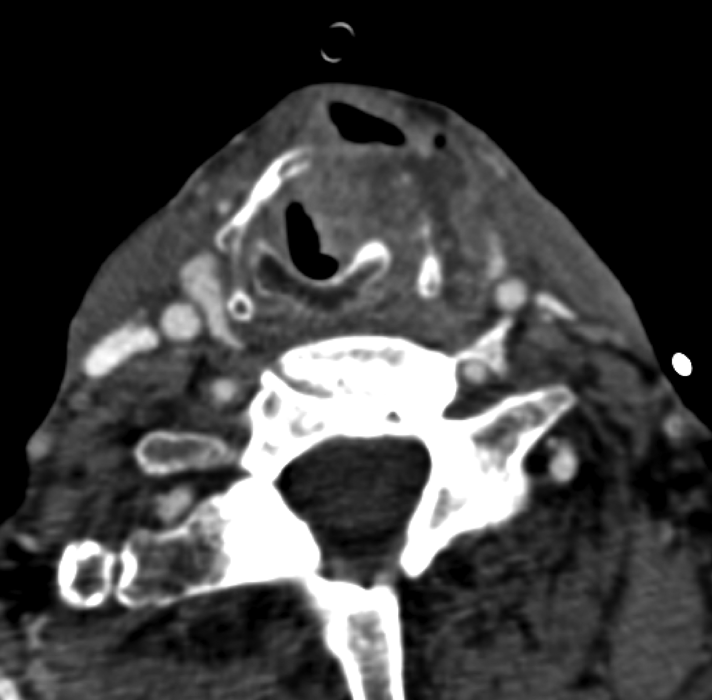
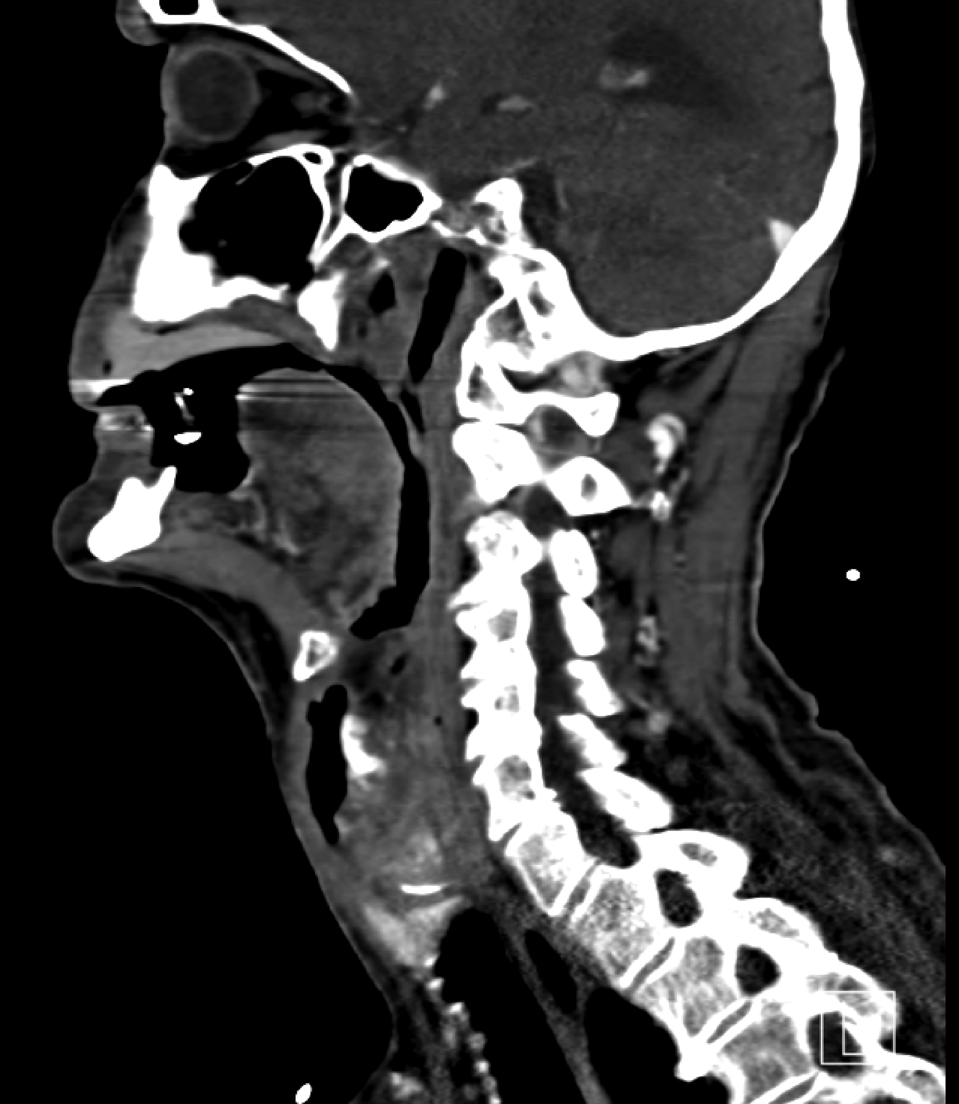
Restaging computed tomography was performed (Figures 1 and 2), which indicated a clinically staged T4N0 transglottic cancer. The cancer had destroyed the thyroid cartilage, allowing air to escape into the subcutaneous tissues whenever there was a high subglottic pressure build-up, when coughing for example.
Axial, contrast-enhanced computed tomogram showing subcutaneous air and left-sided tissue bulk consistent with tumour.

Sagittal, contrast-enhanced computed tomogram showing subcutaneous air and left-sided tissue bulk consistent with tumour.
The patient was treated for aspiration pneumonia, medically optimised (treated with intravenous antibiotics and inhalational therapy) and appropriately counselled for surgery. Two weeks later, the patient underwent total laryngectomy with an elective left neck dissection and pectoralis major flap wound protection. Unfortunately, he had high post-operative troponin levels (0.149 mcg/l (norm <0.014 mcg/l)) and remained in hospital for three weeks. During this time, he underwent a secondary transoesophageal puncture for a voice prosthesis (Provox®).
After discharge, the patient made an uneventful recovery, and had good speech and swallow function. He again refused adjuvant radiotherapy. The patient died just over a year later from a myocardial infarction without clinical or positron emission tomography radiographic signs of disease recurrence.
Discussion
Tracheal and laryngeal injury can occur from an ‘extraluminal’ or ‘endoluminal’ injury, and may present with surgical emphysema. The emphysema may in turn lead to extraluminal compression of the airway and subsequent airway obstruction. Several papers have documented traumaticReference Dissanaike, Shalhub and Jurkovich1–Reference Kolber, Aspler and Sequeira3 or iatrogenicReference Luers, Bovenschulte and Beutner4 injuries to the airway. Neonates are known to be particularly at risk of laryngeal injury from intubation.Reference Wei and Bond5 There is even a case report of iatrogenic laryngeal perforation occurring during the intubation of a pig.Reference Steinbacher, von Ritgen and Moens6 Large, external laryngoceles are also a well described entityReference Dursun, Ozgursoy, Beton and Batikhan7 that can present with an acute neck swelling following coughing; however, these still have an intact epithelial lining and are therefore not associated with emphysema.
• Extraluminal compression, intraluminal changes and/or endoluminal obstruction can cause airway obstruction
• There is no widely accepted standardised approach for evaluating the ‘at risk’ airway
• Patients need to be carefully counselled as to the risks of airway obstruction, especially if they decline definitive treatment
• Despite the risk of tumour seeding from a perforated larynx, our patient declined adjuvant therapy; there was no sign of recurrence
To our knowledge, this is the first case report of tumour breaking through the laryngeal cartilage leading to acute extraluminal airway compromise. Fortunately, the patient did not require immediate intubation to secure his airway: once the extraluminal component had been resolved with direct compression, the intraluminal component was not sufficiently large to cause airway compromise. The patient was monitored in an intermediate care bed; the option of rapid intubation was available should the need have arisen. The appropriate investigations were then carried out and the patient was discussed by our tumour board. The patient was then counselled before undergoing definitive therapy.
Interestingly, despite the patient declining adjuvant radiotherapy, there was no disease recurrence in the neck. One would imagine that the extralaryngeal infiltration and perforation required to present with a ‘pneumo-larynx’ would have meant extensive tumour seeding in the neck. This would normally have presented itself as persistent disease after surgery, yet this was not the case.
Airway emergencies must be handled with great care. Even after the acute stridor had subsided, our patient could not be considered completely stable until the cancer had been definitively treated.
Assessment of the airway at risk is often based on objective markers, such as tachypnoea and oxygen saturation, and subjective factors, such as patient fatigue. Any airway intervention will depend on the acuteness of presentation and available expertise. Several options exist for securing the airway in patients with laryngeal cancer.8, Reference Moorthy, Gupta, Laurent and Weisberger9 These include endotracheal intubation, transoral debulking,Reference Hassani and Bhananker10, Reference Laccourreye, Lawson, Muscatello, Biacabe, Laccourreye and Brasnu11 tracheostomyReference Pezier, Nixon, Joshi, Pang, Guerrero-Urbano and Oakley12 or the now defunct emergency laryngectomy.Reference Moorthy, Gupta, Laurent and Weisberger9, Reference Narula, Sheppard, West and Bradley13 The choice will be physician and unit specific, and may even depend on whether the patient presents out of normal hours.