Introduction
The peculiarly modified northeastern Nearctic species Herniosina voluminosa Marshall, Reference Marshall1987 (Diptera: Sphaeroceridae: Limosininae) was originally described as a limosinine species of “isolated phylogenetic position”, not readily assignable to any named genus (Marshall Reference Marshall1987). It was tentatively included in the genus Herniosina Roháček, Reference Roháček1983 on the basis of its strongly protruding male S1+2 (a distinctive synapomorphy of Herniosina) with the explicit caveat that its male and female terminalia differ essentially not only from the other two described (European) species of this genus, but also from all other known genera of Limosininae (Marshall Reference Marshall1987: 711). Roháček (Reference Roháček1993, Reference Roháček2016) later described three further European species of Herniosina and recently (2016) redefined the genus as a monophyletic Palaearctic group, excluding the Nearctic H. voluminosa and indicating that it should be placed in a new genus. We here describe and name that new genus as Volumosina, redescribe its type species in detail with further consideration of its phylogenetic placement, and provide new information on its natural history and distribution.
Material and methods
Material examined
The specimens used are deposited in collections as follows: Canadian National Collection of Insects, Arachnids, and Nematodes, Ottawa, Ontario, Canada (CNCI); University of Guelph Insect Collection, School of Environmental Sciences, University of Guelph, Guelph, Ontario, Canada (DEBU); Department of Entomology, College of Life Science and Agriculture, University of New Hampshire, Durham, New Hampshire, United States of America (DENH); and Slezské zemské muzeum, Opava, Czech Republic (SMOC).
Abdomens of selected specimens were detached, cleared by boiling several minutes in a 10% solution of potassium hydroxide, then neutralised in a 10% solution of acetic acid, washed in water and subsequently transferred to glycerine. Postabdominal structures were dissected and examined under binocular microscopes; detailed examinations of genital structures were performed with compound microscopes. After examination, all dissected parts were put into glycerine in small plastic tubes sealed with hot forceps and pinned below the respective specimens.
Drawing techniques and photography
Legs were drawn on squared paper using a Reichert (Vienna, Austria) binocular microscope with an ocular screen. Genitalia illustrations were made using Abbe’s drawing apparatus on a compound microscope (JENAVAL; Carl Zeiss, Jena, Germany) at magnifications of 130–500×. Whole specimens were photographed using a Canon (Brampton, Ontario, Canada) DSLR mounted on a Stackshot rail (Cognisys, Traverse City, Michigan, United States of America) and stacked into high depth of field images using Helicon Focus (HeliconSoft, Kharkiv, Ukraine).
Measurements
Six main characteristics of the species were measured: body length (measured from anterior margin of head to end of cercus, thus excluding the antenna), index t 2 :mt 2 (=ratio of length of mid tibia:length of mid basitarsus), wing length (from wing base to wing tip), wing width (maximum width), C-index (Cs 2 :Cs 3 ) (=ratio of length of second costal sector:length of third costal sector) and index rm\dm-cu:dm-cu (=ratio of length of section between rm and dm-cu on discal cell:length of dm-cu).
Presentation of faunistic data
Data from formerly unpublished non-type specimens are standardised and presented in full. Data on type specimens are given in Marshall (Reference Marshall1987) and are not repeated here (except for those of the holotype), but distributional data are summarised. Phenological and other natural history information obtained from the material examined and literature are given in the “Natural history” section.
Morphological terminology
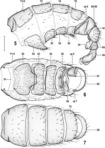
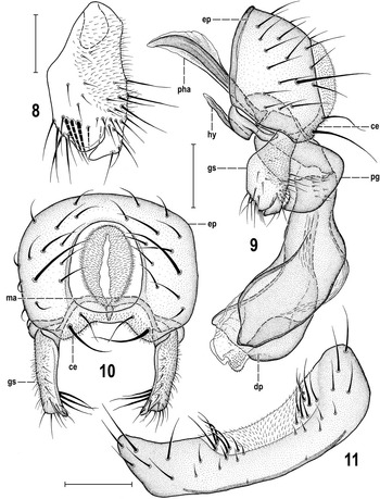
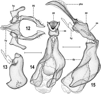
Follows that used for Sphaeroceridae by Roháček (Reference Roháček1998) in the Manual of Palaearctic Diptera, including terms influenced by the “hinge” hypothesis of the origin of the eremoneuran hypopygium (Zatwarnicki Reference Zatwarnicki1996). This differs from recent Sphaeroceridae papers by North American authors, which generally follow the terminology in the Manual of Central American Diptera (Cumming and Wood Reference Cumming and Wood2009). Most significantly, the surstylus sensu Cumming and Wood is referred to as the gonostylus, the ejaculatory apodeme sensu Cumming and Wood is here interpreted as the ejacapodeme, the basiphallus sensu Cumming and Wood is here called the phallophore, and the subepandrial sclerite sensu Cumming and Wood is here referred to as the medandrium. Morphological terms of the male postabdomen and genitalia are depicted in Figures 5–15; those of the female postabdomen in Figures 22–24. Abbreviations of morphological terms used in text and illustrations, along with their widely used synonyms, are listed below.
Abbreviations of morphological terms used in text and/or figures: A 1 —anal vein; ac—acrostichal (seta); ads—additional (setulae) on frons; C—costa; ce—cercus; Cs 2 , Cs 3 —2nd, 3rd costal sector; CuA 1 —cubitus; dc—dorsocentral (seta); dm—discal medial cell; dm-cu—discal medial-cubital (=posterior, tp) cross-vein; dp—distiphallus; ea—ejacapodeme (=ejaculatory apodeme); ep—epandrium (=periandrium); f 1 , f 2 , f 3 —fore, mid, hind femur; g—genal (seta); gs—gonostylus (=surstylus); hu—humeral (=postpronotal) (seta); hy—hypandrium; ifr—interfrontal (seta); M—media; ma—medandrium (=subepandrial =bacilliform sclerite); mt 2 —mid basitarsus; oc—ocellar (seta); occe—outer occipital (seta); occi—inner occipital (seta); ors—fronto-orbital (seta); pg—postgonite (=paramere); pha—phallapodeme (=aedeagal apodeme); pp—phallophore (= basiphallus); pvt—postvertical (=postocellar) (seta); R 1 —1st branch of radius; R 2+3 —2nd branch of radius; R 4+5 —3rd branch of radius; r-m—radial-medial (= anterior, ta) cross-vein; S1–S10—abdominal sterna; sc—scutellar (seta); sp—spiracle; stpl—sternopleural (=katepisternal) (seta); T1–T10—abdominal terga; t 1 , t 2 , t 3 —fore, mid, hind tibia; va—ventroapical seta on t 2; vi—vibrissa; vte—outer vertical (seta); vti—inner vertical (seta).
Volumosina Roháček and Marshall, new genus
Type species
Herniosina voluminosa Marshall, Reference Marshall1987, here designated.
Etymology
The generic name is an abbreviated conjunction of Volu[minosa]+[Li]mosina and also an anagram of “voluminosa”, the name of the type species. This name should be treated as feminine in gender for nomenclatural purposes.
Description
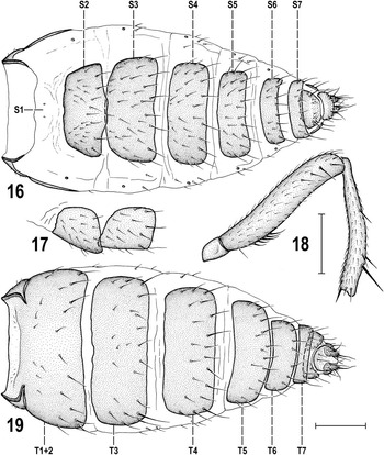
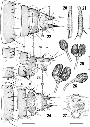
(1) pvt present but reduced to small setulae; (2) three ifr, middle pair strong and cruciate; (3) three to five minutes ads inside and below ors; (4) g small, not longer than anterior peristomal setula; (5) two hu, the internal reduced to microseta; (6) two postsutural dc, the anterior short; (7) ac setulae in six to eight rows on suture, the medial prescutellar ac slightly enlarged; (8) two stpl but only the posterior strong, the anterior small, hair-like; (9) scutellum relatively large, shorter than broad, rounded trapezoidal; (10) t 2 chaetotaxy as in Figures 18, 20–21, dorsally with only one proximal and three to four distal setae; in male ventrally with a row of small spine-like setae and with reduced va (Fig. 18), female ventrally with only fine setulae and va long (Fig. 21); (11) male f 2 ventrally with a short row of about five curved setae subbasaly (Fig. 18); (12) C not extended beyond apex of R 4+5 (Figs. 2–3); (13) R 4+5 slightly sinuate (Figs. 2–3); (14) dm cell long, with small processes of M and CuA 1 beyond dm-cu (Figs. 2–3); (15) alula relatively small, narrow, with relatively acute apex (Figs. 2–3); (16) male abdomen straight (not down-curved), T5 and S8 of normal size (Figs. 1, 5); (17) male S2 enlarged and strongly protruding posteromedially (Figs. 1, 5–6); (18) male S3 with anterior corners slightly lobate (=anteromedially slightly emarginate); (19) S3–S5 with anterior dark-pigmented stripe (Fig. 6); (20) male S5 transverse but of normal size, posteromedially with unpigmented area armed with lateral setae (Fig. 11); (21) epandrium with only one strong and long lateral seta (Figs. 9–10); (22) male cerci reduced, fused medially and fused with epandrium, forming a short subanal plate (see Fig. 10), each with two (one long) setae; (23) gonostylus (Fig. 8) relatively small and simple, setose (with longest setae internally, see Fig. 10), micropubescent externally and with inconspicuous posteroventral process; (24) hypandrium short (Fig. 9), with medial apodeme subtriangular (Fig. 12); (25) aedeagus and postgonites extremely enlarged; phallapodeme of normal size (Figs. 9, 13–15); (26) (enlarged) phallophore elongate but simple, distally movably attached to proximal end of distiphallus (Figs. 14–15); (27) distiphallus extraordinarily enlarged, relatively simple but distally asymmetrical, its lateral sclerites separated by membranous apex distally (Figs. 14–15); (28) postgonite also unusually robust and broad (Fig. 13) but flat (Fig. 12), with a group of short setae on outer side distally; (29) ejacapodeme present and well developed but hidden in cavity of phallophore (Fig. 15); (30) female postabdomen gradually tapered from preabdomen (Figs. 16, 19) telescopically retractible but relatively broad; (31) female T8 dorsomedially undivided but membranous and depigmented (Fig. 22); (32) female T10 semicircular, laterally fused with bases of cerci, medially with a pair of long setae and partly micropubescent (Figs. 22–23); (33) female S8 relatively large, broad and convex, micropubescent and bearing a pair of long setae (Figs. 23–24); (34) female S10 relatively large, simple, flat, and pale-pigmented, microtomentose and setose in posterior half (Figs. 23–24); (35) female genital chamber largely membranous, with spectacles-shaped sclerite (Fig. 27) represented only by pale-pigmented and weakly sclerotised rings laterally to insertion of spermathecal ducts; (36) spermathecae (2+1) cylindrically pyriform (Figs. 25–26), surface with transverse ridge-like sculpturing; terminal sclerotised parts of ducts short and those of paired spermathecae fused just in front of duct insertions; (37) female cerci relatively robust, subconical, and laterobasally fused with T10, each with two long (apical and dorsopreapical), sinuate setae; one ventral subbapical and two lateral setae also relatively long (Figs. 22–23).
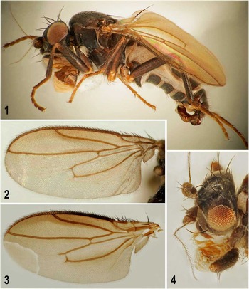
Volumosina voluminosa (Marshall, Reference Marshall1987). 1. Male laterally (Canada: Ontario), body length ~2.2 mm. 2. Left wing (Canada: Ontario), length ~1.6 mm. 3. Left wing (male paratype, partly reconstructed), length ~1.6 mm. 4. Head dorsolaterally (Canada: Ontario).

Volumosina voluminosa (Marshall, Reference Marshall1987), male paratype. 5. Abdomen laterally. 6. Abdomen (genitalia removed) ventrally. 7. Abdomen, dorsally. For abbreviations see the “Morphological terminology” section. Scale bar=0.2 mm.

Volumosina voluminosa (Marshall, Reference Marshall1987), male paratype. 8. Gonostylus, sublaterally, widest extension. 9. Genitalia, laterally. 10. External genitalia, caudally. 11. S5, ventrally. For abbreviations see the “Morphological terminology” section. Scale bar=0.1 mm.

Volumosina voluminosa (Marshall, Reference Marshall1987), male paratype. 12. Hypandrium and postgonites, ventrally. 13. Postgonite, laterally. 14. Aedeagus (phallus), dorsally. 15. Aedeagal complex, laterally. For abbreviations see the “Morphological terminology” section. Scale bar=0.1 mm.

Volumosina voluminosa (Marshall, Reference Marshall1987), paratypes. 16. Female abdomen ventrally. 17. Female S2 and S3, laterally. 18. Male trochanter, f 2 and t 2 , anteriorly. 19. Female abdomen, dorsally. For abbreviations see the “Morphological terminology” section. Scale bar=0.2 mm.

Volumosina voluminosa (Marshall, Reference Marshall1987), female (Figs. 20–21, 25 based on paratypes, others on specimen from Canada; Ontario). 20. t 2 dorsally. 21. t 2 anteriorly. 22. Postabdomen, dorsally. 23. Postabdomen, laterally. 24. Postabdomen, ventrally. 25–26. Spermathecae. 27. Sclerotisation (spectacles-shaped sclerite) of the genital chamber, ventrally. For abbreviations see the “Morphological terminology” section. Scale bar=0.2 mm (Fig. 20–21), 0.1 mm (Figs. 22–24), 0.05 mm (others).

Discussion
The new genus as defined above is established to include a single species, V. voluminosa (Marshall, Reference Marshall1987) distinguished by peculiar gigantism of the male aedeagal complex (all parts except for phallapodeme) and a strongly enlarged protruding male S2 combined with a relatively simple and (surprisingly) unmodified female postabdomen. The new genus can be best recognised by the combination of the following, probably apomorphic (one unique [U] within Limosininae) characters in the male abdomen and female terminalia: (17) male S2 enlarged and (in contrast to Herniosina) strongly protruding posteromedially (Figs. 1, 5–6); (21) epandrium with only one strong and long lateral seta (Figs. 9–10); (24) hypandrium short (Fig. 9), with medial apodeme subtriangular (Fig. 12); (25) aedeagus and postgonites extremely enlarged [U] but phallapodeme normal (Figs. 9, 13–15); (29) ejacapodeme present and well developed but hidden in cavity of phallophore (Fig. 15); (32) female T10 laterally fused with bases of cerci (Figs. 22–23); (36) spermathecae cylindrically pyriform with surface transversely ridge-like sculptured and terminal sclerotised parts of ducts short (Figs. 25–26).
Volumosina seems to belong to the Limosina genus group along with Apteromyia Vimmer, 1929, Gigalimosina Roháček, Reference Roháček1983, Herniosina Roháček, Reference Roháček1983, and Limosina Macquart, 1835, with which it shares three of the four apomorphic characters used to define the group by Roháček (Reference Roháček1982): C not extended beyond apex of R 4+5 , short female postabdomen, and long phallophore (basiphallus). Although it is externally most similar to Apteromyia, its relationships within the Limosina genus group remain uncertain, as further considered in the “General discussion” section below.
Species included
Volumosina voluminosa (Marshall, Reference Marshall1987), from northeastern North America.
Volumosina voluminosa (Marshall, Reference Marshall1987) new combination
Herniosina voluminosa Marshall, Reference Marshall1987: 711 [description, both sexes, illustration]; Cooper and Cumming Reference Cooper and Cumming2000: 103 [list of type specimens in CNCI]; Roháček et al. Reference Roháček, Marshall, Norrbom, Buck, Quiros and Smith2001: 149 [catalog]; Roháček Reference Roháček2016: 75 [notes on relationships].
Type material
Holotype: ♂ United States of America. New Hampshire: Coos County, 3 mi. NE East Inlet Dam, Norton Pool, 12–24.vi.1986, flight intercept trap, D.S. Chandler (CNCI). Paratypes: 1 ♂, 8 ♀ with same locality data as for holotype; for other paratypes see Marshall (Reference Marshall1987: 713). Paratypes are deposited in CNCI, DEBU, DENH, and SMOC.
Other material examined
Unless otherwise indicated this material is in the DEBU. CANADA. Ontario: Algonquin Provincial Park, Swan Lake Research Station, Scott Lake Survey, bracket pan traps on fallen hemlocks near Swan Lake, 29.v–16.vi.1995, S.A. Marshall (4 ♂, 2 ♀); Algonquin Provincial Park, Swan Lake Research Station, Scott Lake Survey, bracket pan traps on fallen hemlocks near Swan Lake, 4–28.v.1995, Marshall and Caloren (5 ♂, 3 ♀; 2 ♂ SMOC); Algonquin Provincial Park, Swan Lake Research Station, Scott Lake Survey, aspirated from rotting debris in maple snag, 5.v.1994, S.A. Marshall (4 ♂, 4 ♀; 2 ♀ SMOC); Algonquin Provincial Park, Swan Lake Research Station, Scott Lake Survey, pan traps on fallen maple, 4–27.v.1995, S.A. Marshall (1 ♂, 1 ♀); Algonquin Provincial Park, Wildlife Research Station, on moose antlers, R. Bonduriansky (1 ♂). Note: coordinates for the Swan Lake Research Station (now a Forest Research Reserve) are 45°29'29"N, 78°43'18"W. Coordinates for the nearby Wildlife Research Station are 45°35'32"N 78°31'20"W.
Other distributional records
A photograph of a headless male V. voluminosa is posted on the barcode of life database (www.boldsystems.org) corresponding to a specimen in the Canadensys database with the following data:
CANADA. Quebec: Mont St-Hilaire, beech-sugar maple forest, 2.vii.2001, E. Fast and S. Hawkins (1 ♂, headless, Lyman Entomological Museum at McGill University (http://data.canadensys.net/explorer/en/resources/lemq-specimens/occurrences/LEM0050414; accessed on 12 April 2017).
Redescription
Male (Fig. 1). Total body length 1.74–2.26 mm; general colour blackish brown with brownish grey microtomentum and subshiny, only sides of T2–T4 more or less glabrous and shiny. Head brown to blackish brown, somewhat higher than long (Fig. 1). Frons (Fig. 4) brown to dark brown but stripes bordering interfrontalia distinctly darker brown (inner stripe) to black (outer stripe) and dull. Occiput blackish brown and similarly as ocellar triangle, orbits, interfrontalia, and frontal lunule brownish grey microtomentose; frontal triangle narrow, delimited by different, distinctly glittering microtomentum, anteriorly acute and almost reaching margin of frons. Cephalic chaetotaxy: pvt present but reduced to small convergent or apically crossed setulae; occe and occi also relatively weak, strongly inclinate, subequal or occe slightly longer and usually less than half length of vti; vti robust and long; vte and oc subequal or slightly shorter than vti; two ors, posterior distinctly longer than anterior and slightly shorter than vti; three ifr, middle pair robust and cruciate (Fig. 4), twice as long as posterior pair; anterior ifr usually small, only half length of posterior ifr; three to five minute ads inside and below ors, one additional microseta also laterally to anterior ifr; g weak, hardly longer than anterior peristomal setula (=subvibrissa); vi longest of cephalic setae, markedly longer than vti; five to six peristomal setulae; a single long row of postocular setulae behind eye margin. Frontal lunule relatively long (equilateral), brown and grey microtomentose. Facial cavities below antennae darker brown, relatively shiny; medial carina well developed, distinctly protruding between antennae. Mouthparts pale brown, clypeus medially darker; palpus often paler, ochreous, relatively long, with one longer apical seta beside small ventral setulae. Gena relatively high (Fig. 1), dark brown and all greyish brown microtomentose. Eye of medium size; longest diameter 2.9–3.0 times as long as smallest genal height. Antennae strongly divergent; scape and pedicel dark brown, 1st flagellomere paler brown and somewhat tapered apically, with shorter pubescence than that of arista. Arista long, about 3.7 times as long as antenna, with relatively long and dense ciliation (Fig. 4).
Thorax blackish brown and dark brownish grey microtomentose, mesonotum subshiny, pleuron with microtomentum denser and hence somewhat duller (Fig. 1). Suturae between pleural sclerites sometimes paler, particularly tubercle between mesopleuron and sternopleuron pale brown to ochreous. Scutellum relatively large, about 1.5 times as wide as long, rather flat, with rounded trapezoidal outline. Thoracic chaetotaxy: two hu but internal reduced to microseta; two postsutural dc; anterior dc small, thin (only two to three times as long as dc microsetae) and situated slightly behind suture, posterior (prescutellar) dc strong, three times as long as anterior dc and about as long as basal sc; only six to seven rows of relatively long ac microsetae on suture; medial prescutellar ac pair slightly enlarged, about twice longer than ac microsetae; sa weak, shorter than posterior npl; posthumeral (presutural) seta well developed, as long as anterior npl; two long sc, basal slightly longer than scutellum, apical about 1.6 times as long as basal; two stpl, but only posterior long, anterior reduced to hair-like microseta), two to three additional microsetae below the latter.
Legs brown to dark brown; mid and hind coxae, trochanters, knees, and tarsi paler brown to ochreous (Fig. 1). f 1 with usual posterodorsal and posteroventral rows of slightly curved setae (those posteroventral in distal half of f 1 longest). t 1 , f 3 , and t 3 without macrosetae, uniformly setulose. f 2 with a short row of five to six curved ventral setae subbasally (Fig. 18) and with three to four (one longer) anterior setae in distal third; t 2 ventrally with a row of small adpressed spine-like setae in distal 2/5ths to 2/3rds, one shortened va seta and one distinct but fine anteroapical seta (Fig. 18); dorsal chaetotaxy of t 2 as in Figure 20, with one short anterodorsal seta in basal third, one long anterodorsal or rather subdorsal seta in apical fourth, and one to two small setae above it (if two then one anterodorsal and one dorsal), and one short posterodorsal seta below apical third. Hind basitarsus short and broad (as usual) and together with 2nd tarsomere densely golden pubescent. Ratio t 2 :mt 2 =1.72–1.87.
Wing (Figs. 2–3) with brown membrane, darker along anterior margin; veins yellowish brown to dark brown. C ending at apex of R 4+5 . R 2+3 sinuate and apically distinctly upcurved to C; R 4+5 slightly sinuate and ending near apex of wing. Discal cell (dm) elongate, relatively long and narrow, distally tapered, with small processes of both M and CuA 1 beyond dm-cu. A 1 slightly sinuate, ending far from wing margin. Anal lobe large, well developed; alula narrow, apically subacute. Wing measurements: length 1.50–1.61 mm, width 0.64–0.67 mm, C-index=1.14–1.34, rm\dm-cu:dm-cu=3.10–3.85. Halter with ochreous to yellow stem and dark brown knob.
Abdomen (Figs. 5–7) with blackish brown sclerites, both dorsally and ventrally. Preabdominal terga (T1–T5, Figs. 5–7) large, partly at fine microsculture, partly glabrous, and largely brownish-grey microtomentose, all sparsely setose; T2–T4 laterally or basally laterally devoid of microtomentum and shiny. T1 only partly (laterally) separate from T2 and basally with a few microsetulae. T1+2 largest and widest, T3–T5 becoming narrower posteriorly; T3 and T4 subequal in length; T5 shorter and more transverse than T4. Preabdominal sterna S2–S4 (Figs. 5–6) also microtomentose and more-or-less microscuptured (most distinctly on S2) but more densely setose than on terga. S1 reduced and membranous but S2 peculiarly enlarged, posteromedially protruding as a robust, terminally slightly bilobed projection (Figs. 1, 5–6); S3 slightly larger than S4, anteriorly somewhat emarginate and, like S4 and S5, with anterior dark-pigmented stripe (Fig. 6). S5 (Fig. 11) transverse but not reduced, with a large posteromedial unpigmented microtomentose area armed by a group of dense thicker sinuous setae on each side. Postabdomen: T6 and T7 absent. S6, S7, and S8 fused left laterally and forming together asymmetrical synsclerite S6–S8 (see Figs. 5–6), situated from ventral (original S6), left lateral (S7) to dorsal (S8) side of postabdomen. S6 (Fig. 6) medially with a central desclerotised area but without setae; S7 with a few (three to four, two of them longer) setae; S8 with about five to six setae at posterior margin (Fig. 7). Left 6th spiracle situated left laterally near fusion line of S6 and S7 (Figs. 5–6). Opposite right side with a distinctive annular sclerite (ring sclerite) probably derived from an enlarged 7th left spiracle (Fig. 6).
Genitalia
Epandrium (Figs. 9–10) of medium length and width, slightly wider than high (Fig. 10), with one long and strong lateral seta in the middle, otherwise uniformly setose. Anal fissure relatively small, suboval. Cerci reduced, short, fused with epandrium and medially to form subanal plate; each cercus slightly projecting ventrally and carrying one long and one shorter seta (Figs. 9–10). Medandrium short (low) and fused with cerci medially and connected by internal arms with posterior corner of gonostyli (Fig. 10). Hypandrium small and short (Fig. 9), with medial subtriangular apodeme and separate lateral arms (see Fig. 12); internally connected with postgonites, externally with anterodorsal part of gonostylus (Fig. 9). Gonostylus (Figs. 8–10) flat, somewhat spatulate, narrower basally than distally, with a posteroventral boot-shaped process closely attached to main body; outer side of gonostylus with fine setae anteroventrally and posteriorly in distal half and micropubescence covering posterodorsal half (Fig. 8); inner side with a series of three to four more robust and longer anteroventral setae (see Fig. 10). Aedeagal complex (Figs. 13–15) peculiarly formed. Phallapodeme of normal size, rod-like but with well-developed, dorsal keel. Aedeagus (Figs. 14–15) and postgonite (Fig. 13) gigantically enlarged and heavily sclerotised. Phallophore elongate, wider proximally, tapered distally, as long as but many-times wider than phallapodeme, articulating with the latter proximally and with distiphallus distally. Distiphallus (Figs. 14–15) extremely voluminous, somewhat asymmetrical, dilated in distal half (Fig. 14) and composed of two crooked lateral lobes proximodorsally coalesced and medially connected by a weakly sclerotised ventral plate (or possibly membrane); apical membranous part finely tuberculate, projecting between lobes. Postgonite robust, much larger than gonostylus, flat (see also Fig. 12) and broad, of somewhat hook-like outline in profile (Fig. 13), with a group of short setae in the middle of distal part of outer side and one seta on an anteroproximal process (attached to hypandrial arm). Ejacapodeme also enlarged but hidden inside (cavity of) phallophore, elongate, with one widened end (see Fig. 15).
Female
Similar to male unless mentioned otherwise below. Total body length 1.74–2.38 mm. f 2 ventrally without subbasal group of curved setae, simply setulose; t 2 ventrally only finely setulose, without specialised setae, apically with one long va seta and also anteroapical seta somewhat longer (Fig. 21); dorsal chaetotaxy of t 2 (Fig. 20) as in male. t 2 :mt 2 =1.68–1.84. Wing measurements: length 1.54–1.79 mm, width 0.66–0.76 mm, C-index=1.13–1.38, rm\dm-cu:dm-cu=2.98–3.44. Abdomen: microsculpture on preabdominal terga and sterna finer than in male. Preabdominal terga shorter, more transverse (Fig. 19) and becoming narrower posteriorly. Not only T2–T4 but also T5 with large (in T4 much larger than in male) lateral areas without microtomentum and shiny. T5 distinctly shorter and also narrower than T4. Preabdominal sterna almost unmodified (Fig. 16). S2 of normal size, slightly posteromedially produced (Figs. 16–17) to almost plain (in specimens from Ontario); S3 largest (longest) sternum; S4 transversely suboblong, shorter than S3 but subequal in width; S5 shorter and narrower than S4; S3–S5 uniformly pigmented, without anterior darkened marginal stripe (Fig. 16), shortly but more densely setose than associated terga.
Postabdomen (Figs. 22–24) narrower than preabdomen at 6th segment (see Figs. 16, 19), telescopically retractible but relatively broad, with transverse S6–S8; T6, T7, S6, and S7 with more-or-less unpigmented anterior margins. T6 wide, transversely suboblong, distinctly longer than S6 and T7, with setae restricted to posterior and lateral margins; T7 somewhat narrower and much shorter (Fig. 22) and more bent on lateral side of abdomen than T6 (Fig. 23), sparsely setose only at posterior margin. T8 dorsomedially undivided but widely depigmented (Fig. 22), thus with only lateral/lateroventral parts dark pigmented (Fig. 23), with dorsolateral setae. T10 semicircular, smaller than S10, laterally fused with cerci, pale pigmented, with a pair of longer but fine dorsomedial setae and with distinct micropubescence posteromedially (Fig. 22). S6 transversely oblong, wider and with longer setae than S7 (Fig. 24). S7 shorter than S6, with two pairs of long setae at posterior margin, remaining setae short (Fig. 24). S8 (Figs. 23–24) narrower than S7 but also transverse, shortly pentagonal to suboval, convex and paler medially, covered by longer micropubescence and with one pair of long, curved or sinuous setae, otherwise with a few small setulae. S10 relatively large and flat but pale pigmented (Figs. 23–24), rounded pentagonal, micropubescent on posterior half and setose in front of posterior margin, with one pair of longer setae. Spermathecae 2+1 (Figs. 25–26) black, somewhat cylindrically pyriform, each with proximal conical part smooth but larger distal part with distinctive transverse ridge-like sculpture and with more-or-less depressed apex; terminal sclerotised and pigmented parts of ducts short (but somewhat variable in length) slightly to distinctly bulbous; paired spermathecae with bases short, fused into single duct close to spermathecal bases. Spectacles-shaped sclerite (Fig. 27) represented only by almost circular, pale-pigmented, and weakly sclerotised rings laterally to insertion of spermathecal ducts. Cerci (Figs. 22–24) relatively robust, subconical, laterobasally fused to T10; each with one long dorsal preapical seta, one slightly longer apical sinuate setae, one ventral subapical seta, and two relatively long curved lateral setae.
Comparative notes
Volumosina voluminosa males are highly distinctive; easily recognised by their enlarged and protruding S2 and disproportionately large and exposed aedeagus (Fig. 1), visible even in air-dried specimens. Females lack obvious corresponding abdominal modifications, and resemble those of some Nearctic species of Apteromyia, Aptilotus Mik, 1898, and Nearcticorpus Roháček and Marshall, Reference Roháček and Marshall1982. Both known species of Apteromyia, i.e., A. claviventris (Strobl, 1909) and A.newtoni Marshall and Roháček, Reference Marshall and Roháček1982, differ from V. voluminosa in the female sex by short, subequal ifr setae, t 2 with distal posterodorsal seta situated near dorsal seta (see Marshall and Roháček Reference Marshall and Roháček1982: fig. 16) and by sclerites of the female postabdomen (T8 divided in two widely separate sclerites, S8 small and narrow, S10 short, cercus elongate and separate from T10). Some macropterous Nearctic species of Aptilotus, i.e., A. concavus (Spuler, 1925) and A. cruciatus Marshall, Reference Marshall1983, have the middle ifr enlarged and crossed as in V. voluminosa but they can be recognised in the female sex by C distinctly overpassing R 4+5 and broad dm cell (see Marshall Reference Marshall1983: figs. 44–45), ball-shaped spermathecae and cerci separate from T10. Finally, the females of both Nearcticorpus species, N. canadense Roháček and Marshall, Reference Roháček and Marshall1982 and N. pecki Marshall and Roháček, Reference Marshall and Roháček1982, can be separated from V. voluminosa by their short subequal ifr, broad dm cell, large (broad) S3, postabdomen long and narrow (markedly narrower than preabdomen at 6th segment), bulbous spermathecae with single spermatheca much larger than paired spermathecae and cerci not fused with T10 (see Roháček and Marshall Reference Roháček and Marshall1982: figs. 3, 65, 73–76).
Natural history
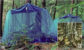
The entire type series (over 30 specimens) was taken in flight intercept traps in old growth forests in New Hampshire in May–October 1985 and 1986 (Marshall Reference Marshall1987). Most other known specimens of this species were collected during an old growth forest insect survey (Fig. 28) in a hemlock-maple (Tsuga canadensis (Linnaeus) Carrière (Pinaceae) and Acer saccharum Marshall (Sapindaceae)) ecotone in Algonquin Provincial Park, Ontario in May–June 1994 and 1995. Almost all were taken in pan traps on large fallen trees (Fig. 29) or hand captured in decaying wood in snags (standing dead trees). Although thousands of sphaerocerids were identified from pan traps and emergence traps placed in the surrounding leaf litter and other habitats as part of the same survey, no specimens of V. voluminosa were found in those traps, nor were any found in Malaise trap and baited trap samples from the same site (40 other species of Sphaeroceridae were collected in those traps). The only V. voluminosa specimen found away from large woody debris was collected on a discarded moose antler at the nearby Wildlife Research Station.
Habitat of Volumosina voluminosa (Marshall, Reference Marshall1987). 28. A 1995 photograph of a tent emergence trap shows the general habitat of the species at the Swan Lake Reserve in Algonquin Provincial Park, Ontario (no V. voluminosa specimens were collected in these traps). 29. Yellow pan traps (15×18 cm), also photographed in 1995, caught several V. voluminosa specimens. Others were taken in pan traps mounted on shelf brackets attached to logs and snags. Photographs by S.A. Marshall.

These data strongly indicate that V. voluminosa is an old growth forest specialist that develops only in large woody debris (snags, fallen trees). It would be interesting to find the larvae to confirm this assumption, and to rear out adults with which to explore the interesting functional morphology (see Discussion). This species appears to be one of the rarest sphaerocerids in North America, and it may prove to be of conservation concern because of its rarity and its apparent dependence on old growth forests with intact, large woody debris. Since species of potential conservation concern usually require common names, we suggest the name Big Wood Sphaerocerid Fly.
Distribution
Volumosina voluminosa is thus far known only from Carr and Coos Counties in New Hampshire, from Mont St-Hilaire in Québec, and from Algonquin Provincial Park in Ontario.
General discussion
Character similarities of Volumosina
Voluminosa belongs in the Limosina genus group (as established by Roháček Reference Roháček1982), along with Apteromyia, Gigalimosina, Herniosina, and Limosina. External characters of this group include a well-sclerotised body with large preabdominal sclerites; strongly reduced to absent pvt setae; g small; the same thoracic macrosetae; wing with C not extended beyond apex of R 4+5 ; both R 2+3 (more) and R 4+5 (slightly) sinuate and the latter ending near apex wing; dm cell long and narrow; male f 2 and t 2 with specialised ventral setae (facilitating grasp of the male on the female during mating); male preabdominal sterna S1+2–S4 (or some of them) modified; relatively short but telescoping female postabdomen (prolonged only in Herniosina). However, the majority of these characters are probably plesiomorphic and do not demonstrate directly the relationships of these genera. Shared genital characters of the Limosina genus group include the following: long (elongate) phallophore without epiphallus; postgonite with distinct setae (reduced only in Herniosina); ejacapodeme hidden inside the phallophore or reduced to absent; spermathecae more-or-less pear shaped and often with surface sculpture. The position of Volumosina in the Limosina genus group remains unclear because Volumosina shares few and conflicting putative characters with other genera. Possible shared characters with other genera include the following:
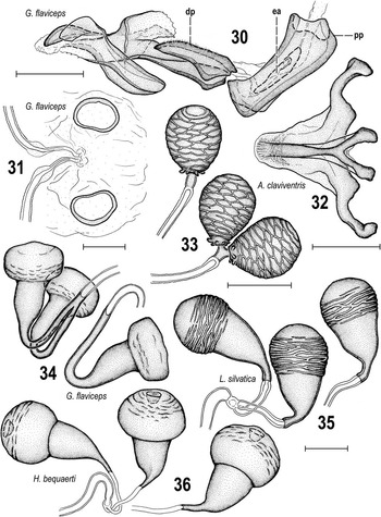
With Limosina: male cerci reduced and integrated with epandrium; female S8 relatively large; spermathecal surface ridged (see Figs, 26, 35).
Structures of terminalia in species of the Limosina genus group. 30. Gigalimosina flaviceps (Zetterstedt, 1847), aedeagus, laterally. 31. Gigalimosina flaviceps, spectacles-shaped sclerite ventrally. 32. Apteromyia claviventris (Strobl, 1909), hypandrium, ventrally. 33. Apteromyia claviventris, spermathecae. 34. Gigalimosina flaviceps, spermathecae. 35. Limosina silvatica (Meigen, 1830), spermathecae. 36. Herniosina bequaerti (Villeneuve, 1917), spermathecae. For abbreviations see the “Morphological terminology” section. Scale bar=0.1 mm (Figs. 30, 32), 0.05 mm (others).

With Gigalimosina: phallphere (basiphallus) enveloping entire ejacapodeme (see Figs. 15, 30); female vaginal sclerotisation including ring-shaped sclerites (see Figs. 27, 31).
With Herniosina: strongly protruding male S2 (or S1+2).
With Apteromyia: male male f 2 ventrally with a short row of curved setae subbasaly (Fig. 18); hypandrium short, with subtriangular apodeme (see Figs. 12, 32); spermathecae with ridge-like surface sculpture, and particularly, short terminal sclerotised part of ducts fused closely to their ends in paired spermathecae (see Figs. 26, 33).
Roháček (Reference Roháček2016) argued that the strongly protruding male S1+2 of Palaearctic Herniosina (=Herniosina sensu stricto) is not homologous with the superficially similar structure in the Nearctic H. voluminosa, and excluded this species from Herniosina for that reason. In Herniosina species the male S2 is bulging in the middle of the sclerite with apex directed ventrally (see Roháček Reference Roháček2016: figs. 1–2, 15, 17); in Volumosina it is projecting posteromedially, with the apex somewhat bilobed and directed posteriorly (Figs. 1, 5–6). The bulging S2 of Herniosina males is probably correlated with the strongly down-curved postabdomen caused by the enlarged T6 and S8. The superficially similar herniate S2 of V. voluminosa males is probably correlated with the tremendous enlargement of the aedeagus. This suggests that these bulging sterna are of different origin in Volumosina and Herniosina, and thus provide no evidence of a close relationship.
Roháček (Reference Roháček2016) also outlined several possible shared characters of the male genitalia that suggest similarities between Apteromyia and Herniosina (sensu stricto): epandrium with series of robust ventral lateral setae (but only one is present in A. newtoni); distiphallus with unpaired ventromedial lobe projecting posteriorly; phallophore anteriorly narrow and projecting, movabably attached to dorsal side of distiphallus; male cerci modified to compact (and often long) processes below anal fissure. However, these male characters are conflicted by putative synapomorphies in the male f 2 , hypandrium and female genitalia of Volumosina and Apteromyia (as listed above), and by a few other characters shared between Herniosina and other genera of the Limosina genus group (see for example the construction of their spermathecae – Figs. 26, 33–36 or structures of the male internal and external genitalia, see Roháček Reference Roháček1982, Reference Roháček1983). The best that we are able to say with confidence is that all of these genera belong to the same group (the Limosina genus group) in which each included genus is highly autapomorphic, with relatively few characters pointing to clear relationships with any other genus. We thus recognise Volumosina as a separate monotypic genus rather than including its only species in Herniosina based on striking, but arguably non-homologous, similarities.
Gigantism of the male genital structures
The tremendous enlargement of the aedeagus and postgonites (but not the phallapodeme) in V. voluminosa must carry a significant cost of development and maintenance, especially considering that the huge intromittent organ greatly exceeds the size of the genital pouch and is therefore permanently exposed. Plausibly, only the apex of distiphallus can be pressed to S6 and the “window” in the latter sclerite (see Fig. 6) might reflect this “rest” position. It is impossible to insert the whole distiphallus into the male postabdomen; this was tested experimentally with a relaxed and cleared abdomen of one male from Algonquin Provincial Park. Consequently, the distiphallus can at most be appressed to the ventral side of the postabdomen (S6–S8 plus epandrium) in the rest position.
It is a reasonable expectation that the unusual volume of the male phallus would lead to some corresponding modifications of the female postabdominal sclerites, especially in those of the eighth segment, but this is not the case (S8 is relatively large and simple, Fig. 24). The distal end of the distiphallus is as wide as (or wider) than the entire female S8, so the distiphallus can hardly be pushed into female postabdomen. Comparing dimensions of these structures it looks like the distal ends of the (lateral) sclerotised lobes of distiphallus have to remain attached externally (to S8) during the copulation and only the membranous apex between them can contact the female genital opening. The enlarged and posteromedially protruding male S2 (Figs. 1, 5–6) is surely involved in copulation as well, and the gigantic aedeagus and the enlarged male S2 probably work in concert to maintain an unsual copulatory strategy in this species. We look forward to the results of future observations of this species in copula to test these hypotheses and clarify the mystery of the massive male organ of V. voluminosa.
Acknowledgements
The research of the senior author (J.R.) was financially supported by the Ministry of Culture of the Czech Republic by institutional financing of long-term conceptual development of a research institution (the Silesian Museum, MK000100595), an internal grant from the Silesian Museum IGS201706/2017. The second author’s (S.A.M.) work is supported by a Natural Sciences and Engineering Research Council of Canada discovery grant. Tiffany Yau helped with specimen photography at the University of Guelph.