Introduction
Head and neck malignancy is the sixth most common cancer in the UK. The propensity of these tumours to metastasise to different regions calls for strict screening techniques during the initial diagnosis to guide optimum patient management. Between 6 and 20 per cent of these patients will have a synchronous or metastatic tumour at initial presentation, commonly within the head and neck itself but occasionally in the lungs or oesophagus.Reference McLeod, Jess, Anand, Tilley, Higgins and Brennan 1 The lungs form the most common site of distant metastases of head and neck tumours.Reference Jones, Morar, Phillips, Field, Husband and Helliwell 2 – Reference Halpern 4 The presence of distant metastases has a major influence on the choice of treatment and prognosis in these patients.
Computed tomography (CT) of the chest has become an integral part of cancer staging to detect pulmonary lesions; it is superior to the conventional chest X-ray because of its higher spatial and contrast resolution and ability to detect small lesions. The prevalence of positive chest CT findings in patients with head and neck cancer ranges from 4 to 37.5 per cent.Reference Halpern 4 , Reference Bisase, Kerawala and Lee 5 It has been the norm to use chest CT routinely in the staging of all head and neck cancers at the Newcastle upon Tyne Hospitals NHS Foundation Trust since 2001. Routine chest CT often picks up multiple pulmonary nodules of less than 1 cm in size, most of which initially may have to be considered as indeterminate, even in patients already diagnosed to have cancer in extrapulmonary regions. Many of these patients are smokers, and these nodules are often too small and inconspicuous to be monitored by chest X-ray. Follow-up CT is necessary to evaluate their significance.
This article aimed to clarify the significance of such incidentally diagnosed pulmonary nodules in head and neck cancer patients, as there are very few studies in the published literature exploring this. The paper also highlights the various other types of lesions detected on routine screening chest CT and their incidence based on the type of primary tumour.
Materials and methods
All patients who underwent CT examination of the chest for newly diagnosed or recurrent head and neck squamous cell carcinoma (SCC) during a two-year period, between April 2001 and March 2003, at the Newcastle upon Tyne Hospitals NHS Foundation Trust were included in the study. Patients with known lung cancer, who were being investigated for vocal fold palsy, or being staged for thyroid, salivary gland, sinonasal or external auditory canal tumours, and other histologically proven tumours such as lymphoma, melanoma, neuroendocrine tumour or sarcoma, were excluded from the study.
All patients were scanned using a four-slice Volume Zoom CT scanner (Siemens, Erlangen, Germany), with 2.5 mm collimation reconstructed at 5 mm slice thickness for soft tissue and 3 mm for lung windows, following intravenous contrast medium administration. The chest CT findings of these patients were documented on a proforma (designed for data collection) at the time of reporting by the first author (IZM) and transferred onto an electronic spreadsheet for further analysis.
Any abnormality detected on the initial chest CT was documented and then classified as benign, equivocal or malignant, based on appearance, size and presence of calcification. The benign-looking and equivocal lesions that resolved or did not show any significant radiographical change on subsequent CT scans were all grouped as benign. These included nodules, inflammatory masses, fibrosis or pleural calcification. Any of the equivocal lesions in patients who had no further imaging performed or died within a two-year period of the initial imaging were considered as indeterminate as there was no long-term follow up. Lesions were considered malignant based on biopsy results or progression detected on subsequent imaging. These included pulmonary metastases, mediastinal nodes or bronchogenic carcinoma.
The cohort was divided into two groups: group one included newly diagnosed patients with head and neck SCC, while group two included established head and neck SCC patients who were being scanned for recurrence. Any subsequent follow-up chest CT scans for all the included patients, up until 2014, were analysed. The electronic medical records on the hospital database, including radiology and pathology findings, were then reviewed to monitor the patients' progress over the years and to identify current status.
Results
A total of 341 patients were scanned during the 2-year period. There were 119 patients with vocal fold palsy or tumours other than SCC who did not meet the inclusion criteria. The final sample consisted of 222 patients who were included for further analysis: 148 patients in group 1 and 74 patients in group 2. The male-to-female ratio was 3:1, with a mean age of 65 years ranging from 43 to 87 years.
Group one
Of the 148 patients with newly diagnosed head and neck cancer, 75 patients (50.7 per cent) had a normal chest CT scan and 73 (49.3 per cent) had an abnormal scan at the time of staging. The eventual abnormal chest CT findings indicated that 45 patients (61.7 per cent) had benign lesions, 6 (8.2 per cent) had indeterminate nodules and 22 (30.1 per cent) had malignant lesions. The distribution of primary sites and the various benign and malignant findings in this group are shown in Table I.
Distribution of benign and malignant lesions in group one patients with abnormal chest imaging*

* Total n = 73.
The predominant lesions detected on chest CT were pleural calcification (24.7 per cent), benign nodules (17.8 per cent) and metastatic lesions (19.2 per cent). Concurrent malignant lesions were seen in patients presenting with oropharyngeal, hypopharyngeal and supraglottic cancers. The malignant lesions detected in patients with hypopharyngeal cancer were all due to pulmonary metastases. Among patients presenting with supraglottic cancer, one-third had bronchogenic primaries, and in those with oropharyngeal cancer, 37.5 per cent had bronchogenic carcinomas.
Group two
This group included 74 patients with recurrent or suspected recurrent head and neck cancer, of which 46 patients (62.2 per cent) had a normal chest CT scan and 28 (37.8 per cent) had an abnormal scan. The distribution of primary sites and the various benign and malignant findings in this group are shown in Table II. The eventual classifications for the 28 patients with abnormal chest CT findings were as follows: 9 patients (32.1 per cent) had benign lesions, 1 (3.6 per cent) had an indeterminate nodule and 18 (64.3 per cent) had malignant lesions.
Distribution of benign and malignant lesions in group two patients with abnormal chest imaging*

* Total n = 28.
Metastatic lesions were the most common chest CT finding in patients with recurrent cancer (35.6 per cent), especially in recurrent hypopharyngeal cancer cases, compared to those where the chest was scanned for newly diagnosed disease. This difference is statistically significant (Fisher's exact test; p = 0.0009). Unlike the findings in group one, patients with recurrent cancers of the glottis, oral cavity and unknown primary demonstrated malignant lesions on chest imaging.
Delayed lung malignancy
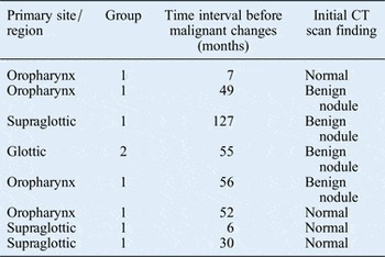
There were four cases of malignant change in a pulmonary nodule that was initially felt to be benign, three patients in group one and one in group two (Table III), accounting for 7.4 per cent of all benign findings. One patient in group 1 developed carcinoma in a benign-looking pulmonary nodule after a period of 127 months, which was probably a scar carcinoma. Two further patients in group 1 developed a bronchogenic carcinoma over a period of time, with no evidence of a pre-existing nodule on the initial scan (a patient with supraglottic cancer 30 months later and a patient with oropharyngeal cancer 52 months later). Two patients in group one, with oropharyngeal and supraglottic primaries, developed pulmonary metastases up to seven months after initial normal CT scans. A patient in group two who developed a cavitating right upper lobe mass suspicious for malignancy was found on open biopsy to have tuberculosis.
Patients with malignant change over time

CT = computed tomography
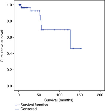
Parameters of diagnostic accuracy were calculated; these are shown in Table IV. The negative predictive value for the whole cohort was 93 per cent; this strongly suggests that benign-appearing nodules will stay benign, especially in the context of a newly diagnosed cancer. Time-to-event analysis indicates that 30 per cent of changes occur within five years, with very few occurring subsequently (Figure 1).
Time-to-event graph showing the conversion of benign nodules during the follow-up period.

Diagnostic accuracy rates in both patient groups

Data represent percentages and 95 per cent confidence intervals (in parentheses). PPV = positive predictive value; NPV = negative predictive value
Discussion
Screening of the chest with CT in patients with head and neck cancer has increased the detection of pulmonary nodules. Since a significant proportion of these patients are smokers, this creates a clinical dilemma, as these nodules may represent metastatic disease, scarring or primary tumours. Many nodules are usually too small to be amenable to fine needle aspiration or bronchoscopy. The rationale for follow up of these nodules is the early detection of a synchronous cancer, but this would be at the expense of increased healthcare costs, patient anxiety and poor utilisation of limited resources. The American College of Chest Physicians recommends CT follow up at 3-, 6-, 12- and 24-month intervals.Reference Tan, Flaherty, Kazerooni and Iannettoni 6 The guidelines given by The Fleischner Society propose follow up of pulmonary nodules based on their size and relative risk to the patient.Reference MacMahon, Austin, Gamsu, Herold, Jett and Naidich 7
In our cohort, 45.4 per cent of patients had some abnormality detected on routine screening of the chest, with 40 per cent of these lesions being malignant. The malignant lesions were more frequent in patients with recurrent cancer. This rate is higher than the figures reported in the literature on thoracic abnormalities, which are present in around 4–37.5 per cent of patients.Reference Halpern 4 , Reference Bisase, Kerawala and Lee 5 , Reference Loh, Brown, Baker, Gilbert, Gullane and Irish 8 , Reference De Bree, Deurloo, Snow and Leemans 9 Seven patients in the total cohort were considered to have indeterminate findings owing to the lack of long-term follow-up data. Given the sample size, this is an acceptable proportion that is unlikely to skew the results and the interpretation.
It is vital to appreciate the different characteristics of these two groups of patients in evaluating the chest CT findings. Although 49.3 per cent of patients in group one had a thoracic abnormality, 61.7 per cent of these were due to benign causes, with only 30.1 per cent being malignant lesions. In contrast, 37.8 per cent of patients in group two had a thoracic abnormality, but 64.3 per cent of these were due to malignant lesions, most commonly pulmonary metastases. The negative predictive value was lower and there was a statistically significantly higher incidence of malignant lesions in the group with recurrent cancer.
Our study showed that over a 12-year surveillance period, only 4 patients (7.4 per cent) with an initially benign-looking pulmonary nodule developed malignant change. Therefore, in the setting of a benign-appearing nodule observed on the chest CT scans of a newly diagnosed patient, a negative predictive value of 93 per cent (identified using the follow-up data from this study) confers a high degree of confidence that the said nodule is benign and should be treated as such.
Unlike other published articles, our study involved chest CT imaging of all consecutive newly diagnosed and recurrent head and neck SCC patients, irrespective of the locoregional spread of the tumour. In group one, the incidence for all thoracic malignancy was 10.1 per cent and that for bronchogenic carcinoma was 0.7 per cent at presentation on the initial staging CT scan. However, in patients with recurrent head and neck cancer, the incidence of thoracic malignancy at the time of recurrence diagnosis rose to 22.9 per cent and the incidence of bronchogenic carcinoma increased to 5.4 per cent. Over time, the total incidence of thoracic malignancy increased to 14.8 per cent in group one and 24.3 per cent in group two.
There is support for the findings of this study in the literature. McLeod et al., in their systematic review of 25 studies, showed a pooled point prevalence of positive chest CT findings of 7.93 per cent.Reference McLeod, Jess, Anand, Tilley, Higgins and Brennan 1 There was a significantly higher point prevalence of positive chest CT findings of 25.32 per cent in those studies examining patients with recurrent head and neck SCC.Reference McLeod, Jess, Anand, Tilley, Higgins and Brennan 1 Other studies in the literature, such as those by Arunachalam et al.Reference Arunachalam, Putnam, Jennings, Messersmith and Robson 10 and Ghosh et al.,Reference Ghosh, Roland, Kumar, Tandon, Lancaster and Jackson 11 have shown incidence rates of 6.7 per cent and 5.7 per cent, respectively, for synchronous primary lung tumours at initial staging of newly diagnosed head and neck SCC.
Our study showed four patients (7.4 per cent) where an initially benign-looking pulmonary nodule developed into a bronchogenic carcinoma on routine surveillance with CT. Another four patients with an initial normal chest CT scan developed pulmonary malignancy on follow up. Four of these patients had a primary tumour in the oropharynx and the other four had tumours located in the larynx; seven of these patients belonged to group one. Malignant change was detected over a wide range of time, from 6 to 127 months. Our data suggest that the vast majority of small pulmonary nodules never progress to more sinister pathology.
In a retrospective review of the chest CT findings at initial presentation for 192 patients with head and neck SCC, Hsu et al. detected an initial malignancy rate of 20 per cent, with progressive change in 44 per cent of nodules initially thought to be indeterminate over a 59.7-month period.Reference Hsu, Chu, Liu, Lan, Chang and Tsai 12 In a retrospective review of the chest CT findings for 239 patients with a new head and neck cancer over an 11-year period, Beech et al. noted that 13 per cent of patients had a small pulmonary nodule.Reference Beech, Coulson, Najran, Olliff and Jennings 13 Nine per cent of these patients were subsequently diagnosed with pulmonary metastases within a 6–36-month period, with all metastases developing at different sites to the initial nodule. A further 1 per cent of patients who initially had a normal chest CT scan went on to develop pulmonary metastases over a 2–39-month period.Reference Beech, Coulson, Najran, Olliff and Jennings 13 Our study showed that 7.4 per cent of patients initially classified as having benign lesions developed malignancy, and 3.3 per cent of patients with an initial normal chest CT scan developed malignant pulmonary lesions on follow up, consistent with the above publications.
Screening of the chest with CT scanning has formed part of the staging protocol in head and neck cancer patients at our centre since 2001. This imaging was conducted irrespective of the tumour–node–metastasis stage and primary tumour subtype, as the superiority of CT scanning over routine chest X-ray has been clearly documented.Reference Ghosh, Roland, Kumar, Tandon, Lancaster and Jackson 11 Our study population was grouped into consecutive but distinct categories of patients: those with newly diagnosed head and neck cancer and those with recurrence. This helps us to understand the difference in the chest CT findings between the two groups. The study also had a long-term follow-up period of at least 12 years from the date the patients were admitted into the cohort.
-
• Most pulmonary abnormalities detected on computed tomography (CT) in staging of newly diagnosed head and neck squamous cell carcinoma (SCC) are benign
-
• Most pulmonary abnormalities detected in staging of recurrent head and neck SCC are malignant
-
• Few benign-looking pulmonary nodules on initial staging CT of head and neck SCC develop malignancy
This study confirms the importance of routine chest CT as part of the imaging protocol in patients with head and neck cancer. With head and neck cancer patients being treated more aggressively and living longer, the importance of a baseline chest CT for future comparison is important because of the increased likelihood of synchronous malignant pulmonary lesions developing, particularly at the time of recurrence. The CT technology now allows 1 mm reconstruction of images as a routine, resulting in the detection of even smaller nodules.Reference Edey and Hansell 14 The advent of computer-aided detection and diagnosis software may assist in the identification and characterisation of such pulmonary nodules. Although these nodules will require monitoring, our study has shown that these should not alter the initial patient management decision.