The window duct differs from a patent arterial duct as the connection between the descending aorta and pulmonary trunk occurs without a distinct tubular structure. Although physiologically similar to an aortopulmonary window, the anatomic location of the window duct occurs at the pericardial reflection on the pulmonary artery, and is therefore extrapericardial in contrast to an aortopulmonary window, which is intrapericardial.
Case report
A 1-month-old girl with a history of a two-vessel cord presented with emesis. Outpatient gastrointestinal evaluation was negative. She then presented emergently with lethargy and hypoxemia and was transferred to our neonatal intensive care unit. Non-reactive dilated pupils were noted and she was determined to have partial aniridia.
An echocardiogram was obtained showing persistence of diastolic flow in the right and left pulmonary arteries, an atrial septal defect, a dilated and hypertrophied right ventricle with septal flattening, and a markedly dilated pulmonary trunk.
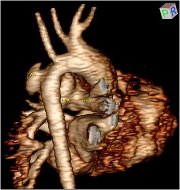
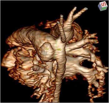
Cardiac magnetic resonance imaging demonstrated a massive window duct measuring 1.7 cm by 2 cm, with a 0.6 cm by 1.2 cm saccular aneurysm from the leftward aspect. The left ventricular size was normal with mildly decreased systolic function.
Surgical repair was performed by median sternotomy. Dissection was challenging because of pulmonary trunk dilation, the large-sized window duct and the saccular aneurysm. After the posterior pericardial reflection was entered at the distal pulmonary trunk, the pulmonary artery appeared fused directly to the descending aorta without the tubular appearance of a patent arterial duct. The right pulmonary artery was circumferentially dissected. During an attempt to control the left pulmonary artery before initiation of bypass, the posterior left pulmonary artery was injured. The area was packed and emergent bypass was initiated followed by cooling to deep hypothermia.
After aortic clamping, cardioplegic arrest and induction of circulatory arrest, the distal pulmonary trunk was opened transversely. On internal inspection, the dilated portion adjacent to the connection with the aorta was friable and had an intimal appearance consistent with the arterial duct tissue. This portion also gave rise to the saccular aneurysm. There was a distinct intimal transition between this tissue and normal appearing pulmonary trunk tissue that gave rise to the right and left pulmonary arteries.
Volume rendering of a contrast-enhanced magnetic resonance angiogram of the chest viewed from a right posterior perspective demonstrates the proximal descending aorta with the aneurysm (An) on the left and the pulmonary trunk anteriorly. The bifurcation of the pulmonary trunk into the right and left pulmonary arteries (RPA and LPA) is shown. Also demonstrated is a “bovine aortic arch” (common origin of the right brachiocephalic and left common carotid arteries).

The left pulmonary artery injury was repaired and the entire duct connection was excised from the descending aorta leaving a 1-cm wide and 2-cm long defect in the aorta extending from opposite the left subclavian to the descending aorta. This defect was patched with a pulmonary artery homograft.
The duct tissue including the aneurysmal segment was resected from the pulmonary trunk, leaving a similarly large defect. This defect was closed with a separate homograft patch to avoid distortion of the pulmonary trunk bifurcation. During rewarming with the heart still arrested, the atrial septal defect was closed. A transthoracic pulmonary artery line was placed and a lung biopsy was obtained because of chest radiograph and magnetic resonance evidence of lung disease.
The patient was weaned from cardiopulmonary bypass on empiric nitric oxide. The pulmonary artery pressure was initially 80% systemic. Post-repair transoesophageal echocardiography demonstrated widely patent right and left pulmonary arteries.
By postoperative day 5, the pulmonary artery pressure was half-systemic.
Her lung biopsy demonstrated alveolar simplification and pulmonary interstitial glycogenosis. Ophthalmology consultants confirmed bilateral aniridia with a normal fundal examination and recommended regular eye examinations to monitor for glaucoma, limbal stem deficiency and cataracts. Genetic workup was negative for aniridia–Wilms tumour syndrome, Loeys–Dietz and Marfan’s syndromes. She was discharged on postoperative day 16 on sildenafil.
The same volume rendering of a contrast-enhanced magnetic resonance angiogram viewed from a left posterior perspective demonstrates the location of the window duct (WD) between the descending aorta and the pulmonary trunk, with the aneurysm (An) shown.

Currently, she is 12 months old and has weaned from all cardiac medications. Echocardiography demonstrates mild septal flattening with normal function. She is growing and developing normally.
Discussion
This anomaly was first described in 1953 in an autopsy report by Maurice Lev as “very wide, but short, with the descending aorta appearing to be the continuation of the pulmonary artery”.Reference Lev 1 The window duct is distinguished from all forms of aortopulmonary window that are intrapericardial. The anatomic location of the window duct is at the reflection of the pericardium on the distal pulmonary trunk. It is therefore extrapericardial.
As the window duct has no tubular length, echocardiography relies on secondary signs of the communication, such as persistence of diastolic flow in the right and left pulmonary arteries. Additional echocardiographic findings in our patient that suggested a window duct rather than a patent arterial duct were right ventricular hypertrophy and pulmonary trunk dilation, as described by Lev.Reference Lev 1
Preoperative diagnosis and differentiation from a patent ductus arteriosus is important. The usual approach to ligation of a patent arterial duct is by left thoracotomy without cardiopulmonary bypass. In 1989, Komeda’s group performed a left lateral thoracotomy on a 6-month-old with an assumed patent arterial duct. This approach was abandoned when appearance of the pulmonary trunk fused with the aortic arch was noted.Reference Komeda, Miki and Kusuhara 2 The patient died of multi-organ failure before reoperation was possible. In 1998, Grünenfelder and his group reported an unanticipated window duct in an 18-month-old boy.Reference Grünenfelder, Bartram and Van Praagh 3 The smaller aortic isthmus was mistakenly occluded with a lethal outcome. The authors reported the case to promote understanding that a window duct requires a unique approach. They proposed patch occlusion on cardiopulmonary bypass.
Successful percutaneous closure of window ducts in adult-sized patients has been reported.Reference Bialkowski, Szkutnik, Kusa and Stein 4 In 2010 and 2013, groups in India reported successful surgical repairs: first, a 12-year-old boy with cardiopulmonary bypass and deep hypothermic circulatory arrest,Reference Agarwal and Mustaev 5 and the second, an 18-year-old with bypass with total circulatory arrest.Reference Talwar, Upadhyay and Ramakrishnan 6
Our patient is the first reported with a window duct diagnosed by magnetic resonance imaging and the youngest with successful repair reported to date. No other patient reported with this anomaly has had aniridia, alveolar simplification and pulmonary interstitial glycogenosis.
The anatomic detail obtained by magnetic resonance imaging was beneficial in orchestrating surgical repair. Intraoperative efforts to define the anatomy would have been challenging and could have led to inaccurate understanding of the lesion, as highlighted by the experience of Grünenfelder and his group. When a window duct is suspected, magnetic resonance imaging is recommended to guide a safe and efficient repair.
Acknowledgement
None.
Financial Support
This research received no specific grant from any funding agency, commercial, or not-for-profit sectors.
Conflicts of Interest
None.
Ethical Standards
This case report did not involve human or animal experimentation.