Introduction
Fibro-osseous tumours are rare and seldom appear in the sinonasal region.Reference Choi, Jeon and Park1–Reference Berlucchi, Salsi, Farina and Nicolai3 Clinically, it is sometimes difficult to differentiate the two subgroups fibrous dysplasia and ossifying fibroma.Reference Mafee, Yang, Tseng, Keiler and Andrus4 Correct differential diagnosis is crucial to enable accurate therapy. Ossifying fibroma should be resected whenever possible. For fibrous dysplasia, however, ‘wait and see’ is the treatment strategy of choice, including frequent, regular clinical follow up.Reference London, Schlosser and Gross5, Reference Post and Kountakis6
Case report
A 14-year-old boy presented with a two-month history of progressive left nasal obstruction and recurrent epistaxis. Clinical examination, including rigid nasal endoscopy, revealed a large tumour obstructing the left nasal cavity. Exophthalmia of the left eye was noted, without visual impairment or double vision. Drainage of the lacrimal sac and duct was normal, as was olfactory function.
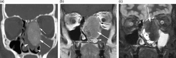
Computed tomography (CT) and magnetic resonance imaging (MRI) were performed. The CT scans (Figure 1a) showed a large, expansive mass arising from the left middle turbinate, with an extensive central ‘ground glass’ appearance surrounded by hypodense, fibrous soft tissue areas. The lesion was sharply delineated by a thin, bony wall, representing the expanded borders of the adjacent sinonasal structures. Magnetic resonance imaging (Figure 1b and c) revealed a well defined mass, with an intermediate to slightly hypointense signal on unenhanced T1-weighted images and an homogenous, high signal intensity on T2-weighted images. Intense homogeneous enhancement was seen after gadolinium contrast injection. The radiological features were considered to be consistent with fibrous dysplasia.
(a) CT-scan. Tumour orginating from the middle turbinate with extensive ground glass ossification. large arrow: tumour with a center of ossification; large arrow head: fibrous component; small arrow head: inferior turbinate; small arrow: displacement of the lamina papyracea; tumour size: 44,5 × 30 × 39 mm. (b) T1-weighted MRI after gadolinium injection. Well defined mass with significant homogeneous enhancement of the ossified portion of the tumour (large arrow); displacement of lamina papyracea (black arrow) and inferior turbinate (arrowhead). (c) T2-weighted MRI. Extensive low signal intensity area due to ossification (large arrow). The soft tissue margin can be characterized with MR as cystic periphery of the mass (asterisk). Note fluid-filled maxillary sinus due to obstruction of the ostium

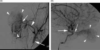
Selective angiography of the tumour vessels was performed in order to delineate the blood supply and also to evaluate the possibility of pre-operative embolisation. Extensive blood supply from both the left internal and external carotid arteries was encountered (Figure 2a and b).
(a) Angiography. Blood supply via external carotid artery. large arrow: external carotid artery; large arrow head: maxillary artery; small arrow: sphenopalatine artery; small arrow heads; tumour vessels. (b) Angiography. Blood supply via internal carotid artery. large arrow: internal carotid artery; small arrow: ophtalmic artery; small arrow heads: tumour vessels

Subsequently, the sphenopalatine artery only was embolised. Branches originating from the proximal part of the ophthalmic artery were spared in order to avoid consecutive visual impairment or even blindness due to obliteration of the centralis retinae artery.
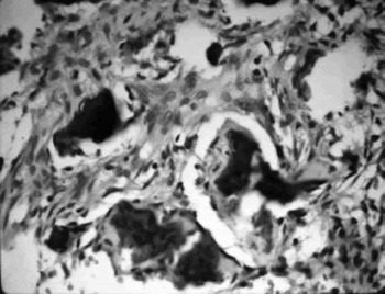
Histological investigation of the tumour revealed randomly distributed bone spicules rimmed by osteoblasts. These distinct histological features are pathognomonic for ossifying fibroma (Figure 3).
Histological specimen (hematoxilin and eosin; ×400). Bony trabeculae composed of immature lamellar bone surrounded by osteoblasts with fibrous stroma.

A complete endoscopic resection was performed under surveillance, using a three-dimensional navigation system. During surgery, the tumour was found to invade the sphenoid sinus, leaving the frontal skull base intact. The lamina papyracea was dislocated but not destroyed.
Over four years' follow up, no clinical or radiological sign of recurrence was diagnosed. At the time of writing, the young man was free of functional or cosmetic impairment, and his visual function was within normal limits.
Discussion
The lesion showed well delineated borders and features of expansive growth, with displacement, thinning and ballooning of pre-formed structures. This pattern usually indicates slow progression, despite the subjectively rapid onset of complaints. Primary sarcomatous tumours are rare in the midface and often occur in younger people. They are characterised by an invasive growth pattern, with irregular borders and bone destruction.Reference Mafee, Valvassori and Becker7 The features seen in this case were strongly suggestive of a benign lesion.
Fibro-osseous lesions are rare, benign tumours of the sinonasal and orbital regions. The term encompasses fibrous dysplasia and ossifying fibroma, including their histological variants. These tumours are often diagnosed incidentally by radiographic examination. If symptomatic, both may present with a painless swelling or deformity of the face and with associated compromised function of adjacent structures.Reference Mafee, Valvassori and Becker7 Fibrous dysplasia may occur in three clinical subtypes: monostotic (70 per cent of cases), polyostotic (27 per cent of cases) and McCune–Albright syndrome (3 per cent of cases).Reference Commins, Tolley and Milford8, Reference MacDonald-Jankowski9 Ossifying fibroma appears most frequently in the third or fourth decade, whereas fibrous dysplasia usually presents in the first two decades. Both entities are more common in females.Reference Mafee, Valvassori and Becker7, Reference Commins, Tolley and Milford8 Fibro-osseous lesions may share clinical, radiological and pathological features.Reference Mafee, Yang, Tseng, Keiler and Andrus4, Reference Post and Kountakis6, Reference Mafee, Valvassori and Becker7, Reference MacDonald-Jankowski9 Only one case of ossifying fibroma of the middle turbinate has been previously published.Reference Caylakli, Buyuklu, Cakmak, Ozdemir and Ozluoglu2
A correct differential diagnosis is crucial in order to plan adequate therapy; an unnecessary tumour resection in such sensitive anatomical areas may cause significant complications and morbidity.Reference Choi, Jeon and Park1, Reference London, Schlosser and Gross5 In the case of ossifying fibroma, complete surgical resection is recommended, as this tumour tends to behave more aggressively than fibrous dysplasia.Reference London, Schlosser and Gross5, Reference Post and Kountakis6 The recurrence rate following incomplete resection is high.Reference Post and Kountakis6, Reference Commins, Tolley and Milford8 Radiotherapy is contraindicated as it is associated with malignant conversion.Reference Post and Kountakis6, Reference Commins, Tolley and Milford8 In contrast, asymptomatic fibrous dysplasia may be followed up by imaging,Reference London, Schlosser and Gross5 and tumour growth usually stops after puberty.Reference Mafee, Yang, Tseng, Keiler and Andrus4, Reference Mafee, Valvassori and Becker7 Moreover, the site of manifestation is an important factor for therapy of both lesions.Reference Post and Kountakis6, Reference Commins, Tolley and Milford8
• This rare case demonstrates the difficulty of establishing a correct diagnosis and treatment plan when a fibro-osseous lesion of the midface is suspected
• When the diagnosis is ‘fibro-osseous lesion, not further specified’, surgery is the treatment of choice whenever possible
• Irregular blood supply of the tumour may add a significant risk to a planned operation and should therefore be evaluated, especially when information about clinical behaviour is sparse
In the present case, the radiographic features were consistent with fibrous dysplasia as well as with ossifying fibroma, making pre-operative diagnosis more difficult. Homogenous ground glass ossification is a typical finding in fibrous dysplasia and may be combined with fibrous, lytic areas. Fibrous dysplasia is characterised by a poorly delineated transition zone to the adjacent bone, whereas ossifying fibroma shows sharply delineated margins resulting from expansive growth. Moreover, osteoblastic rimming is a histological feature normally attributable to ossifying fibroma, but it also may occur in fibrous dysplasia.Reference Mafee, Valvassori and Becker7 However, in the absence of radiological and histopathological correlation, the correct diagnosis might have been fibro-osseous lesion, not further specified.Reference Mafee, Valvassori and Becker7, Reference MacDonald-Jankowski9 In any case, the tumour was resected completely via an endoscopic approach, by a surgeon with considerable experience in endoscopic sinonasal surgery. This surgical approach offers some significant advantages compared with conventional resection, as described elsewhere.Reference Choi, Jeon and Park1, Reference London, Schlosser and Gross5, Reference Post and Kountakis6
Conclusion
Complete surgical resection would seem necessary when a diagnosis of fibro-osseous lesion is uncertain. As information on the clinical behaviour of this rare tumour is sparse, the possibility of extensive, complicated vascularisation must be considered.