Introduction
Increased blood cholesterol levels during the third trimester in pregnancy has long been considered normal and it does not require monitoring or management.Reference Salameh and Mastrogiannis 1 , Reference Potter and Nestel 2 Due to the lack of correlation between maternal and offspring blood cholesterol at term birth in humans, and results in animal experiments indicating that the placenta is impermeable to intact lipoprotein particles,Reference Woollett 3 the effect of transplacental cholesterol transfer has long been neglected. However, a growing number of studies have shown strong correlation between elevated maternal and offspring’s cholesterol levels both in humansReference Napoli, D’Armiento and Mancini 4 and hamsters.Reference Woollett 3 , Reference McConihay, Horn and Woollett 5 Abnormally increased gestational serum cholesterol level was reported to be associated with the cholesterol deposition in the fetal aortas,Reference Napoli, D’Armiento and Mancini 4 maternal and fetal oxidative stressReference Liguori, D’Armiento and Palagiano 6 , Reference Leiva, de Medina and Salsoso 7 and lipid metabolism disorder.Reference Marseille-Tremblay, Ethier-Chiasson and Forest 8 However, it remains little known whether this is due to the direct influence of maternal hypercholesterolemia (HC) on the fetus or through altered placental function.
Formed from the differentiation and invasion of early embryonic trophoblast, placenta drives active exchange of nutrients and oxygen to the fetus. Fetal growth is greatly dependent on energy metabolism of the placenta. The placental nutrient transportation, and some progesterone biosynthesis are highly energy-consuming processes. Mitochondrion is the critical site for oxidative phosphorylation, which yields adenosine triphosphate (ATP) from pyruvate. It was stated that the mitochondrial dysfunction might be involved in the development of preeclampsia.Reference Widschwendter, Schröcksnadel and Mörtl 9 Various animal studies have reported that elevated gestational cholesterol level increased the risk for obesity, diabetes and cardiovascular disease in offspring.Reference Bringhenti, Ornellas, Mandarim-de-Lacerda and Aguila 10 , Reference Kim and Kwon 11 Besides, studies have found that HC has significantly increased the mitochondrial DNA (mtDNA) damage, suggesting that HC might induce the mitochondrial damage and dysfunction.Reference Knight-Lozano, Young and Burow 12 However, the effects of gestational HC on the mitochondrial function of the placenta and long-term health of offspring is unclear.
In this study, placentas from normo- and hypercholesterolemic pregnant women were sampled and genes related to mitochondrial genesis, activities of respiratory chain enzymes were measured to determine the effects of gestational HC on mitochondrial function in turn human placenta.
Materials and methods
Population study procedure
Pregnant women were recruited from Xuanwu Hospital in Beijing during their first prenatal visit (before their 10th week of pregnancy) for this study. All of the human experimental procedures received ethics approval from the ethical committee of Capital Medical University and Xuanwu Hospital. Informed consent was obtained from all subjects before their enrollment in the study. The exclusion criteria were as follows: pregnant women who (1) conceived the fetus using artificial methods including in vitro fertilization, (2) carried more than one fetus, (3) suffered from endocrine or metabolic disorders, pregnancy complications such as gestational diabetes mellitus (GDM), severe infectious diseases as well as previous offspring premature birth or asphyxia, (4) women whose gestational age at delivery was no more than 37 weeks. Pregnant women whose serum total cholesterol (TC) level was higher than 7.25 mmol/L at third trimester (measured at 36–38 weeks) were selected as gestational HC according to Liguori et al.Reference Liguori, D’Armiento and Palagiano 6 Finally, 19 pregnant women with gestational HC and other 19 pregnant women with normal cholesterol level (NC) matched with age, pre-gestational body mass index and the gender of neonate as control were included.
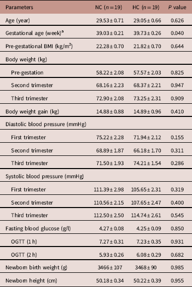
At 20–24, 36–38 weeks of gestation, we performed questionnaires and interviews with the pregnant women recruited, by which information about age, body height and body weight before pregnancy, medical history, family history, ethnicity, smoking history, pregnancy history and dietary situations (food frequency, 24-h dietary recall and dietary intake of supplements) were collected. Relevant characteristics were summarized in Table 1. After delivery, gestation clinical records and neonatal birth records were also collected.
Characteristics of the subjects

NC, pregnant women with normal cholesterol level; HC, pregnant women with gestational hypercholesterolemia; BMI, body mass index; OGTT, oral glucose tolerance test.
a Comparing with the NC group, P<0.05.
Maternal blood samples were collected from pregnant women at 9 and 36 weeks gestation after 12 h overnight fasting and stored in plain tubes. Serum was isolated after clot retraction and centrifuged at 3000 rpm, 4°C for 10 min and was immediately sent to the laboratory for examination. Fasting blood glucose, triglyceride, TC, high-density lipoprotein cholesterol (HDL-C) and low-density lipoprotein cholesterol (LDL-C) levels were measured using the self-dynamic biochemical detection (7170A Hitachi). Non-HDL-C level was calculated from TC minus HDL-C.
Tissues sampling and placental mitochondrial extraction
Placenta tissue was collected as described.Reference Liguori, D’Armiento and Palagiano 6 Fresh placental tissue was obtained immediately after delivery: four pieces (about 2×2 cm for each one piece) of the placental tissue was obtained from the 3, 6, 9, 12 am, avoiding the bleeding and calcification region. The samples were kept in 4°C, and then were transferred immediately to the laboratory of Capital Medical University. Tissues were rinsed in polyphosphate-buffered saline three times to eliminate blood, and then stored at −80°C for further analysis.
Mitochondrial extraction from placenta chorion was performed with Mitochondrial Isolation Kit (CS0201-50T, Leagene) according to the manufacturer’s instructions. About 100 mg of tissue was homogenized in ice-precooled lysis buffer by using a tissue homogenizer. After centrifugation at 800× g , 4°C for 5 min, the clear upper layer was transferred to another centrifuge tube in medium buffer (the clear upper layer, 1:1) and centrifuged at 15,000× g , 4°C for 10 min. Then the clear upper layer was removed, and the deposition was resuspended in wash buffer and centrifuged at 15,000× g , 4°C for 10 min. Again, with the clear upper layer removed, the deposition was resuspended in store buffer. A BCA Protein Assay Kit (Biosinoble, China) was then performed following manufacturer’s instructions. And the rest of the protein was stored at −80°C for further analysis.
Analysis of gene DNA and messenger RNA (mRNA) expression by real-time reverse transcription-polymerase chain reaction (PCR)
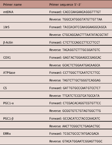
DNA was extracted from placenta tissue (with ethylene diamine tetraacetic acid) using TIANamp Blood DNA Kit (TIANGEN Biotech, DP318, Beijing, China) according to the manufacturer’s instructions. Both DNA yields (nano-grams per microliter) and purity ratios (A260/280 and A260/230) were determined. Extracted DNA was stored at −80°C for the following experiment. The primer sequences (showed in Table 2) for mtDNA and nuclear DNA (nDNA) (18SrRNA gene) were reported by Gianotti et al.Reference Gianotti, Sookoian and Dieuzeide 13 An assay based on real-time quantitative PCR was used for both nDNA and mtDNA quantification using Takara premix Ex Taq II (RR820A). For DNA quantification detection, PCR assays were performed in triplicate in a 20 μl total volume (10 μl SYBP Taq, PCR Forward Primer+Reverse Primer 0.8 μl, ddH2O 8.2 μl and DNA template 1 μl).
Real-time PCR primers

mtDNA, mitochondrial DNA; COX1, cytochrome c oxidase subunit I; ATP6ase, ATP monophosphatase 6; CS, citrate synthase; PGC1-α, peroxisome proliferator-activated receptor-γ (PPARγ) co-activator 1α; PGC1-β, PPARγ co-activator 1β; ERRα, estrogen-related receptor-α.
Total RNA was isolated from placenta using Trizol reagent (Invitrogen) following the manufacturer’s instructions. RNA concentration was quantified by a spectrophotometer (NanoDrop ND-1000; Thermo Fisher Scientific, Waltham, MA, USA), and reversed transcription was performed immediately. For complementary DNA (cDNA) synthesis, RNA reverse transcription was performed by Promega A3500 Reverse Transcription System (catalog no.: A3500, Promega) with a random primer. Relative quantification was performed in a CFX 96 Real-Time PCR Detection System using Takara premix Ex Taq II (RR820A) by using KAPA SYBR FAST qPCR Kit (catalog no.: KK4600, KAPA Biosystems).
Primers were showed in Table 2 and were synthesized by Sangon Biotech (Shanghai). PCR was performed on the CFX Connect Real-Time System (Bio-Rad Laboratories, Hercules, CA, USA). For RNA quantification, PCR assays were performed in triplicate in a 20 μl total volume (10 μl SYBR, PCR Primer F+R 0.8 μl, ddH2O 8.2 μl and cDNA template 1 μl). After an initial incubation at 95°C for 3 min, reactions were cycled 40 times with denaturation at 95°C for 5 s, annealing for 30 s at temperatures specific for each primer pair. Amplification specificity was determined with melting-curve analysis at the end of each cycle. The expression of each target gene was normalized to the expression of reference gene β-actin. The relative mRNA expression levels were calculated as
 $2^{{{\minus}\Delta \Delta C_{T} }} $
.
Measurement of mitochondrial respiratory complex activities
All enzyme complexes were performed according to the instructions of the Complex I, Complex II, Complex III and Complex IV Activities kit (GMS50007-50010, GenMed, Shanghai, China). In brief, the isolated mitochondria were resuspended with the Mito-Cito buffer (Applygen Technologies), frozen at −70°C and thawed at 37°C three times to extract the mitochondrial proteins. The protein concentration in the lysate was determined using the BCA Protein Assay Kit and diluted to 0.1 μg/μl. The absorbance was determined on a spectrophotometer (NanoDrop ND-1000). All measurements were performed in triplicate.
The activity of Complex I-linked nicotinamide adenine dinucleotide (NADH)-ubiquinone reductase was determined by measuring the reduction of ubiquinone to ubiquinol, which leads to a decrease in absorbance of NADH at 340 nm. The specific activity of Complex I was calculated by subtracting the rotenone-nonsensitive activity from the total activity and was expressed as: μm NADH mg/min.
The activity of Complex II-linked succinate-ubiquinone reductase was determined by measuring the reduction of 2,6-dichlorophenolindophenol (DCIP), which was monitored at 600 nm. The activity was expressed as: μm DCIP mg/min.
The activity of Complex III-linked ubiquinol cytochrome c reductase was determined by monitoring the reduction of cytochrome c by the electrons donated from ubiquinol, which was monitored at 550 nm. The specific activity of Complex III was calculated by subtracting the antimycin A-nonsensitive activity from the total activity and was expressed as: μm CoQH2 mg/min.
The activity of Complex IV-linked cytochrome c oxidoreductase activity was determined by measuring the oxidation of cytochrome c, which was monitored at 550 nm. The activity was expressed as: μm Cyt c mg/min.
Statistical analysis
Data were expressed as mean±s.e.m. Statistical analysis was carried out using SPSS V.22.0 and GraphPad Prism 5. All parameters were analyzed by Student’s t-test. P<0.05 was considered to be significant.
Results
Maternal and neonatal characteristics
The subjects were divided into two groups: NC level group (maternal serum TC<7.25 mmol/l) and HC group (maternal serum TC⩾7.25 mmol/l). The gestational age at delivery was significantly higher in HC than that in NC. Differences between HC and NC for the other characteristics were not significant (Table 2).
Maternal serum lipid profiles
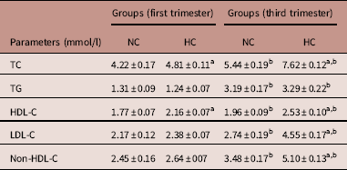
For both groups, maternal serum lipid components were significantly increased at third trimester than that in the first trimester (Table 3). TC and HDL-C levels were significantly higher in HC compared with NC both at first and third trimesters, whereas serum levels of LDL-C and non-HDL-C levels were only significantly increased at third trimester.
Serum lipids profile of maternal blood (mmol/l, n=19 for each group)

NC, pregnant women with normal gestational cholesterol level; HC, pregnant women with gestational hypercholesterolemia; TC, total cholesterol; TG, triglyceride; HDL-C, high-density lipoprotein cholesterol; LDL-C, low-density lipoprotein cholesterol.
a Comparing with the NC group, P<0.05.
b Comparing with the first trimester, P<0.05.
Mitochondrial DNA copy number and mRNA Expression of COX1, ATP6ase, CS, PGC1-α, PGC1-β and ERRα in placenta
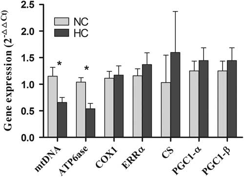
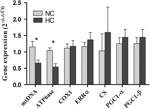
Placental mtDNA copy number of NC and HC was measured as the ratio of nDNA and mtDNA. The placental mtDNA copy number was significantly lower in HC than that in NC (Fig. 1). The placental expression of ATP monophosphatase 6 (ATP6ase) mRNA significantly decreased in HC compared with NC. Expression of cytochrome c oxidase subunit I (COX1), estrogen-related receptor-α (ERRα), citrate synthase (CS), peroxisome proliferator-activated receptor-γ (PPARγ) co-activator 1α (PGC1-α) and PPARγ co-activator 1β (PGC1-β) was higher though the differences were not significant.
Influence of gestational hypercholesterolemia on the mitochondrial DNA (mtDNA) content and expression of messenger RNA level in placenta (
 $2^{{{\minus}\Delta \Delta C_{t} }} $
). Results are expressed as means±s.e.m. * Comparing with the pregnant women with normal cholesterol level (NC) group, P<0.05. HC, pregnant women with gestational hypercholesterolemia; ATP6ase, ATP monophosphatase 6; COX1, cytochrome c oxidase subunit I; EPRα, estrogen-related receptor-α; CS, citrate synthase; PGC1-α, peroxisome proliferator-activated receptor-γ (PPARγ) co-activator 1α; PGC1-β, PPARγ co-activator 1β.
Activities of mitochondrial respiratory complex
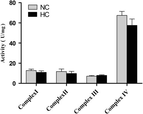
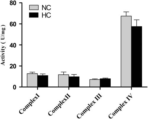
Activities of mitochondrial respiratory Complex I (NADH reducase), II (succinic dehydrogenase), III (coenzyme Q cytochrome C reducase) and IV (cytochrome oxidation) of placenta tissue from HC and NC were compared. There was no significant difference between the two groups (Fig. 2). Except for Complex III, the activities of other complexes in HC group were lower than that of NC group, though no significant differences were observed (Fig. 2).
Influence of gestational hypercholesterolemia on the activities of mitochondrial respiratory complex enzymes. Results are expressed as means±s.e.m. No significant difference was observed. NC, pregnant women with normal cholesterol level; HC, pregnant women with gestational hypercholesterolemia.
Discussion
Pregnancy is an important period for both embryonic development and the formation of various organs, in which fetus develops rapidly, and mitochondrial metabolism carries out actively. In addition to a small amount of the lipids synthetized by fetus, most of the lipids needed in the process of fetal development derived from the mother.Reference Baardman, Kerstjensfrederikse and Berger 14 There was a close relationship between the fatty acids in maternal serum and the development of embryo.Reference Herrera and Ortega-Senovilla 15
Placenta is an important organ for nutrients transport from mother to fetus and excretion of fetal metabolites. A constant and abundant energy source is necessary for rapid growth and maturation of the placenta, and its sound physiological function. Maternal fatty acids are an important source of placental energy metabolism. It has been reported that placentas of obese women had fewer mitochondria.Reference Calabuig-Navarro, Haghiac and Minium 16 , Reference Hastie and Lappas 17 Recently, Calabuig-Navarro reported that placentas of obese women had fewer mitochondria and the decrease of mitochondria in placenta might serve to limit the transfer of maternal lipid to the fetus.Reference Calabuig-Navarro, Haghiac and Minium 16 In the present study, we added new evidence that gestational HC might decrease the placental mitochondria content too. All these results indicated that the placenta mitochondrial content was regulated by the maternal physiological status.
COX is an important constituent of the mitochondrial respiratory chain, and plays a vital role in the process of ATP production by aerobic respiration.Reference Wallace 18 ATP6ase protein is a component of ATP synthase, which converses ATP to ADP in the energy metabolism.Reference Demir, Türkkahraman and Aktaş Samur 19 HumanReference Ling, Poulsen and Carlsson 20 and animal studiesReference Lin, Yang and Tarr 21 have indicated that nutrition may affect the expression of these genes. The expression of COX1 mRNA was found to have decreased in the heart cells of the offspring in dams with copper deficiency.Reference Johnson and Anderson 22 Other animal experiments have also implied that exposure to high-fat diet during pregnancy can result in reduced ATPase transcription in offspring.Reference Liu, Chen and Yao 23 In the present study, mtDNA copy number and ATP6ase expression levels of placental mitochondria were analyzed, and a significant decrease was shown in the HC group when compared with the NC group. Alteration of mtDNA copy number has been supposed as a possible biomarker of mitochondrial dysfunction.Reference Qiu, Sanchez and Hevner 24 Therefore, the decrease of placental mtDNA copy number in the present study may indicate the decline in mitochondrial function. Decreased mRNA expression of ATP6ase may imply the lower level of ATP production and depression of energy metabolism, as reported by Demir et al. previously.Reference Demir, Türkkahraman and Aktaş Samur 19
Mitochondrial genesis is regulated by the mitochondrial genome and nuclear genes, such as CS, PGC1-α, PGC1-β and ERRα. Through the transcriptional regulation of these genes, mitochondria maintain their own fusion and fission balance, ensuring their morphology and function.Reference Wallace 18 CS is commonly used as a quantitative enzyme marker for the presence of intact mitochondria. ERRα and PGC1-α are the key regulators of the transcriptional machinery governing mitochondrial biogenesis and function.Reference Maloyan, Mele and Muralimanoharan 25 As a transcriptional coactivator, PGC1-α transduces many physiological stimuli into specific metabolic activities such as gluconeogenesis, thermogenesis, fatty acid oxidation and mitochondrial biogenesis.Reference Finck and Kelly 26 – Reference Puigserver and Spiegelman 28 PGC1-β is also involved in regulating mitochondrial biogenesis. Though significant increases in mitochondria have been found in tissues where PGC1-α is overexpressed, as the cofactor interacts with other key transcription factors, knockout mice with disrupted PGC1-α are still viable and show normal mitochondrial abundance.Reference Jornayvaz and Shulman 29 – Reference Lin, Wu and Tarr 31 But when compared to mice with normal levels of PGC1-α, these mice show diminished tolerance under physiological stress.Reference Jornayvaz and Shulman 29 – Reference Lin, Wu and Tarr 31 Similarly, in knockout mice with disrupted PGC1-β, the mice exhibited mostly normal levels of mitochondrial function with decreased ability to adapt to physiological stress.Reference Jornayvaz and Shulman 29 , Reference Scarpulla 32 However, a double knockout experiment of PGC1-α/β mice mostly died within 24 h by defects in mitochondrial maturation of cardiac tissue.Reference Lai, Leone, Zechner and Schaeffer 33 These findings suggested that though neither PGC1-α nor PGC1-β alone enabled a cell to start mitochondrial biogenesis, together they were able to complement each other for optimal mitochondrial maturation and function during periods of physiological stress.Reference Jornayvaz and Shulman 29 , Reference Lin, Wu and Tarr 31 , Reference Lai, Leone, Zechner and Schaeffer 33 In this study, CS, ERRα, PGC1-α and PGC1-β mRNA expression levels have not shown a significant difference, but a consistent change, that the expression level in HC was higher than that of NC was exhibited. These findings may be the adaptive response to decreased mtDNA and ATP6ase expression, which could lead to ATP production decline and energy metabolism depression.Reference Demir, Türkkahraman and Aktaş Samur 19 Some researchers have suggested that only in the extreme conditions, mtDNA will be produced in large quantities to provide more mitochondria to supplement the lack of energy in embryonic development.Reference Meldrum, Casper and Diez-Juan 34 In this study, cofounders were controlled as possible as we could. The subjects were strictly selected, pregnant women with preeclampsia, GDM and preterm birth and stillbirth were excluded, and neonatal birth weight are also within the normal range. Mothers who were overweight or obese were not included, either. Therefore, we surmised that though maternal HC in the late stage of pregnancy changed mRNA expression of mtDNA copy number and ATP6ase, maternal HC alone was not able to affect mitochondrial respiratory complex activities.
Electron transport chain (ETC) complex consists of complex I, II, III and IV. Their activities are important for ATP producing. The ETC activities were regulated by pregnant complications. In the IUGR cytotrophoblast cells, researchers found that the mRNA levels of complex II, III and IV were lower but there were no differences at the protein level.Reference Mandò, De and Stampalija 35 In obese pregnant women, it was reported that the activities of complex II, III and IV were not significantly changed, but they were significantly decreased in diabetic pregnant women.Reference Hastie and Lappas 17 The present study observed the decrease of mtDNA and the expressions of ATPase though the ETC activities were not significantly changed. All these results suggested that there might be a post-transcriptional or post-compensatory regulation for mitochondrial function.
In conclusion, our findings suggested that maternal HC in the late stage of pregnancy alone would not have an extreme effect on the placenta, and placental mitochondrial biogenesis may not be influenced seriously by maternal HC in the late stage of pregnancy. This may be due to the adaptive regulation of the placenta, which needs longer follow-up study to make it clear. Meanwhile, further studies, such as the dynamic changes of mitochondria in the process of fetal development to determine the key stages of mitochondrial self-regulation are needed to thoroughly explore the effects of gestational HC on mitochondrial biogenesis and function.
Acknowledgments
The authors are grateful to staff of laboratory and obstetrical department of Xuanwu Hospital in Beijing for their valuable assistance in collecting blood samples and placenta tissues; Department of Epidemiology and Biostatistics of Capital Medical University for the assistance provided during study design and data analysis.
Financial Support
This work was supported by grants from National Natural Science Foundation of China (81273070); the Funding Program for Academic Human Resources Development in Institutions of Higher Learning Under the Jurisdiction of Beijing Municipality of China (Tip-top talent program, CIT&TCD201504092); Support Project of High-level Teachers in Beijing Municipal Universities in the Period of 13th Five-year Plan (CIT&TCD20170323).
Conflicts of Interest
None.
Ethical Standards
The authors assert that all procedures contributing to this work comply with the ethical standards of the relevant national guidelines on human experimentation (Ethical Review of Biomedical Human Research for Trial Implementation, 2007) and with the Helsinki Declaration of 1975as revised in 2008, and has been approved by the institutional committees (Ethics Committee of Xuanwu Hospital, Capital Medical University, No. 2012SY29).