The description of a congenitally malformed heart should ideally give an immediate impression of its morphologic nature. With this principle in mind, it is surely no longer appropriate, in the current era, to use “cor triatriatum”Reference Niwayama 1 to describe an entity unless it has three recognisable and discrete atrial chambers. If translated faithfully, the frequently used terms “cor triatriatum dexter”Reference Ott, Cooley, Angelini and Leachman 2 and “cor triatriatum sinister”Reference Ostman-Smith, Silverman, Oldershaw, Lincoln and Shinebourne 3 also suggest either right or left sidedness of such allegedly triatrial hearts. Therefore, the question is whether a congenitally malformed heart can exist with either three right atriums, or three left atriums, or indeed, whether a heart can exist with three atrial chambers of any kind. Or are the malformed hearts in question better analysed in terms of congenital division of either the morphologically right or the morphologically left atrium? Our suggestion is that the latter is the approach that fosters the best understanding of the malformations, and therefore facilitates optimal communication between professionals. Therefore, in this review, we revisit the evidence that exists regarding the nature of division of the atrial chambers. We begin our review with an account of atrial development, addressing the question as to how best we can recognise the identity of individual atriums, and assessing the role of structures that might, potentially, divide them. We also consider the relationship between the divided atrial chambers and those with obstruction to the atrial outlets.
How do we recognise atrial chambers?
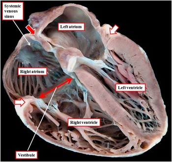
There is no problem in distinguishing the atrial from the ventricular chambers, as these two components are readily recognised as the primary “segments” that provide the basis of segmental analysis.Reference Van Praagh 4 The two segments are separated from one another at the atrioventricular junctions, with the junctions themselves being coincident, in the normally formed heart, with the hinges of the leaflets of the atrioventricular valves (Fig 1).
The cut through a normal heart, replicating the echocardiographic four-chamber section, shows how the atrioventricular junctions (white arrows) separate the atrial and ventricular segments of the heart. Within the atrial segment, the atrial septum separates the right and left atriums, with its superior margin being a fold filled with fat (red arrow). It is noteworthy as to how, on the right side, the pectinate muscles within the appendage extend around the atrioventricular junction to separate the smooth-walled vestibule from the smooth-walled systemic venous sinus. On the left side, the pectinate muscles are confined within the tubular appendage, and thus are not seen in this caudal cut through the heart. The posterior part of the atrium has exclusively smooth walls. Within the ventricles, it is possible to distinguish the coarse trabeculation of the apex of the right ventricle, as opposed to the smoother walls of the left ventricular apex.

Thus, when considering potentially the triatrial hearts, the question is: how to distinguish between the right and left atrial chambers? This question can then be extended to determine whether it is possible morphologically to recognise an additional atrial chamber with its own intrinsic identity. The resolution of this dilemma is provided by the philosophical principle established by Van Praagh et alReference Van Praagh, David, Wright and Van Praagh 5 and dubbed by them as the “morphological method”. When discussing the best means of distinguishing between ventricles, these workers argued cogently that structures within the heart should be identified on the basis of their intrinsic structure and not according to parts that themselves are subject to change. In the setting of the ventricles, this meant that both the atrioventricular valves and the ventricular outlets were disqualified from serving as the final arbiter of ventricular distinction. As was pointed out by Van Praagh et al,Reference Van Praagh, David, Wright and Van Praagh 5 it is the apical trabecular components, specifically the nature of their trabeculations, which provide the best means of discriminating between the morphologically left as opposed to the morphologically right ventricle (Fig 1). When we seek to apply this test in the setting of the atrial chambers, we first need to establish the components that together make up the atriums. Having established these parts, we then need to determine which of the components is most constant and least amenable to change when the heart itself is congenitally malformed. These decisions are now facilitated by the knowledge of atrial development.
Development of the atrial chambers
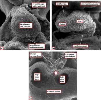
Almost a century ago, Maude AbbottReference Abbott 6 was convinced that the knowledge of cardiac ontology would serve to elucidate the understanding of congenital malformations. Until recently, the evidence relating to cardiac embryology has been insufficiently robust to underscore the analysis of congenital cardiac anomalies. All of this has changed over the last decade. The availability of episcopic microscopyReference Mohun and Weninger 7 now demonstrates with exquisite accuracy how cardiac morphology changes during development. We now have access to a large number of data sets, prepared using this technique, which permit us to analyse the changing morphology of the atrial segment of the mouse heart from the initial stages of formation of the heart tube, occurring during the 9th day subsequent to conception, or E8.5, to term, the latter occurring at E18.5. When first observed, we now know that the structure seen as a linear tube within the confines of the pericardial cavity will give rise, in the definitive heart, to little more than the left ventricle (Fig 2a).Reference Moorman, Christoffels, Anderson and van den Hoff 8 It is the addition of new material from the heart-forming areas of the embryo during E9.5 and 10.5 that sees the emergence of the atrial component of the developing heart, along with the formation of the atrioventricular canal (Fig 2b). At these early stages, the systemic venous tributaries, bringing the blood back to the developing atrial chamber, are relatively symmetrical, although the right-sided tributary is already larger than the left (Fig 2c).
The scanning electron micrographic images show how the heart tube elongates and loops during the 9th day of development in the mouse. ( a ) shows the so-called “straight tube” stage as material begins to be added from the heart-forming areas at the venous and arterial poles. The addition of this new material produces the ventricular loop, with its inlet and outlet parts, as shown in ( b ). At this stage, the atrial component has a solitary lumen, as shown in ( c ), with the systemic venous tributaries entering its caudal margins. The heart is still attached to the pharyngeal mesenchyme in this region through the dorsal mesocardium, with the so-called pulmonary pit (arrow) seen between the reflections of the heart wall with the pharyngeal tissues. Note that, already at this stage, the left-sided tributary is smaller than the right-sided tributary. Images prepared and reproduced by kind permission of Professor Nigel A. Brown, St George's Medical University, London.

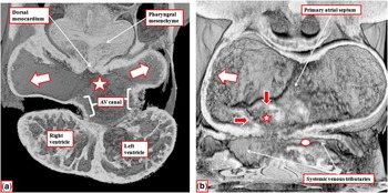
The lungs have not yet formed at E9.5, so that, at this time, there is no pulmonary vein, nor is there any evidence of division within the common atrial chamber. Nonetheless, the atrial chamber is connected dorsally to the pharyngeal mesenchyme. The reflections of the myocardial walls with the pharyngeal tissues at this mesocardial connection produce the boundaries of a pit through which the pulmonary vein will eventually open into the left atrium (Fig 2c). Key changes occur during the 10th day of development in the mouse. Initially, the overall length of the solitary lumen of the heart tube is lined by cardiac jelly. The lumen expands in both the atrial and the ventricular components of the tube by a process known as ballooning. During this period of ballooning, the cardiac jelly itself undergoes a process of endothelial-to-mesenchymal transformation, which produces distinct cushions in the atrioventricular canal and the outflow tract. After the formation of the cushions, the ballooning components are no longer lined by jelly. The expansions occur bilaterally from the atrial component, but develop in series within the ventricular loop. In the atrial component, the parallel expansions become the appendages, whereas in the ventricular loop they become the apical components of the left and right ventricles (Fig 3a).
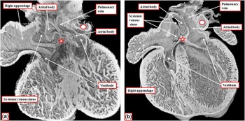
The sections are taken from a mouse heart at E10.5 (10.5 embryonic days after conception) prepared using episcopic microscopy. ( a ) is a “four-chamber” section through the area of the dorsal mesocardium. The atrial appendages (white arrows) have expanded in parallel and bilaterally from the solitary initial cavity of the atrial component of the linear heart tube (star). The atrioventricular (AV) canal drains the atrial segment to the inlet of the ventricular loop, which has ballooned apically to give rise to the apical part of the developing left ventricle. The apical part of the developing right ventricle has ballooned from the outlet of the loop. ( b ) is a short-axis section taken caudally. It shows how, already at this stage, the systemic venous tributaries have shifted so as to open to the right side of the common atrial chamber (star). The primary atrial septum is just beginning to appear in the atrial roof. As the systemic venous tributaries move rightward relative to the atrium, their boundaries become distinguished by the appearance of the venous valves (red arrows). The right appendage expands to the right of the systemic venous sinus (white arrow in ( b )).

Another key change that takes place over this period in the atrial component is the rightward shift of the systemic venous tributaries. As already shown, the left-sided venous tributaries, entering the common atrium through the left sinus horn, are already smaller than their right-sided counterparts at E9.5 (Fig 2c). During the 10th day of murine development, the junction between the tributaries and the body of the common atrium moves rightward, with the left sinus horn then incorporating into the developing left atrioventricular junction (Fig 3b). By this stage of development, it is now possible to distinguish the primordium of the primary atrial septum in the roof of the initial lumen of the atrial component of the heart tube. As the systemic venous tributaries move rightwards, their boundaries with the common atrium also become recognisable owing to the appearance of the venous valves (Fig 3b). Therefore, at the end of E10.5, it is possible to recognise the body of a common atrial chamber, from which the right-sided and left-sided appendages have ballooned. The atrioventricular canal is also recognisable as an entity at this stage, draining exclusively to the developing left ventricle, although its right-sided wall is already providing continuity, in the inner heart curve, between the floor of the right half of the atrial chamber and the roof of the developing right ventricle (Fig 3a). With the rightward shift, however, the systemic venous tributaries now empty into the right half of the still common body of the developing atrial component (Fig 3b).
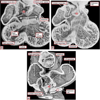
On the 11th day of murine development, the venous valves markedly increase in size, producing clear-cut boundaries between the systemic venous sinus and the other components of the developing right atrium (Fig 4a). Downward growth of the primary atrial septum confines the systemic venous sinus to the right side of the atrial component. At the same time, there is exaggerated growth of the right margin of the dorsal mesocardial connection, which is seen within the atrial cavity as the vestibular spine (Fig 4a). By this stage, the lungs have now appeared within the pharyngeal mesenchyme, and a midline vein has canalised so as to connect the developing intrapulmonary venous plexuses with the left side of the common atrial chamber (Fig 4b). The initial atrioventricular canal myocardium is now becoming sequestrated on the atrial side of the developing atrioventricular junctions. The canal musculature forms the developing atrial vestibules, which at this stage remain as a common structure, as the mesenchymal cap carried out on the leading edge of the primary atrial septum has yet to fuse with the atrial surface of the atrioventricular cushions (Fig 4a). By now, it can be seen that the initial cavity of the heart tube is shared between the dividing atriums to form the atrial bodies, with the ballooned components forming the atrial appendages. Each atrium now also possesses a venous component, although the pulmonary vein is a narrow channel that enters the developing left atrium to the left of the vestibular spine (Fig 4b).
The sections are all from the same episcopic data set, which is prepared from a mouse heart at E11.5. ( a ) is a four-chamber section through the developing primary septum. The atrioventricular canal myocardium becomes incorporated as the atrial vestibules, still forming a common structure at this stage. Note the expanded right margin of the dorsal mesocardium that has become the vestibular spine (star); ( b ) a four-chamber section taken more caudally, shows how the pulmonary vein has canalised and opened to the left side of the initially common atrial chamber; ( c ) in short axis, shows how the appendages have now ballooned in parallel from the atrial body, with the venous valves marking the junction between the systemic venous sinus and the right half of the initially common atrium.

It is the fusion of the mesenchymal cap carried out on the leading edge of the primary atrial septum with the atrial margins of the atrioventricular cushions that now obliterates the primary atrial foramen. The rightward margin of the site of fusion is then reinforced by the continuing growth of the vestibular spine, which now forms the prominent antero-inferior buttress of the atrial septum. Therefore, by E12.5, the initial primary atrial septum itself has become the flap valve of the oval foramen, with a small ridge in the atrial roof marking the site where the primary septum initially broke away from the atrial roof to form the secondary interatrial foramen. The completion of atrial septation, with muscularisation of the vestibular spine to form the antero-inferior buttress of the septum during E13.5, means that by this stage of development it is possible to recognise the definitive atrial components (Fig 5).
These four-chamber sections, taken from episcopic data sets prepared from developing mice at E12.5 ( a ) and E13.5 ( b ), show how the vestibular spine (star) reinforces the rightward side of the site of fusion of the mesenchymal cap of the primary septum with the atrioventricular endocardial cushions ( a ), and then muscularises to form the antero-inferior buttress of the oval foramen ( b ). The caudal section in ( b ) then shows how, subsequent to the closure of the primary atrial foramen, each atrium can be seen to possess an appendage, a vestibular, and a venous component, along with part of the initial lumen of the linear heart tube, which forms the atrial body. The atriums are separated by the atrial septum.

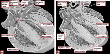
The heart continues to mature to term when expansion of the pulmonary venous component of the left atrium produces a marked fold dorsally. The fold is continuous cranially with the ridge in the atrial roof, which marked the initial break in the cranial margin of the primary atrial septum. It is the fold, together with the cranial ridge and the antero-inferior buttress, which form the right atrial rims of the oval foramen. Therefore, by term, each atrium possesses part of the initial lumen of the primary linear tube. This forms their bodies. On the right side, the venous valves are seen as obvious partitions that mark the boundaries of the systemic venous sinus, which is interposed between the pectinated walls of the appendage and the smooth walls of the tricuspid valvar vestibule. On the left side, in contrast, there are no internal partitions. The pulmonary venous component, the body, and the vestibule together form a confluent smooth-walled atrial component, with the pectinate muscles confined within the tubular appendage (Fig 6).
The sections are taken from an episcopic data set prepared from a developing mouse at E18.5. Both are taken in the four-chamber plane, with ( a ) taken more ventrally relative to ( b ). The sections show how each atrium possesses a body derived from the initial cavity of the linear heart tube, a vestibule derived from the original atrioventricular canal, and an appendage derived by ballooning from the initial common cavity. The right atrium now receives the systemic venous tributaries, with their boundary marked by the prominent venous valves. There is no comparable boundary in the left atrium, with the pulmonary venous component merging smoothly with the body and the vestibule. The vestibular spine (star in ( a )) forms the antero-inferior buttress of the atrial septum, with the caudal margin of the septum formed at term by a fold between the pulmonary venous component of the left atrium and the body of the right atrium ( b ). Note the ridge marking the cranial margin of the oval foramen ( a ).

How, then, can we distinguish between the atrial chambers?
Comparisons between Figure 1 and Figure 6 show that the features of the mouse heart at the completion of intrauterine development are directly comparable to the situation seen in the postnatal human heart, although the mouse continues postnatally to possess a persisting left superior caval vein. The pulmonary venous component of the murine heart is also positioned dorsally, rather than occupying the atrial roof, as is the case of man. The atrial chambers in man, as in the mouse, are separated by the atrial septum. And each atrium can be recognised to possess an appendage, a venous component, part of the body of the initial linear heart tube, and a vestibule. Of these components, it is the appendages that are most constant when the heart is congenitally malformed. Furthermore, it is the extent of the pectinate muscles, again as in the mouse heart, which serve to distinguish between the morphologically right and morphologically left appendages.Reference Uemura, Ho, Devine, Kilpatrick and Anderson 9 The pectinate muscles of the morphologically right appendage extend around the vestibule, interposing between the vestibular smooth wall and the smooth-walled systemic venous sinus. The pectinate muscles of the morphologically left appendage, in contrast, are confined within the tubular appendage so that the majority of the walls of the left atrium, when viewed in four-chamber sections, are entirely smooth (Fig 1). Applying the concept of the “morphological method”,Reference Van Praagh, David, Wright and Van Praagh 5 it is the presence of the atrial appendages that should serve to distinguish the number of atrial chambers present when the heart is congenitally malformed. This framework now permits us to assess the structure of the allegedly “triatrial hearts”.
The anatomic basis of “cor triatriatum dexter”
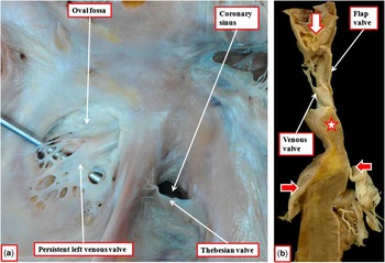
It has long been recognised that the essence of “cor triatriatum dexter” is the persistence of the valves of the embryonic systemic venous sinus.Reference Yater 10 As we have shown, in the mouse heart, these valves are still present at term. They serve to demonstrate the presence, in the right atrium, of its body, which is interposed between the left venous valve and the septum. This component is part of the initial cavity of the atrial component of the linear heart tube. In contrast, in man, these venous valves have usually become attenuated by term, with only small parts of the right valve remaining in the postnatal hearts as the valves guarding the orifices of the inferior caval vein and the coronary sinus, forming the Eustachian and Thebesian valves, respectively. The left valve, in man, usually disappears in its entirety so that it is rarely possible to recognise the precise location of the remnant of the body derived from the linear heart tube. On occasions, remnants of the left venous valve can be recognised in the postnatal human heart, although they do not produce clinical problems. Then they do serve, as in the mouse heart, to demonstrate the location of the body of the right atrium (Fig 7).
The images are from an anatomically normal human heart and show persistence of the left valve of the systemic venous sinus. ( a ) shows the atrial septum as seen from the right side. The probe is placed between the remnants of the left valve of the systemic venous sinus and the floor of the oval fossa. ( b ) is a cut through the septum replicating the four-chamber echocardiographic section. It confirms the separate nature of the persistent left venous valve and the flap valve of the oval fossa. Note the in-folding that forms the superior rim of the fossa (white arrow) and the buttress forming the antero-inferior part of the septum (star). Also note the off-setting of the hinges of the atrioventricular valves (red arrows).

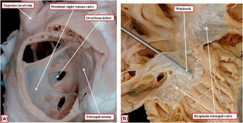
The right venous valve does produce congenital problems when its parts persist in postnatal life. Exuberant persistence of the Thebesian valve, which guards the orifice of the coronary sinus, can totally block its orifice, producing atresia of the coronary sinus.Reference Falcone and Roberts 11 This lesion can often go unrecognised, and thus its frequency is hard to determine. Persistence of the Eustachian component of the right venous valve is more frequently recognised, although still rare.Reference Yater 10 , Reference Trento, Zuberbuhler, Anderson, Park and Siewers 12 It is seen most commonly when there is obstruction of normal right atrial outflow, either because of tricuspid atresia, or in the setting of pulmonary atresia with intact ventricular septum (Fig 8a). The valve can also persist when there has been normal development of the right atrioventricular junction, and can be aneurysmally transformed to form a windsock (Fig 8b).
The images show the features of persistence of the right valve of the systemic venous sinus. ( a ) shows a persistent right valve in the setting of tricuspid atresia owing to the absence of the right atrioventricular connection. The valve serves to direct the flow from the inferior caval vein through the oval foramen, but also obstructs the orifice of the coronary sinus. ( b ) shows a probe placed in a windsock formed from a persistent right venous valve in a heart that also exhibits minimal tricuspid valvar dysplasia.

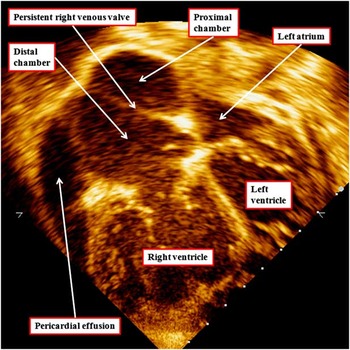
Depending on its extent, the enlarged windsock can obstruct the tricuspid valve, or even extend to produce pulmonary obstruction. However, on occasions, it can be asymptomatic, being observed as a chance finding during echocardiographic examination.Reference Mahy and Anderson 13 Irrespective of its consequences, persistence of the venous valve produces division of the morphologically right atrium into proximal and distal chambers (Fig 9). Although unequivocally dividing the morphologically right atrium, the persistent venous valve does not produce a triatrial heart, as the morphologically right appendage remains a solitary entity and belongs to the distal atrial component. The inferior caval vein, shielded by the persisting Eustachian valve, enters the proximal atrial component. In contrast, the superior caval vein is not always confined within the proximal chamber. As shown in Figure 8a, the superior caval vein in the heart with tricuspid atresia opens lateral to the persisting venous valve. In the presence of tricuspid atresia, this finding is of no significance, as all the systemic venous returns must cross the oval fossa to reach the ventricular mass. In the image from the patient shown in Figure 9, kindly provided by Dr I.B. Vijayalakshmi, the superior caval vein also opens to the distal chamber, as revealed by injection of contrast bubbles into an arm vein. In the patients with the windsock malformations, in contrast, the right venous valve presumably does confine the superior caval vein also within the proximal atrial chamber, thus promoting the formation of the aneurysmal windsock (Fig 8b).
The cross-sectional echocardiogram in four-chamber projection is from a patient suspected of having persistent pulmonary hypertension. As the images show, there is division of the morphologically right atrium by persistence of the right venous valve into proximal and distal chambers. The inferior caval vein opens into the proximal chamber, which also contains the right atrial body and the oval fossa. The superior caval vein, in contrast, opens into the distal chamber, which also contains the appendage and the vestibule. The superior caval venous drainage has been revealed by the injection of agitated contrast material into an arm vein, the bubbles opacifying the distal chamber and the right ventricle. Also note the presence of a pericardial effusion. Image kindly provided by Dr I.B. Vijayalakshmi, Sri Jayadeva Institute of Cardiovascular Sciences and Research, Bangalore, and reproduced with her permission.

What about “cor triatriatum sinister”?
Persistence of the right valve of the systemic venous sinus provides an obvious explanation for division of the morphologically right atrium. The situation is more complicated when considering the hearts that allegedly possess the three left atriums, or so-called “cor triatriatum sinister”. However, in the majority of cases, the morphology is relatively constant, although there are subtle differences in the make-up of the components present on the left side of the atrial septum. It is the very existence of unusual variants that creates problems in providing a universal explanation for their morphogenesis. The first description of the classic variant is credited to Church, and appeared in the middle of the nineteenth century.Reference Church 14 The essence of the lesion is an oblique fibromuscular partition that divides the morphologically left atrium into discrete components (Fig 10).
The image shows an autopsied heart with the “classic” form of so-called “cor triatriatum sinister”. An oblique fibromuscular partition divides the morphologically left atrium into two chambers. The superior chamber received the pulmonary veins, and the oval fossa opens into this chamber. The inferior chamber gives rise to the left atrial appendage and contains the vestibule. The two chambers have been described variously as being “proximal” or “distal”, according to whether this is judged on the flow of blood, or the adjacency of the chambers as viewed by the observer. We will describe the chambers as being pulmonary as opposed to vestibular, as the pulmonary veins always open to the upper chamber in the classical variant of the lesion, and the vestibule is always part of the lower chamber. As we will discuss, however, the situation can be complicated by anomalous locations of the pulmonary veins. Note the presence in the illustrated heart of a persistent right venous valve.

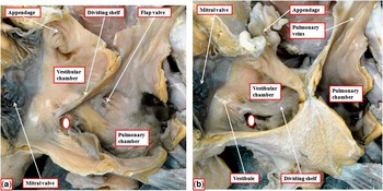
Even within the hearts representing the “classic” variants of the lesion, there can be variations in the specific morphology. The most obvious variation, reflecting clinical presentation, is the size of the hole in the shelf that divides the atrium. This hole can be of medium or substantial size, as seen in Figures 11 and 12, or no more than a pinhole (Fig 10). The constant feature of the classic variants is the drainage of the pulmonary veins into the superior of the divided parts of the left atrium, whereas the vestibule and the atrial appendage are parts of the inferior atrial chamber. In contrast, the body of the atrium can be variously divided between the two parts. This is revealed by the fact that the oval fossa can open either into the superior pulmonary atrial chamber (Fig 11) or into the inferior vestibular compartment (Fig 11).
In this example of a heart with the divided left atrium, the oblique fibromuscular shelf is positioned so as to place the oval fossa into the pulmonary atrial component. Note that the communication between the chambers is of reasonable size (white oval). The vestibular chamber, as is always the case, gives rise to the left atrial appendage.

In this example of an autopsied heart with the divided left atrium, the dividing partition is placed such that the oval fossa, which is deficient, opens to the vestibular chamber. The vestibular chamber also gives rise to the left atrial appendage. Only the pulmonary veins open into the superior atrial compartment. Note that the communication between the atrial compartments is much larger in this specimen (white circle), but the pulmonary chamber is still dilated.

Various theories have been advanced to explain the morphogenesis of the classic variant, none of which is entirely convincing. One theory depends on persistence of the left superior caval vein.Reference Gharagozloo, Bulkley and Hutchins 15 Although cases certainly exist in association with persistence of the left superior caval vein, which then drains to the right atrium through an enlarged coronary sinus and extends obliquely across the dorsal wall of the left atrium, paralleling the location of the dividing shelf, the majority of patients with “classic” variants do not show persistence of the left-sided systemic venous channel. Moreover, in the mouse heart, which retains the left superior caval vein as part of normal development, there is no sign of encroachment on the left atrial cavity during normal development (Fig 5).
The other favoured theories depend on malseptation, malincorporation, or entrapment of the pulmonary vein.Reference Van Praagh and Corsini 16 However, these theories also fail to withstand rigid scrutiny. As we have shown, the pulmonary vein, in manReference Webb, Kanani, Anderson, Richardson and Brown 17 as in the mouse,Reference Webb, Brown, Wessels and Anderson 18 initially develops as a solitary channel formed adjacent to the left atrioventricular junction (Fig 4b). It is only with ongoing development in both man and mouse that there is enlargement of the pulmonary venous component of the left atrium. Only in man does this compartment eventually achieve a location on the atrial roof, with one vein typically opening at each corner of the roof.Reference Webb, Kanani, Anderson, Richardson and Brown 17 Although abnormal incorporation of the vein could well explain those cases in which the oval fossa drains into the vestibular chamber (Fig 12), it is harder to explain the drainage of the oval fossa to the pulmonary chamber on the basis of malincorporation or entrapment. Over and above the developmental evidence, it is also well established that a dividing fibromuscular shelf can divide the left atrial chamber even in the presence of partially or totally anomalous pulmonary venous connection.Reference Thilenius, Bharati and Lev 19 Paradoxically, the very fact that the pulmonary vein originates initially adjacent to the atrioventricular junction does provide a plausible explanation, on the basis of malincorporation, for one of the unusual cases discovered by Thilenius et al.Reference Thilenius, Bharati and Lev 19 The structure of the dividing shelf, as demonstrated by Van Praagh and Corsini,Reference Van Praagh and Corsini 16 is also suggestive of entrapment of the developing pulmonary vein. However, the embryological concepts advanced by both these groups of authorsReference Van Praagh and Corsini 16 , Reference Thilenius, Bharati and Lev 19 have little in common with the changes shown in our developing mice.
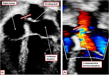
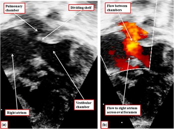
Although the morphogenesis of the lesions has still to be established, there is no question but that the lesions are readily diagnosed with modern-day techniques, which show all the variations in morphology (Figs 13 and 14). Once identified, it is then relatively straightforward for the dividing shelf to be removed surgically, thus restoring the morphology of the morphologically left atrium.
The echocardiographic images, in four-chamber projection, show an example of divided left atrium with the oval fossa (double-headed arrow) communicating with the pulmonary chamber. The right-hand panel ( b ) shows the acceleration of flow across the hole in the dividing shelf.

The echocardiographic images, again in a four-chamber projection, show an example of the divided left atrium in which the oval fossa is committed to the vestibular chamber. Flow across the oval foramen from the left atrium to the right atrium is shown by the colour Doppler signal in ( b ).

What about supravalvar mitral stenosis?
One of the lesions producing obstruction of the left atrium at its outlet could be considered to be part of the spectrum of the divided left atrium, in that it obstructs the vestibular atrial component. The lesion (Fig 15), however, is fundamentally different in its structure from the shelf that divided the morphologically left atrium.
The images show the presence of a supravalvar shelf obstructing the exit from the left atrium. In ( a ), the left atrioventricular junction has been joined together and is photographed from above. The shelf extends around the parietal margin of the left atrial vestibule. Note the presence of a defect within the oval fossa. In Figure 15b, the left atrioventricular junction has been opened and is photographed from the left side. The obstructing shelf is a fibrous ridge that encircles the parietal component of the left atrial vestibule, separating the vestibule from the opening of the left atrial appendage.

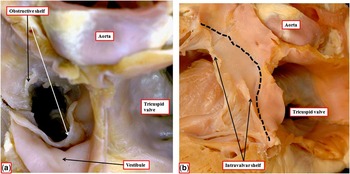
The shelf, which is fibrous rather than fibromuscular, is located on the surface of the parietal component of the left atrial vestibule, rather than dividing the vestibule from the remainder of the atrial chamber. In more extreme forms, the shelf can also extend to form a diaphragm that includes the septal component of the vestibule. The vestibular obstructive lesion (Fig 15) is itself then fundamentally different from the comparable fibrous shelf found within the funnel created by the leaflets of the mitral valve, and often considered to be a variant of “supravalvar stenosis” (Fig 16).
The images show an obstructive intravalvar mitral lesion. At first sight, when viewed from above ( a ), the obstructive lesion is comparable to the supravalvar vestibular lesion (compared with Fig 14a). Opening the atrioventricular junction, as shown in Figure 15b, shows that the fibrous shelf is formed within the funnel created by the mitral valvar leaflets and is adherent to the atrial surfaces of both the aortic and mural leaflets of the valve. The obstructive lesion is distal to the atrioventricular junction (black dashed line). Therefore, it is intravalvar rather than supravalvar.

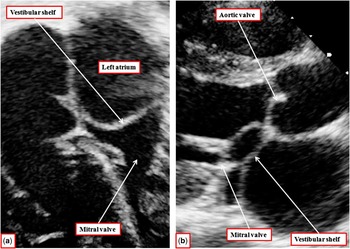
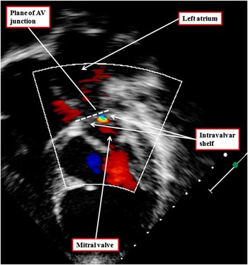
As with the supravalvar lesion, the obstructive shelf is made up of fibrous tissue. Moreover, again as with the vestibular lesion, it can extend variably around the circumference of the left atrioventricular junction. Unlike the vestibular lesion, however, the second entity is formed within the funnel created by the leaflets of the mitral valve. As emphasised by the group from Rome, the lesion is intravalvar rather than supravalvar.Reference Toscano, Pasquini and Iacobelli 20 Nevertheless, the fibrous nature of both the lesions is almost certainly indicative of an acquired aetiology, even if the lesion may be acquired during foetal development. The association of the lesions as part of the Shone syndromeReference Shone, Sellers, Anderson, Adams, Lillehei and Edwards 21 also points to the likelihood of disturbances of flow being associated with their development. As with division of the left atrium, modern-day clinical techniques now permit the diagnosis and differentiation of the lesions obstructing the inflow to the left ventricle (Figs 17 and 18), hopefully providing the impetus for timely surgical removal.
The cross-sectional echocardiographic images, shown in four-chamber ( a ) and parasternal long-axis ( b ) planes, reveal the presence of an obstructive shelf in supravalvar location, occupying the vestibular component of the left atrium.

The cross-sectional echocardiographic image with colour Doppler shows the presence of a fibrous shelf within the funnel created by the leaflets of the mitral valve. The obstructive lesion is below the level of the atrioventricular junction and hence is intravalvar rather than supravalvar in location.

Conclusions
Perhaps the most important principle to have been put forward relative to cardiac anatomy is the so-called “morphological method”. The essence of this principle is that structures within the heart should be identified and described on the basis of their intrinsic morphology, and not by reference to components that are themselves susceptible to change. When applied to the atrial chambers, the principle dictates that the appendages should be the arbiters of morphologically rightness as opposed to leftness. On the basis that there are only two atrial appendages, either morphologically right or left, it then follows that there can only be two atrial chambers. Each of the chambers can be divided. As we have shown, this produces discrete entities recognisable on the basis of their intrinsic morphology. Hence, it is our opinion that the lesions frequently described as representing “cor triatriatum” are better considered as showing divided atrial chambers. As we have also demonstrated, either the morphologically right or the morphologically left atrium can be congenitally divided.
It is then an easy matter to explain the division of the morphologically right atrium on the basis of persistence of the valves of the embryonic systemic venous sinus. It is much harder to provide a unifying explanation to account for the various forms of divided left atrium, with none of the currently popular theories withstanding rigorous scrutiny. The fibromuscular partition dividing the left atrium is also different in terms of structure and histology from the lesions that obstruct the inflow to the left ventricle at either vestibular or intravalvar level. These latter two entities are themselves readily distinguished depending on their relationship with the left atrioventricular junction, and only the vestibular variant should be described as being supravalvar. All of the various entities can now be readily diagnosed and differentiated using cross-sectional echocardiography, meaning that once identified they can efficiently be removed at surgical operation.
Acknowledgements
We are indebted to Dr I.B. Vijayalakshmi from the Sri Jayadeva Institute of Cardiovascular Sciences and Research, Bangalore, who kindly shared with us her recent experience with two patients thought to have persistent pulmonary hypertension, but who proved to have persistence of the right venous valve. She also kindly permitted us to reproduce one of her as yet unpublished images demonstrating the lesion (Fig 9). We also thank Professor Nigel A. Brown, St George's Medical University, London, for permission to publish the images making up Figure 2.