Introduction
To date, commercial neuroprosthetic implants use metallic electrodes to directly inject electrical charge and stimulate neural tissue.[Reference Cogan1] This process involves moving from electron flow in the device to ion flow in the tissue. It is the ions in the biologic environment which enable depolarization and generation of action potentials in the neural tissue. As a result, the target tissue, which is often injured or diseased, is subjected to electrical fields that raise the extracellular voltage.[Reference McCreery, Yuen and Bullara2] This process has significant impacts on the local biologic environment, including cell electroporation, generation of undesirable chemical species, localized pH changes, and subsequent inflammatory reactions, as well as electrode degradation.[Reference Cogan1, Reference McCreery, Yuen and Bullara2] The consequences of this mode of operation is that chronic frustration of the wound-healing process produces a scar tissue reaction, which encapsulates the implant and electrically isolates it from the target tissue.[Reference Woolley, Desai, Steckbeck, Patel and Otto3, Reference Gilmour, Woolley, Poole-Warren, Thomson and Green4] As a result, the amount of charge required to activate the target tissue often increases over time and the implant loses efficacy.[Reference McCreery, Yuen, Agnew and Bullara5, Reference Onnela, Takeshita, Kaiho, Kojima, Kobayashi, Tanaka and Hyttinen6]
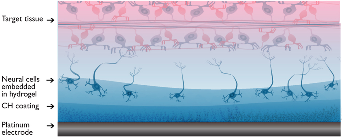
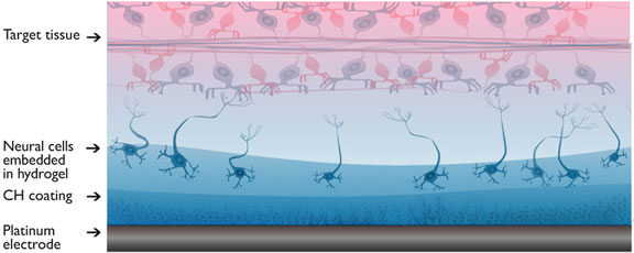
Rather than relying on unwieldy metal electrodes and direct charge injection, tissue-engineered bioelectronics will use cells embedded within devices to provide a natural mode of physiologic tissue activation.[Reference Aregueta-Robles, Woolley, Poole-Warren, Lovell and Green7] This “living electrode” concept, shown in Fig. 1, is designed to seamlessly integrate living neural cells with electrodes through modification of electrode coating technologies. This approach to creating living bioelectronics draws upon the knowledge from implantable neurostimulators and bioelectrode research and introduces principles of tissue engineering. There are a number of challenges to producing a cell laden electrode. One of the initial hurdles is in providing a carrier for the encapsulation of the neural cells at the electrode surface. The fabrication technique and carrier must be cytocompatible, but the carrier is also required to support stable integration of the coating technology with the underlying electrode.
Schematic representation of synaptic interfaces between an electrode and target tissue produced from integrating living neural cells into tissue-engineered hydrogel coatings.

Integration of living cells within electronics was first explored in 1980s for application to biosensors.[Reference Ochiai, Shibata, Sawa and Katoh8] Studies by Ochiai et al.[Reference Ochiai, Shibata, Sawa and Katoh8] demonstrated that living blue-green algae could be embedded within an alginate gel to create a photoconverter that was stable but unfortunately had low conversion efficiency.[Reference Ochiai, Shibata, Sawa, Shoga and Ohta9] Campbell et al.[Reference Campbell, Hodgson and Wallace10] investigated red blood cells embedded in a conductive polymer (CP), to present surface antigens as the sensing element. It was found that surface antigen integrity was maintained, but cell viability and function was not studied. More recently, Richardson-Burns et al.[Reference Richardson-Burns, Hendricks, Foster, Povlich, Kim and Martin11] polymerized the CP poly(ethylene dioxythiophene) (PEDOT) around primary mouse neural cells cultured on metal electrodes. While the cells were found to survive initial short-term exposure to chemical reagents, cell death occurred within a few days. It was suggested that focal adhesions may have been disrupted or electroporation may have occurred as a result of the applied voltage during fabrication.[Reference Richardson-Burns, Hendricks, Foster, Povlich, Kim and Martin11] The fundamental issues that have hindered the development of living electrodes include toxicity and damage to the cells caused by exposure to monomers and DC voltages during the fabrication process[Reference Richardson-Burns, Hendricks and Martin12, Reference Ouyang, Green, Feldman, Martin, Schouenborg, Garwicz and Danielsen13] and the capacity to provide a three-dimensional (3D) environment that is supportive of cell survival and growth within the electroactive and spatially confined environment of a microelectrode device.[Reference Mahoney and Anseth14]
In the current study, a layered approach was proposed, as depicted in Fig. 1. This layered format provides significant advantages over prior cell-based electronics. Firstly, while non-conductive hydrogels, such as alginate and agarose, have been shown to dampen the mechanics of stiff electrode arrays, the swelling properties of these materials displace the target tissue from the device interface. Homogenous hydrogels are also prone to delamination unless they are chemically grafted to the electrode surface.[Reference Baek, Green, Granville, Martens and Poole-Warren15] Conversely, conductive hydrogels (CHs), being interpenetrating hybrids of CPs and hydrogels,[Reference Green, Hassarati, Goding, Baek, Lovell and Martens16] have been shown to be stable under implant conditions,[Reference Hassarati, Dueck, Tasche, Carter, Poole-Warren and Green17] and swelling can be controlled through design of the hydrogel and CP components.[Reference Goding, Gilmour, Martens, Poole-Warren and Green18] By encapsulating cells in an overlying hydrogel layer above a CH coating, the cells can be protected from the solvents and high DC voltages required for CH (or CP) fabrication. Since the biologic environment exclusively passes charge in ions, this design and specifically the CH layer will also protect encapsulated cells from high-voltage capacitive charging that occurs on metallic electrodes. The hydrogel used for the upper layer must be a degradable hydrogel, which not only provides both biochemical and mechanical support for neuroprogenitor cells, but also enables them to grow and differentiate, producing active neural networks based on their own extracellular matrix (ECM). The degradation rate must be carefully matched to the development of ECM such that encapsulated cells continue to support their own survival and development over chronic implant periods.
Prior studies by the authors have shown that degradable poly(vinyl alcohol) (PVA) forms a hydrogel that is cytocompatible and can be tailored for both degradation time frame and bioactivity.[Reference Green, Lim, Henderson, Hassarati, Martens, Lovell and Poole-Warren19, Reference Lim, Alves, Poole-Warren and Martens20] The PVA in this study was modified with phenol cross-linking groups (PVA-ph) that enabled incorporation of biomolecules, including gelatin and sericin. Studies on cell encapsulation within PVA have shown that small amounts of gelatin greatly improve cell survival through provision of attachment cues that enable cell spreading.[Reference Lim, Alves, Poole-Warren and Martens20] Sericin is used to support cells during fabrication by providing protection from initiators and light exposure.[Reference Lim, Kundu, Reeves, Poole-Warren, Kundu and Martens21] This initial proof-of-concept was shown to enable encapsulation of the neural-like PC12 cell and neuroglial Schwann cells.[Reference Green, Lim, Henderson, Hassarati, Martens, Lovell and Poole-Warren19, Reference Aregueta-Robles, Lim, Martens, Lovell, Poole-Warren and Green22] In an effort to move toward a more complex and clinically relevant cell approach, primary neuroprogenitors are incorporated within the PVA-ph and applied to platinum macroelectrode constructs. These studies investigate the impact of the cell laden degradable layer on the electrode performance. Layered living electrode constructs were fabricated and characterized across electrochemical, mechanical, and physical properties. Furthermore, the ability for encapsulated neuroprogenitor cells to produce ECM within the electrode surface was established.
Materials and methods
Fabrication of living electrode constructs
To enable characterization of bulk properties, large Pt disc electrodes (10 mm diameter, 100 µm thick) were used. Pt discs were first deposited with a pre-coat of PEDOT. This adherence layer is produced by galvanostatic electropolymerization from a monomer precursor solution consisting of 0.1 M EDOT and 0.05 M paratoluene sulfonate in 50% deionized (DI) water and 50% acetonitrile. Deposition was carried out at 1 mA/cm2 for 1 min. The non-degradable hydrogel was then formed from a macromer solution of 10 wt% methacrylate and taurine-modified PVA (PVA-Tau), using methods established by Goding et al.[Reference Goding, Gilmour, Martens, Poole-Warren and Green18] The hydrogel film was cross-linked to form a thin layer across the electrode surface with ultra-violet light (70 mW/cm2, 365 nm) for 180 s. PEDOT was deposited through this gel from an aqueous solution of 0.03 M EDOT at 0.5 mA/cm2 for 30 min. The thickness of the CH layer was ~10 µm, in line with prior studies using this technique and similar fabrication parameters.[Reference Green, Hassarati, Goding, Baek, Lovell and Martens16–Reference Goding, Gilmour, Martens, Poole-Warren and Green18] The final layer was produced from a macromer solution of 8 wt% phenol-modified PVA, 1 wt% gelatin, and 1 wt% sericin (S5201 Sigma Bombyx mori) using the methods developed by Lim et al.[Reference Lim, Alves, Poole-Warren and Martens20] Neuroprogenitor cells (described below) were added to the macromer solution at 5 × 10⁶ cells/mL. The initiators ruthenium (2 mM) and sodium persulfate (20 mM) were included. A silicone mold was placed over the CH coating such that the cell laden layer was produced with a uniform thickness across the sample and between batches. A coverslip was placed over the macromer solution, and the cell laden construct was exposed to visible light using a BlueRay light source with visible spectrum filter (15 mW/cm2) for 180 s to cross-link the final hydrogel layer. The silicone mold in these studies was 250 µm thick for all studies with the exception of the mechanical compression and degradation studies, where a 1-mm-thick mold was used to increase the thickness of the degradable cell laden layer. While this is substantially thicker than desired for an implantable electrode, it should be noted that in these studies, the electrode size was deliberately scaled up to enable characterization of the construct using techniques that are commensurate to other polymer and hydrogel literature. The greater thickness is necessary for obtaining accuracy in compression studies and for yielding data within the detection limit of the microbalance used for physical characterization studies.
Electrochemical characterization
Cyclic voltammetry (CV) was used to characterize the electrochemical activity of living electrodes at each stage of fabrication. A three-electrode cell was formed with a large Pt counter electrode (1 cm2) and an isolated Ag/AgCl reference electrode immersed in nitrogen-purged Dulbecco's phosphate-buffered saline (DPBS). The voltage between the reference and working electrode (living electrode sample) was cycled between −600 and 800 mV at 150 mV/s for 20 cycles using an eDaq potentiostat and eCorder unit (eDaq, Sydney, Australia). The charge storage capacity (CSC) was calculated by integrating the resulting current waveform relative to time on the 20th cycle.
Charge injection limit was determined using protocols previously established by Cogan[Reference Cogan1] in the same three-electrode cell format used for CV. The limit was defined as the voltage required to reach the reduction potential for water. An in-house biphasic stimulator was used to deliver constant-current, charge-balanced pulses. A phase length of 0.2 ms was chosen, based on standards from the prior literature,[Reference Beebe and Rose23] and the current was increased until the residual interphase voltage (E mc) reached−600 mV versus Ag/AgCl (see Green et al.[Reference Green, Matteucci, Dodds, Palmer, Dueck, Hassarati, Byrnes-Preston, Lovell and Suaning24] for definition and schematics of E mc relative to the applied biphasic waveform). The charge delivered across a single phase at this point was regarded as the charge injection limit.
Finally, frequency-dependent electrochemical impedance spectroscopy (EIS) was determined using an eDaq impedance analyzer. The same three-electrode cell was used to record the impedance of samples exposed to 30 mV sinusoids (versus Ag/AgCl) delivered from 1 Hz to 10 kHz.
Compression analysis
The compression modulus of the construct was measured on an Instron 5543P3124 mechanical tester with a 50 N load cell, at loading speed of 0.5 mm/min. The reactive force of the sample was measured as a function of the plate separation and compression was progressed until the hydrogel was fractured. The compressive modulus was determined by fitting a line through the initial linear component of the force versus strain curve (5%–15% strain) and calculated for stress across the electrode surface area. The compressive modulus was calculated as Y = (F/A)/(∆L/L). Compressive tests were performed at 0, 1, 7, 14, and 21 days on the construct both with and without cells encapsulated within the upper degradable hydrogel layer.
Degradation and swelling behavior
The samples were prepared for the swelling and mass loss studies on the macroelectrode Pt discs as detailed above. Directly after polymerization of the final layer, all samples were weighed for the initial wet mass (m initial,wet) and three samples were immediately lyophilized to obtain their dry mass (m initial,dry). The remaining samples were then submerged in a sink of DPBS, and incubated at 37 °C. Samples were removed from the incubator at different time points (1, 7, 14, 21 days), blotted dry, and weighed (m swollen). The swollen samples were then freeze-dried and weighed again (m dry). The swelling ratio was calculated from the equation:
 $$q = \displaystyle{{m_{{\rm swollen}}} \over {m_{{\rm dry}}}}. $$
Comparison of the wet and dry weight at each time point was used to determine q, using three samples of each variant. The full electrode construct was studied, both with and without cells encapsulated within the upper layer.
The dry weight of the samples was used to track degradation of the upper layer over the study period. It is noted that this technique is a modification of conventional mass loss and swelling to enable measurement of construct properties where each component has different properties and behavior in a hydrated environment. The mass of samples produced without the upper layer was used as a control to establish the baseline contribution from the non-degradable CH and Pt base.
Neuroprogenitor harvest, encapsulation and ECM production
All procedures were approved in by the Animal Care and Ethics Committee at the University of New South Wales, and in accordance with the Australian Research Council guidelines for animal experimentation. Primary mixed glial cells were derived from post-natal day 1 Sprague Dawley rat pups using established methods.[Reference Gilmour, Goding, Poole-Warren, Thompson and Green25] Rats were euthanized with gaseous isoflurane before decapitation. The forebrain was explanted and the meninges removed before dissecting the cerebral cortices. The cortices were placed in HBSS (−Mg2+, −Ca2+) and the tissue was mechanically dissociated then digested using two 20 min incubation periods in trypsin ethylenediaminetetraacetic acid (0.25%) and collagenase (1%). Trypsin was deactivated using a stop digestion mix (soybean trypsin inhibitor and DNase). Cells were triturated using a 21-gauge needle followed by a 23-gauge needle. The 3 mL of fresh media (DMEM + 4.5 g/L glucose + 10% fetal bovine serum (FBS) + 1% penicillin streptomycin) was added to the cells then centrifuged for 5 min at 1200 RPM. The supernatant was decanted and replaced with 5 mL of fresh media before plating cells and placing in a T75 tissue culture flask (PLL coated using a 15 min incubation in 134 µg/mL solution in DI water). Glial cells were expanded in T75 cell culture flasks and then cryogenically stored until required for encapsulation.
Neural progenitor cells were harvested using the methods described by Thomson et al.[Reference Thomson, McCulloch, Sorenson, Barnett, Seed, Griffiths and McLaughlin26] and Sorensen et al.[Reference Sorensen, Moffat, Thomson and Barnett27] Briefly, fetal spinal cords were harvested from gestational age day 15 Sprague Dawley rat fetuses, enzymatically digested into a single-cell suspension. The glial cells were revived 3 days prior to harvesting neural progenitor cells, the two cell populations were combined in the ratio 30% glial cells and 70% neural progenitors. Cell suspensions consisting of either glial cells alone or both glial and neural progenitors were encapsulated within the living electrode construct as described above in the construct fabrication method. Encapsulated cells were cultured in differentiation medium consisting of DMEM (4500 mg/L glucose), 0.5% hormone mix (described in Thomson et al.[Reference Thomson, McCulloch, Sorenson, Barnett, Seed, Griffiths and McLaughlin26]), 10 ng/mL biotin, and 50 nm hydrocortisone. Glial cells alone were cultured in the DMEM with FBS media described above.
Encapsulated glia and mixed glia and neural progenitor cells were assessed for cell viability at 24, 48, 72, and 168 h. Cell viability was assessed with Calcein-AM and dead cells were counterstained with propidium iodide (PI). Encapsulated cells were rinsed with 1× PBS, then immersed in Calcein-AM and PI solution in DPBS for 10 min, and then immediately imaged on an epifluorescence microscope. Acquired images were loaded into ImageJ (NIH software) viable (Calcein-AM stained) and non-viable (PI stained), and viable cells with damaged membrane (Calcein-AM and PI staining) cells were counted manually.
Encapsulated glial and neural precursor cell constructs were fixed at 7, 14, and 21 days in 4 wt% paraformaldehyde to assess the production of ECM. Constructs were stained for collagen, laminin, and cell nuclei and imaged with a 10× objective on a Zeiss 780 confocal microscope. Z-stack images were analyzed using Imaris software (Bitplane, Oxford Instruments, Abingdon, UK). Surface projections were formed and the locations of laminin and collagen were visualized.
Results and discussion
Electrical properties of the living electrode construct (without cells) were compared with those of the CH and Pt immediately following fabrication. This initial time is when the degradable hydrogel is at its thickest and has the most potential to impact on the electrical properties of the device. CV, EIS, and biphasic stimulation regimes provided the CSC, electrochemical impedance, and cathodic charge injection limit of electrodes, respectively. Figure 2(a) shows the average CSC of the living electrode construct at 38.0 mC/cm2, which was higher than that of the CH at 30.5 mC/cm2. It was also significantly higher than the Pt control electrodes (P < 0.05, student t-test), which had a CSC of 4.2 mC/cm2. The electrochemical impedance of the living electrode, shown in Fig. 2(b), was similar in magnitude to the CH across all frequencies. In the biologically relevant region of 1–1000 Hz, the construct exhibited a lower impedance magnitude than Pt with a near zero degree phase lag. Finally, the charge injection limit at each fabrication stage is shown in Fig. 2(c) and confirms that the addition of the degradable hydrogel layer did not negatively impact on the charge transfer properties of the electrode. For this metric, a significantly higher electrochemical charge injection limit was recorded for the living electrode when compared with both the CH and Pt. The electrochemical properties for the CH coatings are in line with literature, demonstrating a substantial drop in impedance and increase in both CSC and injection limit.[Reference Green, Hassarati, Goding, Baek, Lovell and Martens16–Reference Goding, Gilmour, Martens, Poole-Warren and Green18, Reference Cheong, Lim, Jakubowicz, Martens, Poole-Warren and Green28]
Electrical properties of full living electrode (LE) construct compared with bare Pt- and CH-coated electrodes. (a) Charge storage capacity (CSC), (b) electrochemical impedance spectroscopy (showing impedance and magnitude), and (c) electrochemical charge injection limit for 0.2 ms biphasic stimuli. Error bars represent 1 SD, *P < 0.05 (n = 6).

In prior studies, the lamination of the CH with the additional PVA layer to create a living electrode was found to double the CSC (from 25 mC/cm2 for the CH to 50 mC/cm2 for the living electrode), but had minimal impact on charge injection limit. In these studies, only a small increase in CSC was observed (27%) and a greater increase (50%) was found for charge injection limit. The earlier studies used a different CH formulation, being PEDOT grown through PVA-heparin for creating living electrodes. When heparin is used to produce a CH, the sulfonated polysaccharide chain provides doping molecules for balancing the charge across the PEDOT backbone.[Reference Green, Hassarati, Goding, Baek, Lovell and Martens16] Since the heparin chains are only included at 2 wt% within the non-degradable hydrogel component, their availability during polymerization of the PEDOT may vary substantially, impacting on the electrochemical properties of the CH. In the current study, a recently designed CH, with PEDOT grown through PVA-Tau, was employed.[Reference Goding, Gilmour, Martens, Poole-Warren and Green18] This new CH has tailored placement of sulfonate doping groups (i.e., taurines) along the PVA backbone, which enables more controlled electrodeposition of PEDOT. It is expected that this CH system improves the stability and reproducibility of the CH component and subsequently the living electrode.
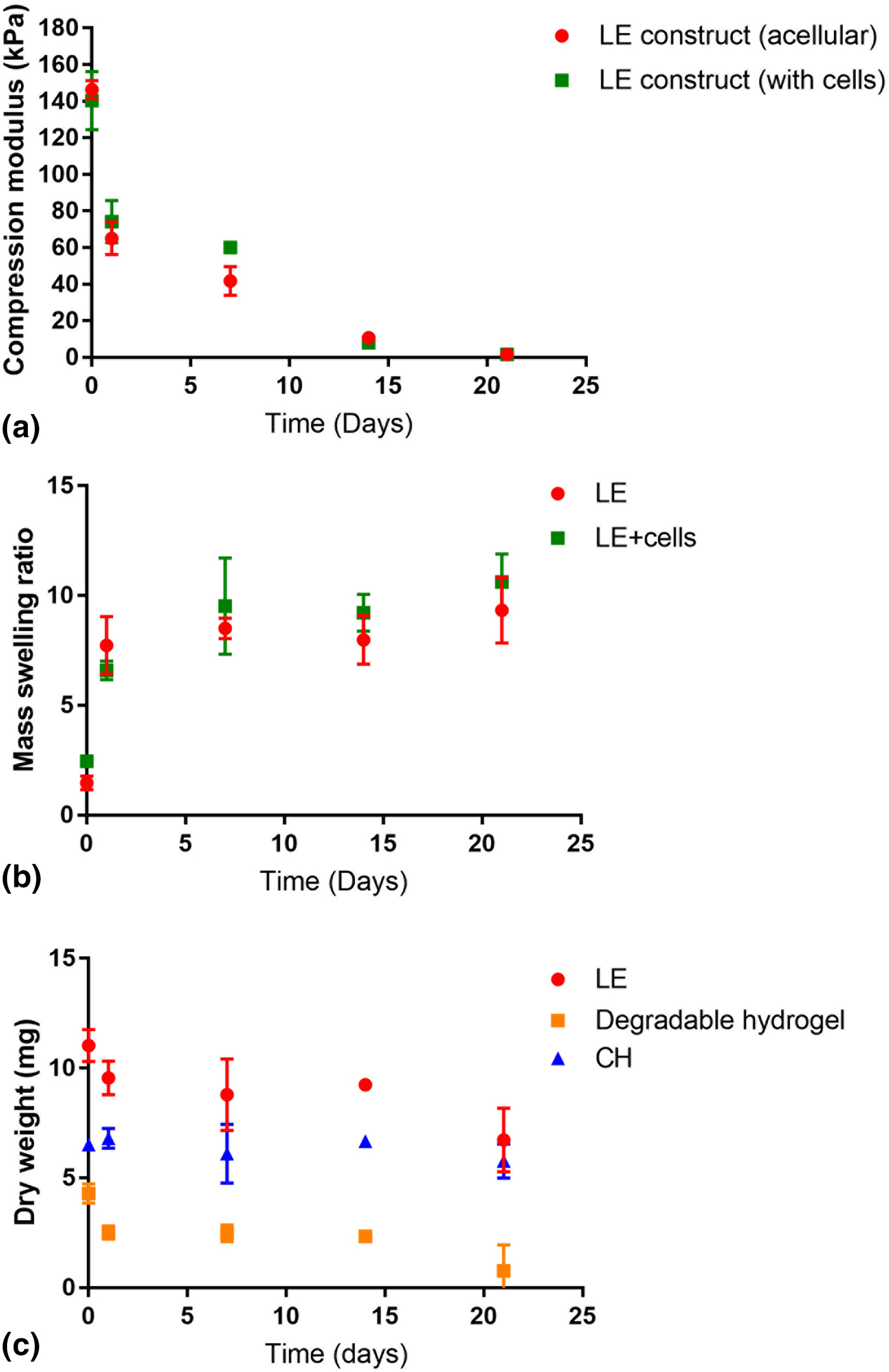
Mechanical compression, shown in Fig. 3(a), was used to understand the role of the coating technologies on the bulk modulus of the electrode. It is desirable to dampen the mechanical mismatch between the underlying metallic electrode and the target tissue.[Reference Aregueta-Robles, Woolley, Poole-Warren, Lovell and Green7, Reference Kozai, Catt, Li, Gugel, Olafsson, Vazquez and Cui29] While it is known from prior studies that CHs can significantly reduce metal electrode modulus (from >100 GPa to approximately 2 MPa), the presence of the CP within these materials prevents matching to the tissue modulus.[Reference Green, Hassarati, Goding, Baek, Lovell and Martens16] The use of the additional cell laden hydrogel layer in these studies revealed that the addition of the biosynthetic PVA produced a construct with a dynamic modulus. Initially, the construct had a relatively high modulus of 140 kPa, well above literature values for neural tissue.[Reference Aregueta-Robles, Lim, Martens, Lovell, Poole-Warren and Green22, Reference Lippert, Rang and Grimm30] However, this rapidly halved within the first 24 h, which coincided with placement of the construct in a hydrated aqueous environment. During this time, any unreacted polymer product will leave the system, and the hydrogel components will soften as they swell to incorporate water.[Reference Goding, Gilmour, Martens, Poole-Warren and Green18, Reference Guo, Finne-Wistrand and Albertsson31] By day 14, the electrode constructs had a modulus below 10 kPa with no significant difference between samples incorporating cells and controls produced without cells. At this stage, the construct had a compression modulus within the range of literature values reported for cortical tissues, 1–10 kPa.[Reference Lippert, Rang and Grimm30] At the final time point, the modulus of both sample types was near 1.5 kPa. This progression of modulus loss over time is well aligned with the implant and neural recovery period.[Reference Woolley, Desai, Steckbeck, Patel and Otto3, Reference Gilmour, Woolley, Poole-Warren, Thomson and Green4] It is known that an implant requires a modulus higher than that of the neural tissue to facilitate an accurate and low damage insertion; however, the ongoing presence of stiff materials will impart a frustrated immune response and hence generate scar tissue.[Reference Woolley, Desai, Steckbeck, Patel and Otto3] It should be noted that the living electrode construct is not designed to be implanted on the day it is fabricated. The cells encapsulated within the surface of the device must be placed in an incubator for 24–48 h post-fabrication. This allows any leachant products from fabrication to diffuse out of the system and also enables recovery of the cells from photo or chemical damage that can occur as a result of the encapsulation process.[Reference Lim, Alves, Poole-Warren and Martens20] Therefore, the living electrode will retain enough mechanical integrity for implantation, but will quickly soften and eventually match the surrounding neural tissue, enabling both recovery of the target tissue and development of the encapsulated tissues.
Physico-mechanical properties of living electrode (LE) constructs. (a) Compression modulus of constructs with and without cells incorporated within the upper degradable hydrogel; (b) swelling ratio of construct over 21 days with and without cells; and (c) mass change in electrode over 21 days showing non-degradable and degradable component contributions. All data sets are n = 3 with error bars being 1 SD.
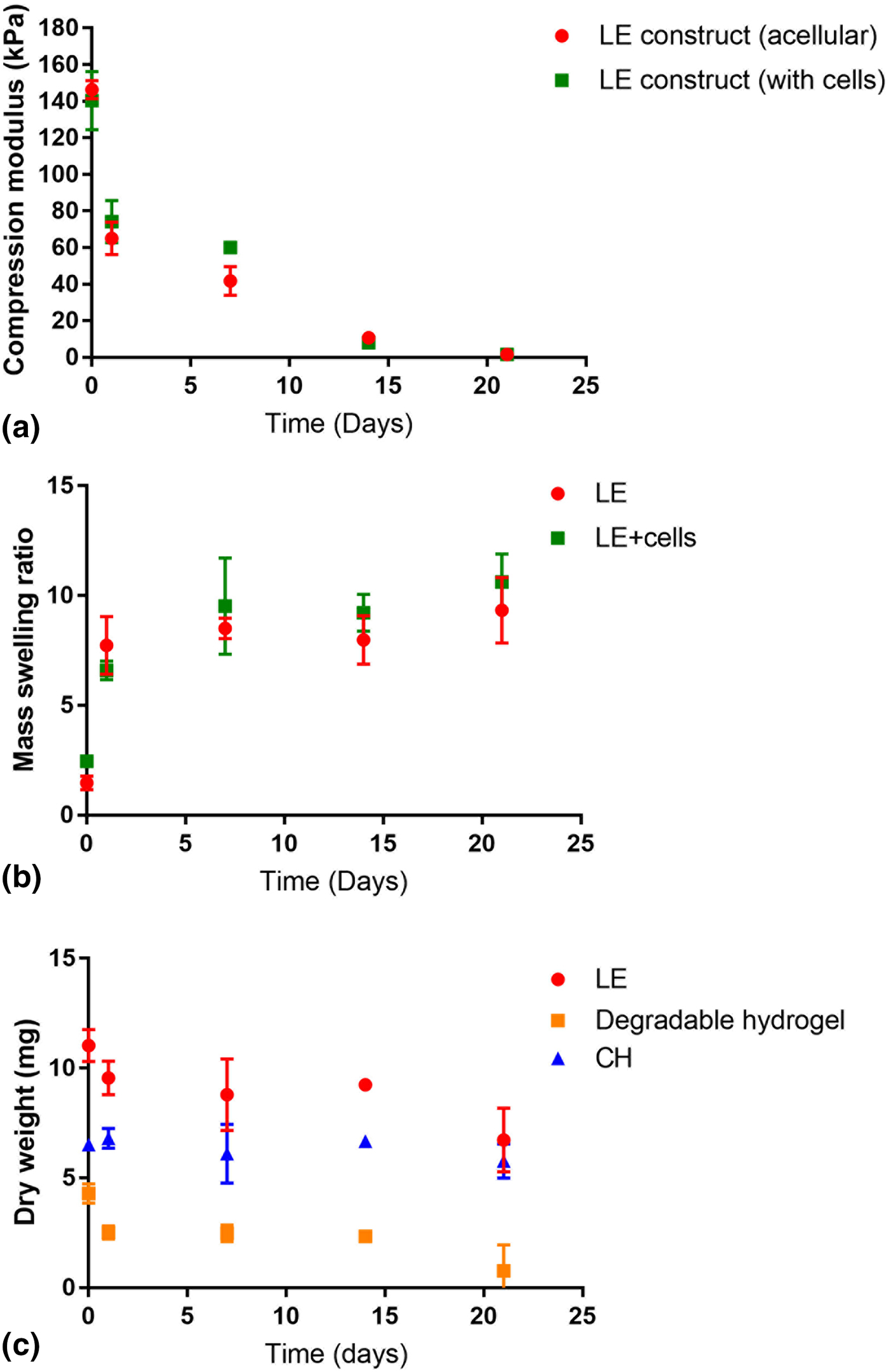
Another important consideration when using hydrogels in bionic implants is the impact of swelling. Prior studies have shown that hydrogel coatings with high swelling ratios can actually push the target tissue further away from the device, creating an inferior connection or total loss of recording capacity, rather than the intended improvement.[Reference De Faveri, Maggiolini, Miele, De Angelis, Cesca, Benfenati and Fadiga32] Physical characterization of the living electrode construct [Figs. 3(b) and 3(c)] demonstrated that the mass of the non-degradable components, being the CH and underlying Pt electrode, hold steady across the 21 days of analysis (at an average of 6.4 mg), while the degradable component undergoes hydrolysis and mass loss. This results in a physical swelling of the construct at the surface. The relative change in degradation and swelling was studied for the living electrode construct both with and without cells. It was shown that initial swelling within the first 24 h imparted a seven times increase in mass, as a result of hydration of the polymer construct. Over the subsequent 20 days, polymer mass was lost from the construct and as a result there was a small increase in swelling ratio to 10. This was substantially lower than swelling ratios for alginate hydrogels applied to cortical electrodes reported in the literature.[Reference De Faveri, Maggiolini, Miele, De Angelis, Cesca, Benfenati and Fadiga32, Reference Kim, Wiler, Anderson, Kipke and Martin33] When comparing the swelling behavior with and without cells, there is no significant difference between these variants. It was hypothesized that as the cells produced ECM, they would slow the degradation of the overlying polymer, reducing swelling behavior. However, this was not the case, and a converse hypothesis was proposed, suggesting that the presence of the cells created pores within the hydrogel enabling increased access for water to hydrolyze the degradable ester bonds.[Reference Lim, Alves, Poole-Warren and Martens20] Ultimately, it is recognized that this is an in vitro analysis of a macroelectrode system that is intended to be translated to microelectrodes and placed in vivo. As such both hypotheses may have relevance, but further development is required to understand the impact of cells on degradation and mechanical properties of living electrodes.
Furthermore, it should be noted that over the first 5 days, the degradable hydrogel dominates the data in both compression and degradation studies. However, as time progresses and the uppermost layer continues to lose material and as a result, the non-degradable components of the living electrode dominate the mass swelling and dry weight measurements. The CH-coated platinum becomes a much greater and unchanging component when compared with the upper degradable hydrogel. In contrast, the degradable surface still dominates the compression measurements from day 5 onwards, as this analysis measures the compression between 5% and 15% strains. At this level of compression, the increasingly softer degradable hydrogel will dominate the measurement. In summary, these studies have provided evidence that the components chosen for living electrodes have mechanical and physical properties that are suited to the application.
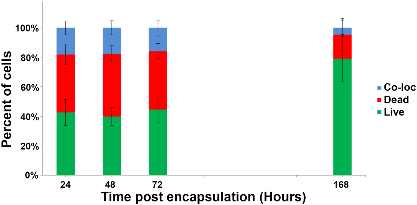
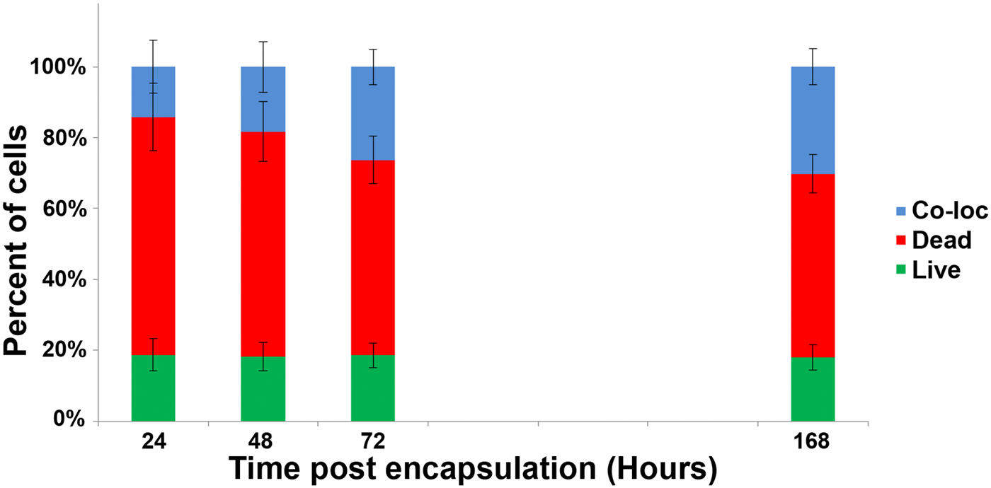
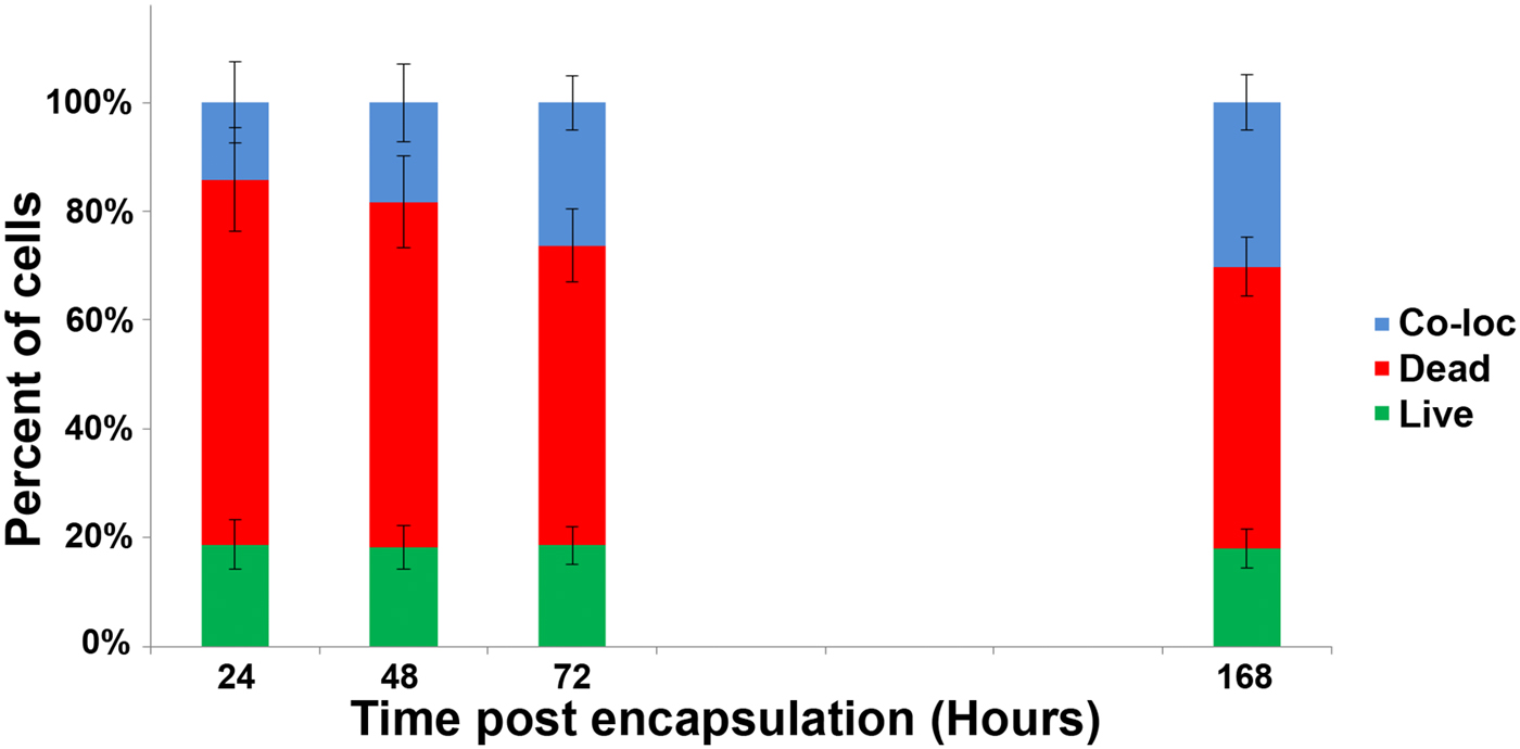
To assess cell survival, the living electrodes were cultured for 7 days. Figure 4 shows quantification of survival of glial cells alone and Fig. 5 shows survival of a combination of neural progenitor cells and glial cells encapsulated within the electrode construct. It is expected that both cell types are required to produce a successful living electrode, with the neuroprogenitors needing the glial support cells to promote successful differentiation into electroexcitable phenotypes.[Reference Aregueta-Robles, Woolley, Poole-Warren, Lovell and Green7, Reference Sorensen, Moffat, Thomson and Barnett27, Reference De Faveri, Maggiolini, Miele, De Angelis, Cesca, Benfenati and Fadiga32] Immediately following fabrication, the combined glial and neural progenitor cell population was found to have low viability (20%), which did not substantially recover over the 7-day growth period. This low viability will impact on degradation as cells that do not recover ultimately fragment (or in the body will be phagocytized) and leave pores behind. It was thought that the low viability was largely related to the neuroprogenitor cells being encapsulated immediately after removal from the tissue source, and hence cells that were disrupted or damaged as a result of the harvesting process were encapsulated along with the viable cells. This could result in a toxic environment where cell debris and apoptotic cells then produce an environment in which the undamaged cells cannot survive. In contrast, the glial cells that were first plated onto tissue culture plastic, and then encapsulated after expansion had a higher viability of 42% immediately after encapsulation and recovered under culture conditions until viability reached over 80% at 7 days. By initially plating the glial cells, the dead cells and debris resulting from the harvest were removed from the cell population, supporting the significant difference in cell survival between encapsulated cell populations. Prior studies in the literature have used clonal cell lines, which do not suffer from the impact of tissue harvesting. As a result, these prior studies have shown higher viability immediately following encapsulation.[Reference Green, Lim, Henderson, Hassarati, Martens, Lovell and Poole-Warren19, Reference Aregueta-Robles, Lim, Martens, Lovell, Poole-Warren and Green22] However, at 7 days post-encapsulation, the viability of the primary glial cells was comparable to that of the clonal cell studies. As a result, to improve the viability of the neuroprogenitor cells in future studies, harvesting and encapsulation time lines will be modified. This will enable the dead cells to be removed and encourage the neuronal population to form a more physiologically relevant structure.
Fraction of glial cells which are either live, dead, or stained with both propidium iodide and Calcein-AM, indicating viable cells with damaged membranes (referred to as co-loc). Error bars ±1 SD.
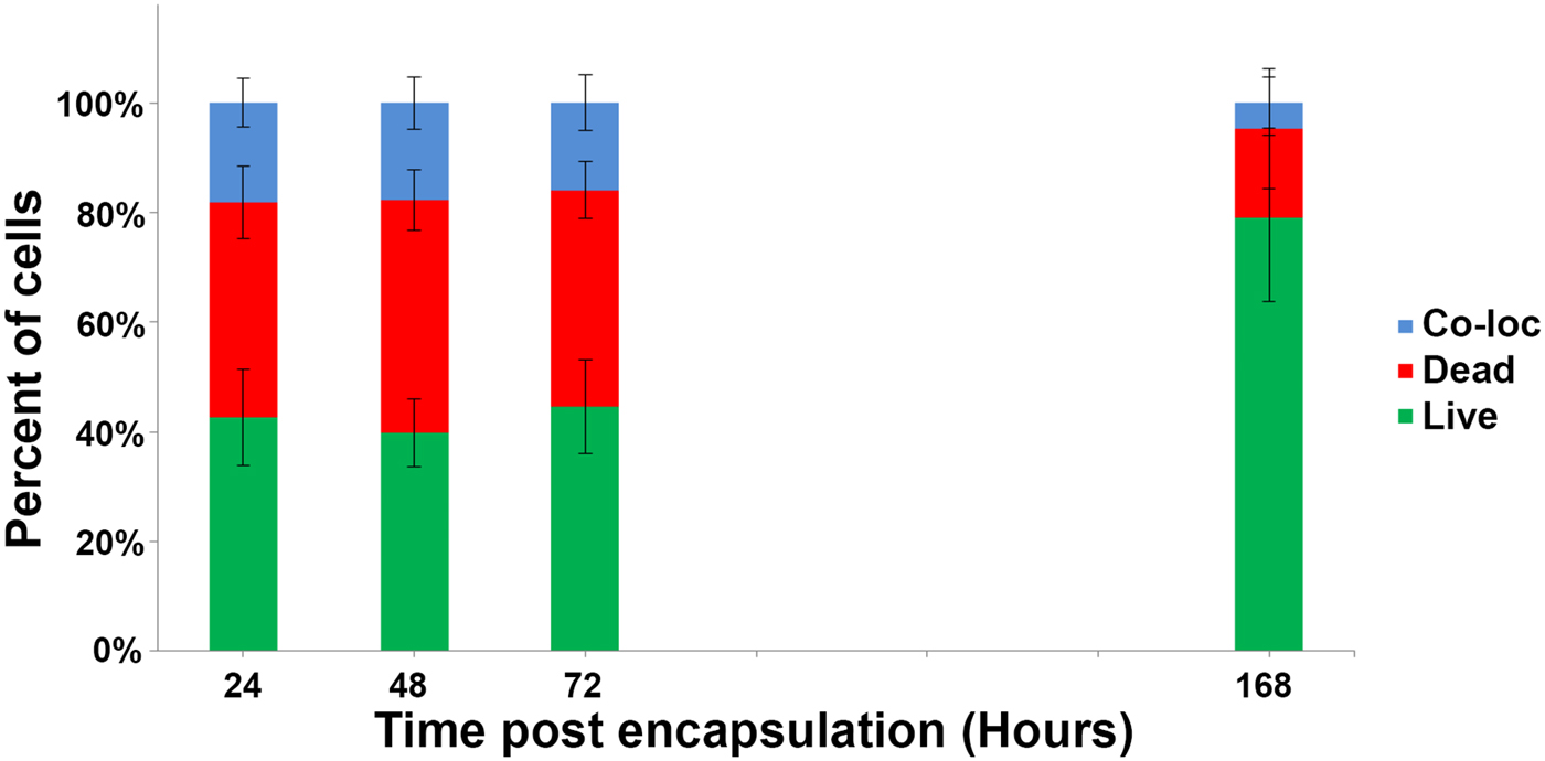
Fraction of neural precursor cells with supporting glia, which are either live, dead, or stained with both propidium iodide and Calcein-AM, indicating viable cells with damaged membranes (referred to as co-loc). Error bars ±1 SD.
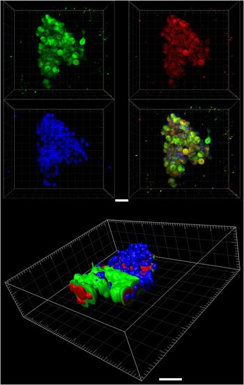
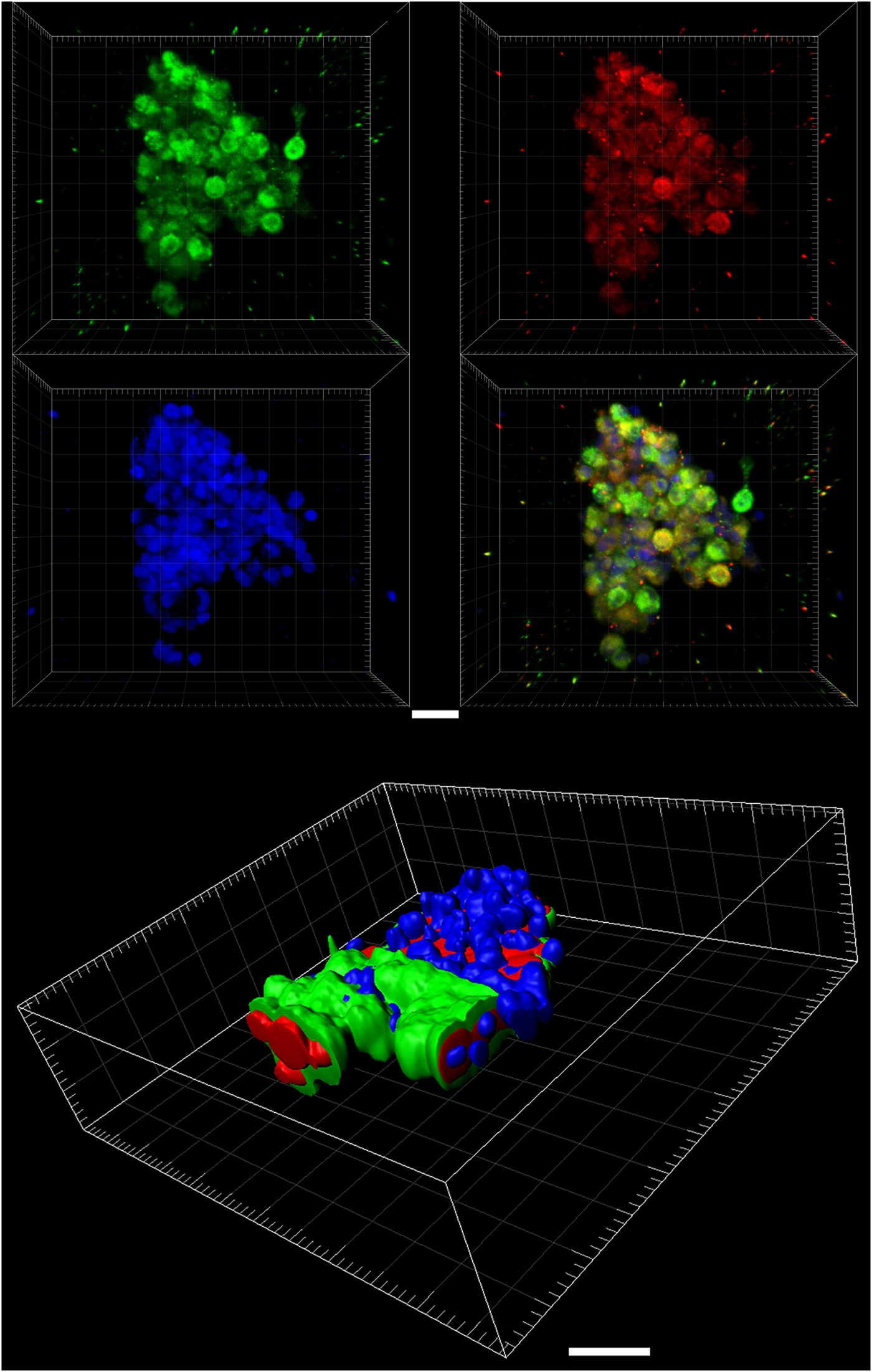
An additional cell study was used to assess the production of the ECM molecules laminin and collagen within the upper hydrogel layer, as shown in Fig. 6. Surface projections of the fluorescent antibody tags were used to assess the location of the collagen and laminin within the hydrogels and cell clumps. Imaging of the hydrogel depth revealed that cells are uniformly distributed but tend to clump together as the hydrogel degrades. Laminin was the dominant protein expressed within the 3D hydrogel volume, and while collagen was produced, it was presented at the cell surface only. This may provide some indication as to why degradation rate and compression modulus is unchanged when cells are added to the construct. While the laminin was secreted, it remains within the immediate vicinity of the cells and is not deposited throughout the hydrogel volume. The collagen, widely known as a structural support protein, is closely associated with the cell membranes, and as a result does not interact with or support the bulk hydrogel. It is likely that the collagen is providing support within the cell aggregations. A significant challenge in future work is to drive development of neural processes beyond these cell aggregations and throughout the supporting hydrogel matrix.
ECM production by glia and neuroprogenitors within the living electrode construct at 14 days post-encapsulation, showing laminin (green), collagen (red), and cell nuclei (blue). Three-dimensional surface reconstruction of the merged channels (bottom) demonstrated that the majority of the collagen (red) was expressed closer to the nuclear envelope (blue) compared with laminin (green). Scale bar = 30 µm.
The living electrode design seeks to provide communication, such as recording or stimulation, through formation of functional synapses between devices and cells. This approach has the potential to provide a safer mechanism for neural cell stimulation, which is more compatible with neural cell survival and regeneration, while mitigating scar tissue reactions. New cell harvest and encapsulation protocols are required to improve cell survival. It is also necessary to understand the translation of this construct from the macroelectrode to the clinically relevant microelectrode scale and into the living neural tissue environment. A self-supporting living electrode will incorporate cues for differentiation of encapsulated cells and drive development of functional, targeted synapses. Studies that incorporate neurophysiological analysis will be necessary to bring a greater understanding of this technology to the field.
Conclusions
A living electrode construct was developed and characterized across a range of properties relevant to performance in an implantable electrode. Macroelectrode constructs were used to understand the impact of a degradable and cell-loaded hydrogel produced as a laminar structure on electrode performance. Electrical, mechanical, and physical metrics were used to demonstrate that the electrical properties were not diminished by the additional non-conductive layer and that the entire construct was significantly softened by the presence of the hydrogel. As the cell-loaded hydrogel degraded, the modulus of the construct was reduced to a range matching that of neural tissue. Glial cells were found to have high viability within the construct, but harvesting techniques impacted survival of the neural progenitor cells. It was confirmed that ECM proteins were produced by the cells within the living electrode constructs. Future work will focus on techniques to improve neural progenitor viability and quantification of the ECM formation relative to hydrogel degradation. These approaches will then be adapted to microelectrode formats for in vivo proof-of-concept studies.
Acknowledgments
The research reported here was supported by the Defense Advanced Research Projects Agency (DARPA) under contract number HR0011-16-C-0032.