INTRODUCTION
Carcinoma of cervix is the commonest malignancy in India in women yielding an incidence of 19.4–43.5 per 100,000.Reference Gault and Asirvadharam1–Reference Vizcaino, Moreno, Bosch, Muñoz, Barros-Dios and Parkin3 In India, most patients present in advanced stages and the prognosis is directly related to the stage at presentation. The important prognostic factors of carcinoma of cervix includes tumour stage according to the International Federation of Gynecology and Obstetrics (FIGO) classification, initial tumour volume, tumour extent within the vagina, histological grading and lymphatic involvement.Reference Davis, Stanhope, Garton, Atkinson and O'Brien4,Reference Kirkbride, Fyles, Rawlings, Manchul, Levin, Murphy and Simm5 To improve the local control in the treatment of advanced stage of carcinoma of cervix, many therapeutic modalities have been used.Reference Davis, Stanhope, Garton, Atkinson and O'Brien4,Reference Kucera, Langer and Smekal6 Among them, irradiation is regarded to be the standard treatment for all tumour stages,Reference Davis, Stanhope, Garton, Atkinson and O'Brien4, Reference Kucera and Vavra7–Reference Nanavati, Fanning, Hilgers, Hallstrom and Crawford9 which includes external beam radiation therapy (EBRT) and brachytherapy (BT), or a combination of the two. BT is used primarily in the cases of early tumour stages, whereas a combination of EBRT and BT is used mainly for advanced stages of the tumour.Reference Davis, Stanhope, Garton, Atkinson and O'Brien4 It is well documented in the literature that there is a positive relationship between the total dose delivered to the tumour and the local tumour control.Reference Perez, Camel, Galakatos, Grigsby, Kuske, Buchsbaum and Hederman10,Reference Fine, Piver, McAuley and Driscoll11 At the same time, the complication rate also has a positive correlation with dose received by surrounding normal tissue/critical organ.Reference Kehwar12–Reference Kehwar and Akber15 Inadequate dose delivery to the treated volume is frequently identified as a possible cause for local failure.Reference Leung and Sexton8 Numerous articles discuss the use of low-dose-rate (LDR) BT, but very few studies have been done for high-dose-rate (HDR) BT techniques for carcinoma of cervix.Reference Davis, Stanhope, Garton, Atkinson and O'Brien4, Reference Nanavati, Fanning, Hilgers, Hallstrom and Crawford9, Reference Schafer, Micke and Prott16, Reference Kucera, Mock, Knocke, Kucera and Pötter17
At M. D. Oswal Cancer Treatment & Research Foundation, Ludhiana (Pb), India, carcinoma of cervix is traditionally treated by primary LDR BT using Selectron–LDR remote controlled after loading BT unit with Cs-137 sources with or without EBRT. Introduction of the Microselectron–HDR remote controlled after loading BT unit in May 2004, in the Department of Radiation Oncology, offered new dimensions in the BT treatment with an advantage of dwell time optimisation.
Very few cancer centres, in India, have advanced three-dimensional (3D) treatment planning systems and computed tomography (CT) simulators while most of the centres have only two-dimensional (2D) planning systems and conventional simulators and treat HDR patients using traditional methods.
In this study, 2D treatment planning system and orthogonal films were used to evaluate the International Commission on Radiation Units and Measurements report-38 (ICRU-38) recommendations18 and were used to define reference points of bladder and rectum, and different points of lymphatic trapezoid on orthogonal radiographs for the patients treated with HDR intracavitory BT (HDR ICBT) and EBRT, and the ICRU reference treatment volume, for each patient, was determined using ICRU-38 definition18 with respect to point A dose, defined per Manchester dosimetry system.Reference Peterson and Parker19,Reference Pérez20 The calculated reference volume was subsequently used to calculate tumour control probability (TCP). The doses and biologically effective dose (BED) values were determined at different points to correlate complication rates and tumour control.
METHODS AND MATERIALS
Radiobiological model
Using basic expressions of the linear quadratic (LQ) equation,14,15,21–25 the cumulative BED at any reference point for combined treatment of EBRT and HDR ICBT can be written by

where BEDEBRT = DEBRT [1 + dEBRT /(α/β)], BEDHDR = DHDR [1 + dHDR /(α/β)], and PCF = –[{0.693/(αTp)}(T + G – Tk)] = proliferation correction factor. In proliferation correction factor, the α, Tp, Tk, T and G are the coefficient of lethal damage in LQ equation, potential doubling time of proliferating tumour cells, kick off time in proliferation after starting irradiation, treatment time in days and any gap in days between two modalities of radiotherapy or between any one therapy, respectively.
The TCPReference Kehwar, Akber and Passi14,Reference Kehwar and Akber15 for ICRU reference volume was calculated using following equation

where ρ is the clonogenic cell density of tumour cells, V is the ICRU reference volume of HDR ICBT and CBED is the cumulative biologically effective dose at point A of the patient.
The values of α/β for acute and late complications were taken 10 Gy and 3 Gy, respectively.Reference Fowler22 For TCP calculations, the values of clonogenic cell density of ρ = 10Reference Leung and Sexton8 (Brenner et al.Reference Brenner, Geard and Hall26), α/β= 10 Gy (FowlerReference Fowler22), α = 0.35 Gy-1 (FowlerReference Fowler22), Tp = 6.6 daysReference Tsang, Fyles, Kirkbride, Levin, Manchul, Milosevic, Rawlings, Banerjee, Pintilie and Wilson27 and Tk = 28 days (FowlerReference Fowler22) were used.
The CBED for bladder and rectum points were calculated using the following equation

where n = the number of fractions of EBRT treatment, d = EBRT fraction size (Gy), i is the i th fraction of HDR ICBT and i = 1, 2, 3 and r = bladder/rectal dose (Gy) for each insertion.
Patients
Forty-nine patients have been treated with EBRT and HDR ICBT treatment for carcinoma of cervix between September 2006 and December 2007 were included in this study. At the time of initial diagnosis, the median age of the patients was 50 years (range 30–75 years). The pre-treatment tumour stage was determined clinically by physical examination and classified according to the FIGO classification.Reference Benedet, Bender, Jones, Ngan and Pecorelli28 In all cases, the histological diagnosis was obtained by biopsy, or partial tumour excision. Clinical manifestation of distant metastasis and lymph node status were evaluated for each patient. Patients with carcinoma of cervix, who had not undergone surgery, were included in the study. All the patients had received chemotherapy as part of the treatment, as a common factor. The BT planning, for all patients, was undertaken by obtaining orthogonal radiographs using the conventional simulator available in the department. The bladder and rectum complications were recorded weekly during treatment and at 1-month intervals after treatment during first year. Thereafter, it was done every alternate month in second year and then six monthly or whenever the patient had any complaint. The complications which appeared during and within 6 months of starting radiotherapy were taken as acute/early complications and thereafter late complications.
Radiation therapy
Radiation treatment consisted of EBRT with weekly gemcitabine followed by HDR ICBT. Initially, the EBRT was delivered to whole pelvis of the patient by Co–60 teletherapy unit when the anterior and posterior parallel opposed fields or box field technique when anterior–posterior (AP) separation was more than 20 cm. After an EBRT dose of 45 Gy/25 fractions delivered in 5 weeks, the HDR ICBT was performed using an Ir–192 Microselectron–HDR remote after loading unit at 1-week intervals. Isoeffective HDR ICBT doses for two and three fractions were calculated using Equation (1) and above-mentioned parameters of the LQ model. The patients were randomly divided into two groups. Group 1 consisted of patients who received two fractions of HDR ICBT with 9.5 Gy per fraction and Group 2 consisted of patients who received three fractions of HDR ICBT with 7.5 Gy per fraction to point A, respectively. Planned total dose to Point A (EBRT + HDR ICBT) was 64 Gy and 67.5 Gy, in Groups 1 and 2, respectively. Corresponding planned BED10 with proliferation correction for EBRT + HDR ICBT, for Group 1 and Group 2, were 84.45 Gy and 84.67 Gy, respectively.
The HDR ICBT doses were delivered using a Nucletron applicator consisting of a central tandem and two ovoids (colpostats). The length of the central tandem was varied from 4 to 6 cm and the diameters of the ovoids from 1.5 to 2.5 cm, based on the patient’s individual anatomy. During each insertion, the posterior and anterior of the vagina were packed with radio-opaque gauze to reduce bladder and rectal doses and to improve visualisation of the posterior vaginal septum. A rectal marker was inserted into the patient’s rectum to visualise the rectum. The rectal marker was made of 1.0 cm diameter plastic tube in which light weight radio-opaque balls of 1.0 cm diameter were inserted at 1.0 cm apart. After implantation of the applicator and rectal marker, orthogonal films were taken using conventional simulator. These films were used to define point A and point B, the bladder and the rectal points (including ICRU reference points) and the lymphatic trapezoid points. The Point A was defined as 2 cm cephalad and 2 cm lateral to the cervical orifice, along the plane of the tandem.Reference Pérez20 Five bladder points were defined on the balloon. On AP radiograph, these points were as follows: point ‘1’ at the centre of the balloon, point ‘2’ at the superior surface, point ‘3’ at the left surface, point ‘4’ at the inferior surface and point ‘5’ at the right surface of the balloon. On the lateral radiograph, point ‘1’ at the posterior surface, point ‘2’ at the superior surface, points 3 and 5 at the centre of the balloon and point ‘4’ at the inferior surface of the balloon. The ICRU rectal reference point was determined according to the guidelines given in ICRU-38.18 Along with ICRU rectal reference point, four more points were also defined on anterior wall of the rectum. Of these points, two points lie superior and two inferior to the ICRU rectal reference point, and have 1.0 cm separation to each other. Similarly, lymphatic trapezoid points were defined using ICRU recommendations on both radiographs.
In the source positioning within the central tandem and ovoids, recommendations of the Manchester systemReference Peterson and Parker19,Reference Pérez20 were adopted to simulate the dose distribution with LDR BT. The active treatment length of the tandem was depended with related on the individual sounding of the uterine cavity. The distance between each source dwell positions on tandem was 0.5–1.0 cm. The standard dose weighting for tandem was approximately two-third and that for ovoids was one-third. Therefore, the duration of source dwelling through tandem occupied nearly two-third of the total dwell time.
Dosimetry
For the patients treated with two-field techniques, the EBRT dose was calculated at midplane, whereas the dose for box field technique was calculated at the cross point of the fields. In all the cases, the superior border of the field was at the junction of fourth lumbar vertebrae (L4) and fifth lumbar vertebrae (L5) body. The HDR ICBT planning was done using two orthogonal films obtained just after each insertion, as mentioned in earlier section. The HDR ICBT isodose curves were reviewed by two physicians to ensure that the residual tumour lie well within the prescribed dose area. The HDR ICBT doses to Point A and point B, the ICRU bladder and rectal points (including ICRU reference points) and different points of lymphatic trapezoid were calculated using the Plato treatment planning system (Nucletron Plato System, Version 2, The Netherlands). To obtain the total doses from a combination of EBRT and HDR ICBT, it was assumed that there is a homogeneous dose distribution from EBRT.
Analysis of tumour control
Pelvic examination of all patients was performed under general anesthesia, just before the time of the applicator insertion, before starting first BT session. Tumour response to EBRT was recorded on a subjective basis as follows:
1. No Gross Residual Tumour (NGRT) response: complete or nearly complete regression of the pelvic tumour, non-specific fibrosis or granulation in the cervix.
2. Gross Residual Tumour (GRT) response: gross tumour or palpable nodularity in the cervix, and/or palpable induration of the parametrium.
Post treatment (EBRT + HDR ICBT) failure (control) rates were classified as local (within the BT reference volume), loco-regional (including all types of recurrent tumour manifestations within the pelvis) and distant failure. The potential correlation between applied total dose and the development of loco-regional relapses or with local control was evaluated. This was done by estimating the total dose at different reference points and corresponding BED and TCP values.
Complications
Bladder and rectal complications, and non-rectal gastrointestinal sequelae (small bowel complications) were scored according to the late effects in normal tissues subjective, objective, management and analytic (LENT SOMA) grading scale.
Statistics
Patient failure (control) was measured from the date of the initiation of radiation therapy to the date of the last follow-up examination. The failure (control) rates were determined using the Kaplan–Meier survival method. The statistical significance between the failure (tumour control), complication rates and the factors affecting treatment response was calculated by the Student’s t-test and Chi-square test.
RESULTS
Patient characteristics
According to the FIGO classification, four patients (8.16%) had tumours of stage II, where as stage IIB was observed in 26 (53.06%) cases, stage IIIB in 19 (38.77%) patients, 47 (95.92%) patients had histologically proven squamous cell carcinoma, whereas two (4.08%) patients had adenocarcinoma. The histological distribution of the subtypes was as follows: 5 patients showed a well-differentiated, 13 moderately differentiated and 11 poorly differentiated carcinoma of the uterine cervix. Another 18 cases had no classification assigned to their lesions.
Applied mean total dose at the different reference points
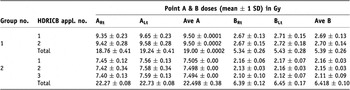
Table 1 includes the doses at point A and point B for each HDR fraction and entire HDR course. In the patients of Group-1, the mean dose per fraction at point A (points ARt and ALt) varied from 8.91 to 10.09 Gy (9.50 ± 2.78 Gy), and at point B (points BRt and BLt) from 2.28 to 3.02 Gy (2.69 ± 1.52 Gy). In entire course of HDR ICBT, the mean total dose ranged from 18.05 to 19.95 Gy (19.00 ± 0.47 Gy) at point A, and 4.61 to 5.82 Gy (5.39 ± 0.27 Gy) at point B. In the patients of Group-2, the mean dose per fraction at point A (points ARt and ALt) varied between 6.88 and 8.11 Gy (7.50 ± 2.12 Gy), and at point B (points BRt and BLt) ranged from 1.83 to 2.43 Gy (2.14 ± 0.91 Gy). The mean total dose in the course of HDR ICBT ranged from 21.36 to 23.64 Gy (22.50 ± 0.41 Gy) at point A, and 5.93 to 6.88 Gy (6.42 ± 0.20 Gy) at point B.
Point A and Point B doses in Gy

The CBED10 values at point A for tumour response, without proliferation correction, were 90.15 ± 0.0006 Gy and 92.47 ± 0.094 Gy, for Group-1 and Group-2 patients, respectively, and with proliferation correction were between 72.15 and 80.55 Gy (76.59 ± 2.31 Gy) and 72.38 and 78.98 Gy (76.41 ± 2.15 Gy) for Group-1 and Group-2 patients, respectively. The CBED10 (with proliferation correction) and CBED3 (without proliferation correction) values at point B were in the range from 42.26 to 50.59 Gy (46.38 ± 2.26 Gy) and from 80.17 to 83.33 Gy (82.23 ± 0.721Gy), respectively, for Group-1, and from 40.64 to 47.82 Gy (45.03 ± 2.11 Gy) and from 82.17 to 84.02 Gy (82.89 ± 0.44 Gy), respectively, for Group-2.
Table 2 includes the doses per fraction, total doses for the courses, BED10 and BED3 for the courses at different points of lymphatic trapezoid. Tables 3 and 4 listed the same, as in Table 2, for bladder and rectum points including ICRU reference points. Tables 2–4 include the data for HDR ICBT applications only, and in the calculations of BED10 no proliferation correction has been taken into account.
Doses in Gy at different points of lymphatic trapezoid and BED10 and BED3 at these points for total doses in respective groups
Doses in Gy at different bladder points and respective BED10 and BED3 at these points for total doses in respective groups
Doses in Gy at different rectal points and respective BED10 and BED3 at these points for total doses in respective groups
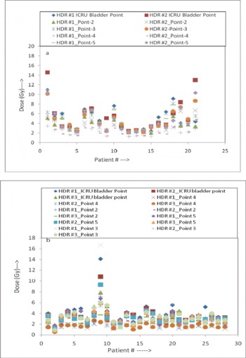
Figure 1a, b shows the variation in dose at different point of bladder and rectum in different HDR ICBT applications.
Variation in the doses at different bladder points with respect to the application in (a) Group-1, and (b) Group-2, patients.

Intracavitory BT treatment volume and TCP
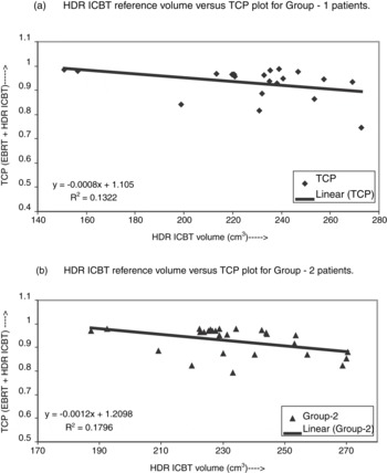
The HDR ICBT reference volume, determined according to the ICRU-38 definition with respect to point A dose, varied from 149.16 to 340.31 cm3 (233.47 ± 27.30 cm3) and from 144.84 to 281.02 cm3 (227.83 ± 32.35 cm3) in Group-1 and Group-2 patients, respectively. The TCP values for average volume, for each patient, were calculated for the total reference dose (EBRT plus HDR ICBT dose) at point A. The clonogenic cell density was taken as 10% of the total cells per cm3 (109 cells/cm3). The mean TCP values calculated without cell proliferation correction factor were 100 ± 0.01% and 100 ± 0.002% in Group-1 and Group-2 patients, respectively whereas with proliferation correction factor, it varied between 74.51–98.65% (93.0 ± 6.4%) and 79.26–97.82% (92.75 ± 5.75%), respectively, in Group-1 and Group-2 patients, respectively, and are shown in Figure 2a,b. Figure 2a,b is the plot between ICRU reference volume and TCP for this volume for combined dose of HDR ICBT and EBRT at point A. The slope, intercept on Y axis, and R2, of the best fit regression lines obtained using the least square fit, –0.0008, 1.105, and 0.1322, respectively, for Group-1, and –0.0012, 1.2098, and 0.1796, respectively, for Group-2. The results of an unpaired Student’s t-test revealed that there was no statistically significant difference between the TCP values of two groups (p = 0.87).
Plot between HDR ICBT reference volumes calculated with respect to the point A dose and corresponding TCP for EBRT + HDR ICBT dose and point A in (a) Group-1, and (b) Group-2, patients. EBRT, external beam radiation therapy; HDR ICBT, high-dose-rate intracavitory brachytherapy; TCP, tumour control probability.

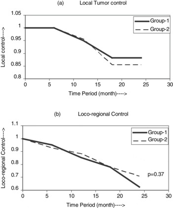
The local and loco-regional control calculated using the Kaplan–Meier survival method for the period of 2 years are shown in Figure 3a,b. The local control at 2 years were found to be 88.21 and 85.58% for Group-1 and Group-2 patients (p = 0.32), respectively, which is well within the limits of the calculated values of TCP whereas the loco regional control at 2 years were 62.49 and 70.94% for Group-1 and Group-2 patients (p = 0.37), respectively. As per the Chi-square distribution, there was no statistically significant difference between local control and loco-regional control for both the groups (p > 0.05).
Plots of (a) local tumour control, and (b) loco-regional control, in Group-1 and Group-2 patients for the period of 2 years.

In this work, the calculated TCP for each patient and clinical tumour control calculated for 2 years at a fixed time interval of 6 months, compared with unpaired Student’s t-test and found that there were no statistical differences between calculated TCP and clinical local control (p = 0.70 for Group-1 and p = 0.83 for Group-2).
Complications
Early and late complications in the bladder, rectum and small bowel were recorded as mild (G1), moderate (G2) and severe (G3 and G4). In Group-1 patients, the overall early treatment-related complications of 61.90% (23.81% of G1, 19.05% of G2 and 19.05% of G3), 90.48% (33.33% of G1, 33.33% of G2 and 19.05% of G3) and 85.71% (47.62% of G1, 28.57% of G2 and 9.52% of G3) were occurred in bladder, rectum and small bowel, respectively whereas in Group-2 the complications were observed in 46.43% (10.71% of G1, 21.43% of G2 and 14.29% of G3 and G4), 46.43% (21.43% of G1, 21.43% of G2 and 3.57% of G3), and 64.29% (32.14% of G1, 21.43% of G2 and 10.71% of G3) patients in bladder, rectum and small bowel, respectively. There were no late complications observed in the bladder and small bowel, and 10.71% (mild) in rectum in Group-1 patients whereas in Group-2 no complications in bladder, 7.14% (3.57% mild and 3.57% moderate) in rectum and 7.14% (3.57% moderate and 3.57% severe) in small bowel were observed during a 2-year period. Student’s t-test statistical analysis reveals that there were no statistical differences in early/late complication rates of the bladder, rectum and small bowel in two groups (p > 0.05).
DISCUSSION AND CONCLUSION
A combination of EBRT to whole pelvis and intracavitory BT [intracoronary radiation therapy (ICRT)] has been considered to be an effective treatment. Several published reportsReference Brenner, Huang and Hall29–Reference Okawa, Sakata and Kita-Okawa33 demonstrate that a combination of EBRT and HDR BT provides comparable efficacy to that of EBRT and LDR BT.Reference Pérez20, Reference Brenner, Huang and Hall29–Reference Selke, Roman, Souhami, Freeman, Clark, Evans, Pla and Podgorsak35 However, the main concern with HDR treatment is to use optimal dose fractionation scheme. Many dose fractionation schemes have been suggested in the literature.Reference Petereit and Pearcey36,Reference Nag, Erickson, Thomadsen, Orton, Demanes and Petereit37 In the cancer centres of developing countries, such as in India, fewer fractionation schemes are more feasible and favourable due to economic reasons. Hence, in M. D. Oswal Cancer Treatment & Research Foundation, we studied two fractionation scheme of 9.5 Gy × 2 fractions and 7.5 Gy × 3 fractions to evaluate their equivalency in the patients of Northern part of India and to get future directions.
Our study has limits in several aspects, such as, a relatively small sample of patients, all the patients received chemotherapy as a common factor which might have interfered in the analysis of the different parameters on late toxicity rates. The volume analysis suggested by ICRU-38 was performed using orthogonal 2D planning with respect to point A doses, EBRT planning is not done with 3D planning system so the assumption of homogeneous dose within the treated volume of EBRT, etc.
The statistical analysis of overall early and late complications reveals that there was no statistically significant difference (p > 0.05) in the incident of early and late complications, in both the groups. When bladder and rectal complications were analysed with the absorbed dose, and corresponding BED10 and BED3, at different points (including ICRU reference points) defined as above-mentioned in foregoing section, and found that the complications have significant correlation with total dose and BED values at the points of bladder and rectum which has received higher doses. No correlation was found between the doses and BED at different point of lymphatic trapezoid and small bowel complications.
It is clear from Figure 2a,b that the points in these figures are fairly close to the best fit regression lines hence can be fairly described with the straight lines. Similarly with the clinical data, it is clear that the local tumour control significantly correlates with ICRU reference volumes, whereas no correlation was found between loco-regional control and ICRU reference volume. The patients who recurred outside the reference volume but within the EBRT-treated volume, in both the groups, have their reference volumes closer to or smaller than the mean reference volume, whereas the patients with larger reference volume had showed up with no recurrence during a 2-year follow-up. It has also been demonstrated that there was a significant difference in reference volumes of any two consecutive fractions of the same patient. This indicates that either applicator positioning was not reproduced for following fractions or there might be difficulty in applicator insertion to get positional reproducibility.
Figure 3a,b reveals that the local control/loco-regional control in both the groups are statistically indifferent, and can be considered isoeffective.
Per applicator variability in the position relative to the first insertion, the calculation of the cumulated dose to points A, B (HDR ICB only) changed (Table 1). This variability in the applicator position changed the doses at different points of lymphatic trapezoid (Table 2), and at the points of bladder (Table 3) and rectum (Table 4). In most of the cases, the bladder and rectum points were not at the same place as were in the first insertion. Figure 1a,b shows the magnitude of dose variation at different bladder points in HDR insertions in Group-1 and Group-2, respectively. In some cases, this variation was very high and cannot be ignored. It is also seen in these figures that ICRU reference point does not necessarily have maximum dose which can be correlated with complications. Hence, BED at reference points did not significantly correlate with bladder and rectal complications, whereas it has positive correlation with total dose and BED at the point which received higher doses. Because of the change in the position of the applicator, the corresponding values of BED had changed. Because TCP is a function of dose at point A and corresponding reference volume, hence have changed accordingly. But in calculations, we have taken an average value of reference volume. As a whole, the clinical local control for both groups falls well within the limits of the calculated values for the parameters of LQ model used in this study.
Several researchers have estimated the rectal dose by calculation at a single or multiple points using either the definitions of rectal reference point according to ICRU-38 reportReference Perez, Fox, Lockett, Grigsby, Camel, Galakatos, Kao and Williamson38–Reference Van Lancker and Storme40 or the barium contrast method.Reference Orton41–Reference Stryker, Bartholomew, Velkley, Cunningham, Mortel, Craycraft and Shafer43 Stryker et al.Reference Stryker, Bartholomew, Velkley, Cunningham, Mortel, Craycraft and Shafer43 and Clark et al.Reference Clark, Souhami, Roman, Evans and Pla44 have found a significant correlation between ICRU rectal point dose and the incidence of late rectal complications. However, in our study, the calculated ICRU rectal reference point dose and BED do not correlate with complications. The same pattern is followed for ICRU bladder reference point, which is supported by the results of other investigators.Reference Van Lancker and Storme45
When we examined the literature to compare our results in terms of local control and complications, studies have been undertaken using ICRU-38 recommendations for LDR. In the phase III randomised trial, Lambin et al.Reference Lambin, Gerbaulet, Kramar, Scalliet, Meder, Malaise and Chassagne46 had studied 204 cases of stage IB and IIB cervical carcinoma those were treated with two different LDRs of BT followed by surgery. The cumulative incidences of local relapse at 2 years were 4.2 and 10.4%, that is, the local control of 95.8 and 89.6% for the two groups. The grade 3 or 4 complications were reported in 22 (10.7%) patients. In the study by Esche et al.,Reference Esche, Crook and Horiot47 they included 338 patients of stage I to III of cervical cancer. The overall grade 3 complication rate was reported in 34 (10.1%) patients. If the results of our study are compared with these published reports, there seems to be no contradiction.
This study revealed that ICRU bladder and rectum reference points in HDR ICBT of carcinoma of cervix do not have any role as a predictor of bladder and rectal complications. The calculated doses or BED values at different points of trapezoid also do not reveal any significant correlation with complications or tumour control. The ICRU reference volume with respect to point A dose has significant correlation with calculated TCP and local control (i.e., local failure). Therefore, it can be considered as a good pretreatment predictor of tumour control. Therefore, 2D orthogonal X-ray-based planning can be considered a good predictor of local control because calculated ICRU bladder and rectal reference point doses did not correlate with complications. It can be concluded that in 2D orthogonal planning, multiple reference points or in CT/MRI-based, 3D planning volume calculations must be adopted.
Acknowledgement
We express our sincere thanks to Dr. J. V. Yakhmi (Physics Division BARC) for his continuous guidance and support in this work.