Implications
α-1 acid glycoprotein (AGP) is an acute phase protein that is negatively correlated at birth with the early postnatal growth rate in swine. This study demonstrates that AGP reduces insulin-stimulated glucose oxidation, protein synthesis and protein breakdown in the C2C12 muscle cell line. Runt pigs and normal birth weight pigs that underperform are born with higher plasma AGP levels than littermates with higher growth rates. This suggests that AGP may interfere with insulin stimulation of protein synthesis and inhibition of protein breakdown, thus contributing to poor early growth rates and intra-litter variability in birth and weaning weights in swine.
Introduction
α-1 acid glycoprotein (also known as orosomucoid) is an acute phase protein that has some known anti-inflammatory and immunomodulatory properties. Increased serum concentrations of AGP at birth are highly correlated with lower postnatal growth rates in swine (Caperna et al., Reference Caperna, Shannon, Blomberg, Stoll and Ramsay2013 and Reference Caperna, Shannon, Stoll, Kahl, Blomberg, Vallet and Ramsay2017), suggesting that AGP may negatively affect some component of the growth process in swine. Previous research has demonstrated that AGP is primarily secreted by the liver in the pig (Lampreave and Piñeiro, Reference Lampreave and Piñeiro1984; Stone and Maurer, Reference Stone and Maurer1987) and this secretion is regulated by several hormones, peptides and other factors in the pig’s liver (Caperna et al., Reference Caperna, Shannon, Stoll, Blomberg and Ramsay2015). Kuranda and Aronson (Reference Kuranda and Aronson1983) reported that skeletal muscle is a major site of AGP accumulation following injection of 125I-AGP, suggesting that skeletal muscle may be a significant site for peripheral AGP action.
As skeletal muscle is a major site for glucose uptake and metabolism, examination of the possible interaction of AGP with glucose utilization by myofibers is needed. This has recently been supported by in vitro and in vivo studies demonstrating that increasing AGP concentrations in cell culture medium or plasma are associated with increased muscle glycogen accumulation (Lei et al., Reference Lei, Sun, Luo, Yourek, Gui, Yang, Su and Liu2016; Qin et al., Reference Qin, Wan, Sun, Wang, Su, Lei and Liu2016). Previous work has suggested that AGP may interact with insulin to enhance glucose utilization in mouse 3T3-L1 adipocytes (Lee et al., Reference Lee, Choi, Hwang, Lee, Lee, Kim, Huh, Koh, Koh, Son, Masuzaki, Hotta, Alfadda and Kim2010), which suggests that AGP may function to counteract the impaired glucose tolerance and reduced insulin sensitivity of tissues during an inflammatory response. Correspondingly, hyperglycemia has been demonstrated to induce serum AGP concentration independent of a specific inflammatory insult (Ling et al., Reference Ling, Mueller, Smith and Bistrian2003), which if Lee et al. (Reference Lee, Choi, Hwang, Lee, Lee, Kim, Huh, Koh, Koh, Son, Masuzaki, Hotta, Alfadda and Kim2010) are correct, function to enhance glucose disposal in tissues.
The impaired growth in the runt and normal birth weight pig that underperforms is characterized by elevated plasma or serum AGP (Caperna et al., 2013 and Reference Caperna, Shannon, Stoll, Kahl, Blomberg, Vallet and Ramsay2017) and reduced muscle mass (Bauer et al., Reference Bauer, Walter, Hoppe, Gaser, Lampe, Kauf and Zwiener1998; Mitchell et al., Reference Mitchell, Ramsay, Caperna and Scholz2012). We hypothesize that AGP inhibits protein synthesis and increases protein breakdown and inhibits glucose oxidation (thus sparing it for glycogen synthesis) or indirectly through an interaction with insulin. Therefore, the present study was designed to examine the effect of AGP on glucose oxidation and tyrosine incorporation into and release from proteins in skeletal muscle myotubes by using the mouse C2C12 cell line as a model.
Material and methods
Culture conditions
Mouse C2C12 myoblasts (American Type Culture Collection #CRL-1772; American Type Culture Collection, Manassas, VA, USA) were used as an in vitro model for skeletal muscle. Cells were seeded into six-well plates (0.5×10⁵ cells/well) for protein synthesis and degradation experiments, 12-well plates for glucose uptake analysis and 12.5 cm2 flasks for glucose oxidation experiments. Plating medium contained Dulbecco’s Modified Eagle medium (DMEM) (ThermoFisher, Grand Island, NY, USA) with 10% fetal bovine serum (Sigma-Aldrich, St Louis, MO, USA) and antibiotics (100 units penicillin/ml, 100 μg streptomycin/ml and 250 ng amphotericin/ml; Sigma-Aldrich). The C2C12 myoblasts were grown to confluence in this medium. Confluent myoblasts were fused into myotubes using DMEM medium containing 5% horse serum (Sigma-Aldrich) and the same antibiotics as in the plating medium (fusion medium). Cultures containing myotubes were used for glucose oxidation or tyrosine incorporation/release experiments 4 days after fusion began. Greater than 80% of the cells were fused by the 4th day post-fusion based on the microscopic analysis.
Glucose metabolism
The first experiment was designed to determine if AGP affects basal glucose oxidation by C2C12 myotubes. The incubation medium was comprised of DMEM supplemented with 10 mM 4-(2-hydroxyethyl)-1-piperazineethanesulfonic acid (HEPES), 2% horse serum and mouse AGP (serum purified protein; Life Diagnostics Inc., West Chester, PA, USA) at concentrations of 0 or 100 µg/ml. Mouse AGP was used to maintain species specificity of this highly glycosylated protein (homology among species is <70%). The concentration of AGP used in this experiment was based upon the physiological concentration in the mouse (Luo et al., 2015). Duplicate 12.5 cm2 flasks were incubated with 2 ml of incubation media for 20 h before the measurement of glucose oxidation. At 20 h of incubation, the medium was replaced with 2 ml of fresh medium containing AGP but also containing 1.0 μCi D-[U-14C]-glucose/ml (Moravek Biochemicals, Brea, CA, USA) with or without 1 µM bovine insulin (Sigma-Aldrich). Flasks were incubated with these incubation media for 4 h; thus, the cultures were exposed to supplemental AGP for a total of 24 h.
Following addition of radiolabeled medium, flasks were gassed for 1 min with 95% air, 5% carbon dioxide and then capped with rubber stoppers containing center wells, according to procedures previously described (Ramsay et al., Reference Ramsay, Blomberg and Caperna2016a). Following 4 h of incubation, 0.5 ml 1 N H2SO4 was injected into the medium to kill the metabolic activity of the cells. After 10 min, flasks were placed vertically and 250 μl of methylbenzythonium hydroxide (Sigma-Aldrich) were injected into the center wells. Carbon dioxide was captured during a 30-min incubation. Stoppers were then removed and the center wells were transferred to scintillation vials for counting. The experiment was repeated across three trials.
Glucose uptake
Glucose uptake was measured by [3H] 2-deoxy-d-glucose (2DOG) uptake as previously described (Nedachi and Kanzaki, Reference Nedachi and Kanzaki2006; Zhang et al., Reference Zhang, Wu, Li, Guo and Ding2010). C2C12 myoblasts were grown to confluence in 12-well plates and induced to differentiate as described above; 4 days following induction of fusion with 5% horse serum, myotubes were incubated for 24 h with or without 100 mouse µg AGP/ml medium and 0 or 1 µM bovine insulin. The medium was comprised of DMEM with 2% horse serum, 20 mM HEPES and no antibiotics. After 24 h of incubation, cells were washed three times with Krebs-Ringer-phosphate-buffered saline (KRPBS) (137 mM NaCl, 4.7 mM KCl, 1.2 mM MgSO4, 1.2 mM KH2PO4, 2.5 mM CaCl2, 2 mM pyruvate, 20 mM HEPES, pH 7.4). The cells were then incubated for 20 min in KRPBS with 0.1% bovine serum albumin. At the end of 20 min, 0.1 mM 2DOG was added in combination with 0.5 µCi of [3H] 2DOG per well and incubation conditions were maintained for an additional 20 min. Incubations were terminated by three washes with ice-cold phosphate-buffered saline. The myotubes were lysed with 0.1 N NaOH containing 0.1% SDS and then 3H was quantified by scintillation counting. Non-specific glucose uptake was measured in control samples following 20 min of pretreatment with 10 µM cytochalasin B before incubation with 2DOG and was subtracted from the results. Protein concentrations were determined using a bicinchoninic acid assay kit (Pierce Chemical Co., Rockford, IL, USA), and radioactivity was normalized to protein concentration. The experiment was repeated across four trials.
Protein synthesis
Protein synthesis was measured by monitoring the incorporation of 3H-tyrosine into cell proteins in the presence of treatment media according to the procedures of Alvarez et al. (Reference Alvarez, Quinn, Busquets, López-Soriano and Argilés2001). Cultures in six-well culture dishes were exposed to fusion medium from confluence until 4 days following fusion with medium changes each day. Next, cultures were washed with three changes of serum-free DMEM. Cells were then exposed to treatment media for 22 h, followed by the addition of 0.25 μCi/ml 3H-tyrosine and another 2 h of incubation. Treatment media were comprised of DMEM, 2% horse serum, 0 or 100 µg mouse AGP/ml (serum purified protein) and 0 or 1 µM bovine insulin.
Following exposure to 3H-tyrosine for the last 2 h of incubation, plates were washed three times with ice-cold Hank’s balanced salt solution supplemented with 2 mM tyrosine. Then, 1 ml of ice-cold 10% trichloroacetic acid (TCA) was added and permitted to sit overnight. Cells were removed from the plates using a rubber policeman and transferred to microfuge tubes. Wells were washed with 0.5 ml 10% TCA and the wash was transferred to the tubes. Tubes were spun for 5 min at 16 000×g. The TCA was removed and the pellet was washed with 1 ml of 10% TCA and spun. The TCA wash was removed and 1 ml of 0.5 M NaOH+0.1% Triton X was added to each microfuge tube. The tubes were then heated in a 50°C water bath for 1 h. A 500 μl sample was transferred to a scintillation vial and neutralized with 1 N acetic acid before scintillation counting. Protein concentrations were determined using a bicinchoninic acid assay kit and radioactivity was normalized to protein concentration. The experiment was repeated across six trials. Data were calculated as nanomoles tyrosine incorporated/2 h per mg protein. Data were converted then to percentages of basal activity to account for culture-to-culture variation across trials. Synthetic activity from cultures incubated with 0 ng/ml mouse AGP represented basal activity and was defined as 100%.
Protein degradation
Protein degradation as measured by 3H-tyrosine release was performed according to Alvarez et al. (Reference Alvarez, Quinn, Busquets, López-Soriano and Argilés2001). Cultures in six-well culture dishes were exposed to fusion medium from confluence until 4 days following fusion with medium changes each day. Fusion medium was supplemented with 0.5 µCi/ml of 3H-tyrosine for 24 h from days 3 to 4 post-fusion. The radioactive medium was then removed and wells were washed three times with serum-free DMEM containing 2 mM tyrosine (chase medium). Cultures were incubated with the third wash for 2 h at 37°C before rinsing again with two changes of chase medium. Test medium was then added for 24 h. Test media were comprised of DMEM, 2% horse serum, 2.0 mM tyrosine and 0 or 100 µg AGP/ml±1 µM bovine insulin. Following 24 h of incubation, a 500 µl aliquot of medium was collected and counted. Another 250 µl aliquot was precipitated with 10% TCA and the radioactivity in the pellet was measured by scintillation counting. Any remaining medium was removed from the cultures and the wells were washed twice with DMEM. Then, 0.5 M NaOH + 0.1% Triton X was added to each well to solubilize the cells overnight. Contents of the wells were then transferred to microfuge tubes and the tubes were then heated in a 50°C water bath for 1 h. A 500 μl sample was transferred to a scintillation vial and neutralized with 1 N acetic acid before counting. Protein concentrations were determined using a bicinchoninic acid assay kit and radioactivity was normalized to protein concentration. The experiment was repeated across six trials. Data were calculated by dividing the radioactivity in the free tyrosine fraction released into the medium by the initial radioactivity per milligram protein. Initial radioactivity was calculated as the sum of the radioactivity in the TCA soluble fraction of the medium, the non-TCA-soluble fraction in the medium and the radioactivity in the cell fraction. Data were calculated as nanomoles tyrosine released relative to total protein incorporating tyrosine/mg protein per 24 h. Data were converted then to percentages of basal activity to account for culture-to-culture variation across five trials. Activity from cultures incubated with 0 ng/ml mouse AGP represented basal activity and was defined as 100%.
Statistical analysis
Data were analyzed by ANOVA using SigmaPlot 12 software (SPSS Science, Chicago, IL, USA) to test for the effects of treatment. Mean separation was analyzed using Student–Newman–Keuls test. Means were defined as different at P<0.05.
Results
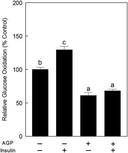
Incubation of C2C12 myotubes with 1 µM insulin resulted in a 29.4±4.9% increase in glucose oxidation (P<0.05, n=3; Figure 1). Treatment with 100 µg mouse AGP/ml reduced glucose oxidation from basal rates by approximately 38.9±4.4% (n=3; P<0.05). Supplementation of both 1 µM insulin and 100 µg AGP/ml medium reduced glucose oxidation by 32.0±2.4%, relative to control (n=3; P<0.05) and 61.1% relative to insulin alone.
Relative glucose oxidation in C2C12 myotubes in response to incubation with α-1 acid glycoprotein (AGP) while in the presence of insulin. Myotube containing C2C12 cultures were incubated in a medium containing mouse AGP (0 or 100 µg/ml medium) for 24 h. The medium was replaced in the last 4 h with fresh medium containing 1.0 μCi D-[U-14C]-glucose/ml and mouse AGP (0 or 100 µg/ml medium) and 1.0 µM bovine insulin, followed by a collection of 14CO2 for analysis of glucose oxidation. Data are expressed relative to cultures incubated without AGP or insulin. a,b,cGroups not sharing a common superscript letter are different (P<0.05, n=3 trials).

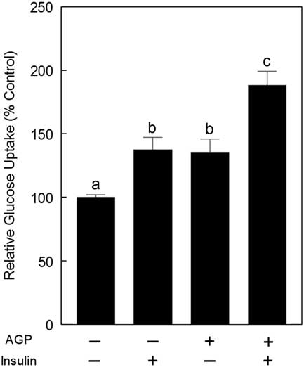
Incubation of cultures for 24 with insulin (1 µM) stimulated glucose uptake by 37.6±9.7% relative to basal medium (P<0.05, n=4; Figure 2). Incubation with 100 µg AGP/ml produced a 35.5±10.5% increase in glucose transport (P<0.05, n=4). Addition of 100 µg AGP/ml increased insulin-stimulated glucose uptake by 88% relative to basal medium (P<0.01, n=4) and by 45% relative to insulin-stimulated media without AGP (P<0.05).
Relative percent glucose uptake into C2C12 myotubes in response to incubation with α-1 acid glycoprotein (AGP). Cultures of C2C12 myotubes in 12-well plates were incubated with 0 or 100 µg mouse AGP/ml medium (Dulbecco’s Modified Eagle medium, 2% horse serum, 20 mM 4-(2-hydroxyethyl)-1-piperazineethanesulfonic acid, antibiotic free) with or without the addition of 1 µM insulin for 24 h. The medium was then removed and cells were washed three times with Krebs-Ringer-phosphate-buffered saline (KRPBS). The cells were then incubated for 20 min in KRPBS with 0.1% bovine serum albumin. At the end of 20 min, 0.1 mM 2-deoxy-d-glucose (2DOG) was added in combination with 0.5 µCi of [3H]-2DOG per well and incubation conditions were maintained for an additional 20 min. Non-specific uptake was measured in control incubations pretreated for 20 min with 10 µM cytochalasin B before 2DOG addition. Radioactivity was normalized to protein concentration. Data are expressed relative to cultures incubated without AGP or insulin. a,b,cGroups not sharing a common superscript letter are different (P<0.05, n=4 trials).

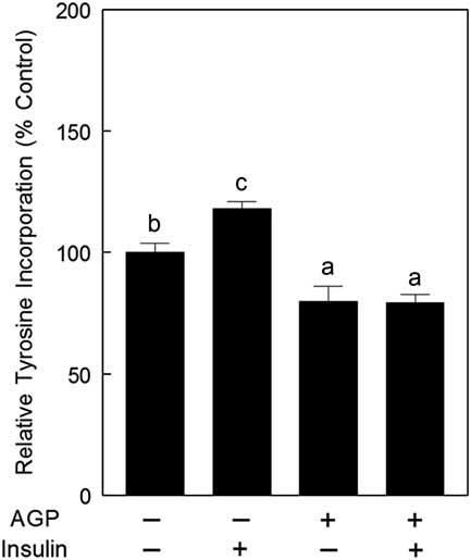
Exposure of C2C12 myotubes to 1 µM insulin for 24 h increased 3H-tyrosine incorporation into protein by 18±2.8%, relative to basal medium (P<0.05, n=6; Figure 3). Incubation with 100 µg AGP/ml medium reduced 3H-tyrosine incorporation by 20.3±6.4%, relative to basal medium (P<0.05, n=6). Addition of 100 µg AGP/ml to medium containing 1 µM insulin reduced 3H-tyrosine incorporation into protein by 20.6±3.3% (P<0.05, n=6), similar to inhibition by AGP alone.
Relative percent tyrosine incorporation into C2C12 myotubes in response to incubation with α-1 acid glycoprotein (AGP). Cultures of C2C12 myotubes were incubated with 0 or 100 µg mouse AGP/ml medium (Dulbecco’s modified Eagle medium, 2% horse serum, 20 mM 4-(2-hydroxyethyl)-1-piperazineethanesulfonic acid, antibiotic free) with or without the addition of 1 µM insulin for 24 h. Protein synthesis was measured during the last 2 h following introduction of 3H-tyrosine into the medium. Radioactivity was normalized to protein concentration. Values are expressed as a percentage of the tyrosine incorporation of cultures incubated without AGP or insulin. a,b,cGroups not sharing a common superscript letter are different (P<0.05, n=6 trials).

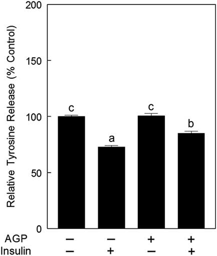
Protein degradation as measured by the free 3H-tyrosine release in response to 1 µM insulin was reduced by 27.1±1.2% relative to control incubations (P<0.05, n=6; Figure 4). Incubation with 100 µg AGP/ml medium had no effect on 3H-tyrosine release (P>0.05, n=6). Combining 1 µM insulin and 100 µg AGP/ml medium resulted in a 14.9±1.8% decrease in 3H-tyrosine release (P<0.01, n=6).
Relative percent free tyrosine released into the medium of C2C12 myotubes in response to incubation with α-1 acid glycoprotein (AGP). Cultures of C2C12 myotubes were incubated with 0.5 µCi/ml of 3H-tyrosine for 24 h beginning on day 4 post-fusion. Following several washes, the cultures were incubated with 0 or 100 µg mouse AGP/ml medium (Dulbecco’s modified Eagle medium, 2% horse serum, 2.0 mM tyrosine, 20 mM 4-(2-hydroxyethyl)-1-piperazineethanesulfonic acid, antibiotic free) with or without the addition of 1 µM insulin for 24 h and then media samples were collected for scintillation counting. Radioactivity was normalized to protein concentration. Values are expressed as a percentage of the tyrosine released from cultures incubated without AGP or insulin. a,b,cGroups not sharing a common superscript letter are different (P<0.05, n=6 trials).

Discussion
Inhibition of insulin-stimulated glucose oxidation, protein synthesis and degradation by AGP were not expected based upon previous research on the interactions of insulin and AGP using the mouse 3T3-L1 cell line (Lee et al., Reference Lee, Choi, Hwang, Lee, Lee, Kim, Huh, Koh, Koh, Son, Masuzaki, Hotta, Alfadda and Kim2010; Lei et al., Reference Lei, Sun, Luo, Yourek, Gui, Yang, Su and Liu2016) and in vivo administration of bovine AGP to diabetic mice that demonstrated an enhancement of insulin sensitivity (Lee et al., Reference Lee, Choi, Hwang, Lee, Lee, Kim, Huh, Koh, Koh, Son, Masuzaki, Hotta, Alfadda and Kim2010). These data indicate that our hypothesis has proven false as mouse AGP reduced the response to insulin in murine C2C12 myotubes. However, these data agree with our previous study which demonstrated that porcine AGP reduces insulin-stimulated glucose oxidation and lipogenesis in porcine adipose tissue (Ramsay et al., Reference Ramsay, Blomberg and Caperna2016a).
Incubation of C2C12 myotubes with insulin increased glucose oxidation, but insulin could not overcome the inhibition of glucose oxidation by AGP. The inhibition of glucose oxidation does not appear to be the consequence of a reduction in insulin-stimulated glucose transport as determined in the present study. Rather, AGP may promote a shift in intermediary metabolism toward glycogen synthesis at the expense of energy utilization (Lei et al., Reference Lei, Sun, Luo, Yourek, Gui, Yang, Su and Liu2016; Qin et al., Reference Qin, Wan, Sun, Wang, Su, Lei and Liu2016). The increase in glucose uptake in response to physiological concentrations of AGP (100 µg/ml medium) was unexpected as previous studies using mouse 3T3 adipocytes did not detect a specific effect of AGP on glucose uptake (Lee et al., Reference Lee, Choi, Hwang, Lee, Lee, Kim, Huh, Koh, Koh, Son, Masuzaki, Hotta, Alfadda and Kim2010), but this would support a role for AGP in promoting glucose utilization for glycogen synthesis, since glucose oxidation was reduced by AGP in the present study. The AGP stimulation of glucose uptake was additive with the effect of insulin which supports the observations of Lee et al. (Reference Lee, Choi, Hwang, Lee, Lee, Kim, Huh, Koh, Koh, Son, Masuzaki, Hotta, Alfadda and Kim2010) in adipocytes, suggesting AGP is not blocking the interaction between insulin and the receptor.
Metabolic syndrome is characterized by inflammation and insulin resistance in skeletal muscle in swine (Zhang and Lerman, Reference Zhang and Lerman2016). An increase in serum AGP has been reported to occur with metabolic syndrome as well (Bogdański et al., Reference Bogdański, Chyrek, Pupek-Musialik and Jabłecka2006). Reductions in insulin-stimulated glucose oxidation, protein synthesis and protein degradation in response to treatment with AGP in the present study suggest that an increase in serum AGP may contribute to the insulin resistance observed in response to systemic inflammation with metabolic syndrome in swine, but this has not been verified.
Use of a 24-h incubation period with AGP permits production of cellular proteins that could interact with insulin signaling and inhibit its actions. Obvious candidates include tumor necrosis factor (TNF) α, interleukin 1 (IL1), IL6 and monocyte chemotactic protein (MCP), as they are typically elevated with an inflammatory response associated with insulin resistance (Hotamisligil, Reference Hotamisligil2006). Previous work with the mouse 3T3-L1 cell line did not report an increase in TNF, IL6 or MCP with AGP treatment, but rather decreases in messenger RNA abundance and/or protein and in vivo treatment with AGP resulted in a reduction in serum IL1, TNF and IL6 (Lee et al., Reference Lee, Choi, Hwang, Lee, Lee, Kim, Huh, Koh, Koh, Son, Masuzaki, Hotta, Alfadda and Kim2010). We have observed either no change or a decline in the secreted TNF and IL6 quantities produced by primary cultures derived from porcine adipose tissue in response to porcine AGP (Ramsay et al., Reference Ramsay, Stoll, Blomberg and Caperna2016b). Therefore, an AGP induction of these cytokines in C2C12 myotubes is unlikely. Further research is necessary to determine if the function of AGP to reduce insulin efficacy includes intracellular responses, which we suspect, as 4-h incubations produced no discernible change in insulin response with AGP treatment (data not presented).
As previously mentioned, plasma or serum AGP in newborn swine can be used to predict growth rate to weaning and beyond (Caperna et al., Reference Caperna, Shannon, Blomberg, Stoll and Ramsay2013 and Reference Caperna, Shannon, Stoll, Blomberg and Ramsay2015). Normal birth weight pigs with elevated plasma or serum AGP levels at birth will grow more slowly than littermates of similar birth weight who have much lower levels of plasma or serum AGP, and these slower growing pigs will have less muscle mass (Mitchell et al., Reference Mitchell, Ramsay, Caperna and Scholz2012). Similarly, runt pigs have much higher plasma AGP levels than littermates at birth and grow slower with less muscle mass (Bauer et al., Reference Bauer, Walter, Hoppe, Gaser, Lampe, Kauf and Zwiener1998). The present in vitro experiments with mouse C2C12 myotubes provide a potential mechanism that may contribute to the reduction in muscle accretion in animals with elevated blood AGP through an inhibition of insulin-stimulated protein synthesis and degradation. The inhibitory effect of AGP on insulin-stimulated protein synthesis and degradation are perhaps greatest in the fetus and newborn when protein synthesis and degradation are relatively high compared with preweaning (Davis et al., Reference Davis, Fiorotto, Nguyen and Reeds1989 and Reference Davis, Fiorotto, Nguyen and Reeds1993); and this late fetal to newborn transition is when AGP levels are highest in swine (Martin et al., Reference Martin, Tesouro, González-Ramón, Piñeiro and Lampreave2005). However, Clapperton et al. (Reference Clapperton, Bishop, Cameron and Glass2005 and Reference Clapperton, Diack, Matika, Glass, Gladney, Mellencamp, Hoste and Bishop2009) have reported that plasma AGP levels are negatively correlated with growth in 4- to 6-month-old pigs selected for high and low growth potential and health status; therefore, plasma AGP may contribute to differences in muscle accretion in older pigs also. Further studies are necessary to verify the effects of AGP on protein turnover in skeletal muscle of swine in vivo and the contribution of insulin to the response.
Acknowledgments
The authors thank Margo Stoll for maintaining the C2C12 cell cultures that were used in this research.
Declaration of interest
The authors have no conflicts of interest.
Ethics statement
Not relevant to this research as no animals were used.
Software and data depository resources
None of the data were deposited in an official repository.