Dilated cardiomyopathy is the most common cardiomyopathy in children and its idiopathic form constitutes the majority of children with this disease.Reference Towbin, Lowe and Colan 1 In turn, the 5-year prognosis is poor, leading to death and/or transplantation in 40–50% in this subset of patients.Reference Towbin, Lowe and Colan 1 , Reference Tsirka, Trinkaus and Chen 2 The current accepted definition by the World Health Organization is a “dilated left ventricle with systolic dysfunction as its hallmark” without mention of right ventricular function or appearance.Reference Richardson, McKenna and Bristow 3
A number of studies have examined left ventricular systolic function and size in the setting of idiopathic dilated cardiomyopathy; nevertheless, the right ventricle remains difficult to interpret secondary to its unique morphology. Therefore, a number of surrogate markers of right ventricular function by echocardiography, both systolic and diastolic, have been proposed and studied, such as tricuspid pulsed wave inflow Doppler, Doppler tissue imaging, right ventricular fractional area change, right ventricular myocardial performance index, and tricuspid annular plane systolic excursion.Reference Rudski, Lai and Afilalo 4 Recent published guidelines suggest an important role for right ventricular functional assessments in all patients.Reference Rudski, Lai and Afilalo 4 , Reference Jurcut, Giusca, La Gerche, Vasile, Ginghina and Voigt 5 However, no prior study has fully examined their role in children with idiopathic dilated cardiomyopathy.
Cardiac magnetic resonance imaging is considered the reference standard for assessment of right ventricular size and function in children and adults.Reference Rudski, Lai and Afilalo 4 , Reference Lai, Gauvreau, Rivera, Saleeb, Powell and Geva 6 , Reference Pavlicek, Wahl and Rutz 7 However, echocardiography remains the most utilised diagnostic bedside tool for the assessment of ventricular function in the critically ill children, in whom obtaining cardiac magnetic resonance imaging can be challenging. We compared children with idiopathic dilated cardiomyopathy with age-matched control subjects to determine which, if any, surrogate markers of right ventricular function were abnormal. In addition, we attempted to determine as our primary end point whether published right ventricular function parameters are independently predictive of cardiac death, which we defined as transplantation or death within 1 year of presentation.
Methods
This retrospective chart review was approved by the institutional review board. Data were extracted from an existing echocardiographic database and clinical charts. Inclusion criteria were new onset of dilated cardiomyopathy and the presence of Doppler tissue images and apical four-chamber views that had the right ventricle included. Patients were excluded if they had other forms of cardiomyopathy, known metabolic disease, genetic syndrome, muscular dystrophy, primary arrhythmia, congenital heart disease, or known infectious aetiology. All effort was made to select the patient's first echocardiogram upon presentation. If the requisite images were not available, the first study that met inclusion criteria within 2 weeks of presentation was selected. All patients who were coded for idiopathic dilated cardiomyopathy from January, 2001 to December, 2010 were reviewed. A total of 115 individual patients were identified. Of these, 84 had their initial presentation after January, 2001 and 30 met the selected inclusion criteria. The most common reasons for exclusion were metabolic, genetic, or muscular dystrophy (17), followed by presence of congenital heart disease (8), tachycardia-induced cardiomyopathy (4), foetal dilated cardiomyopathy (3), and Kawasaki disease (1). An additional 21 patients were excluded because the necessary images were not available. All patients had at least 1 year of follow-up. Control patients for Doppler tissue imaging, myocardial performance index, and tricuspid annular plane systolic excursion consisted of 60 age- and body surface area-matched patients without cardiac disease who were selected from our echocardiographic database.
Echocardiographic images were recorded on commercially available Siemens (Sequoia C512; Siemens, Mountain View, California, United States of America), Phillips (IE33; Phillips, Andover, Massachusetts, United States of America), and General Electric (Vivid 7; GE, Milwaukee, Wisconsin, United States of America) equipment using transducer frequencies and settings appropriate for patients’ sizes. Off-line analysis of data was carried out on syngo Dynamics workstations (Siemens, Mountain View, California, United States of America). Investigators were blinded to the underlying clinical history.
Doppler tissue imaging S′, E′, and A′ waves were recorded in centimetres per second at the tricuspid valve, septal and mitral valve annulus. The isovolumic acceleration time was recorded in metres per second squared. The right ventricular myocardial performance index was calculated from the tricuspid valve Doppler tissue imaging. Tricuspid valve pulsed wave Doppler, tricuspid annular plane systolic excursion, and right ventricular fractional area change were calculated from an apical four-chamber view in a two-dimensional image as described.Reference Rudski, Lai and Afilalo 4 Left ventricular ejection fraction was calculated by 5/6 area length method, Simpson's biplane method, or Teichholz method depending on the age of the patient and available images. The left ventricular end-diastolic dimension was measured from m-mode and the accompanying z-score was then calculated. The left ventricular ejection fraction z-score and end-diastolic dimension z-score were compared with those of age-matched controls.
The study group of 30 patients was then divided by the primary end point. The function variables of the two ventricles mentioned above were compared between the two groups.
Statistical analysis was performed using commercially available software (SAS 9.2, Cary, North Carolina, United States of America). Continuous variables with varied distribution were expressed as median and ranges. The echo parameters of the study subjects were compared with the normal population using Student t-test of equal or unequal variance where appropriate. Categorical variables were analysed by chi-square test. A p-value less than 0.05 was considered statistically significant.
Results
Baseline demographics
The study group comprised 30 patients as outlined in the “Methods” section. The mean age at presentation was 3.9 years – with a median of 1.25 years and a range from 0.1 to 17.8 years. There were a total of nine patients (30%) who reached the primary end point of cardiac death. The median age at transplantation was 1.9 years – with a range from 0.2 to 14 years. There was a male to female ratio of 0.76:1. Following admission, inotropes were started in 20 patients (70%) and a ventricular assist device or extracorporeal membrane oxygenation was placed in three patients (10%).
Patients versus controls
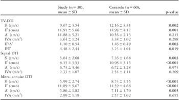
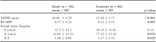
Tricuspid valve Doppler tissue imaging values are shown in Table 1. Tricuspid valve Doppler tissue imaging S′ and E′ were available in all 30 patients and isovolumic acceleration was available in 29 of 30 patients. There were three patients who were noted to have fused E′ and A′ waves. Therefore, A′ and E′/A′ could not be obtained in these individuals. As compared with controls, patients with idiopathic dilated cardiomyopathy had significantly reduced tricuspid valve S′, E′, E′/A′, but not A′, and isovolumic acceleration as shown in Tables 1 and 2.
Comparison of DTI-derived indices: IDCM versus age- and sex-matched controls.

DTI = Doppler tissue index; IDCM = idiopathic dilated cardiomyopathy; IVA = isovolumic acceleration; SD = standard deviation; TV = tricuspid valve
Statistically significant p-values <0.05 in bold
Comparison of RV function parameters: IDCM versus age- and sex-matched controls.

IDCM = idiopathic dilated cardiomyopathy; MPI = myocardial performance index; RV = right ventricle; SD = standard deviation; TAPSE = tricuspid annular plane systolic excursion
Statistically significant p-values <0.05 in bold
Left ventricular function parameters septal and mitral valve annular Doppler tissue imaging S′, E′, A′ and isovolumic acceleration were reduced as expected and are shown in Table 1 as well.
Survivors versus cardiac death
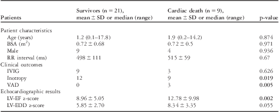
Comparison between 1-year survival and the primary end point yielded a relatively homogeneous grouping as shown in Table 3. The two subgroups did not differ with regard to age, body surface area, heart rate, left ventricular end-diastolic dimension z-score, and administration of intravenous immunoglobulin. They were, however, statistically different in their rate of cardiac catheterisation, need for inotropic support, and left ventricular ejection fraction z-scores. Left ventricular ejection fraction of less than minus 8 z-scores yielded a sensitivity of 70% and specificity of 90% in predicting the primary end point.
Comparison of all study patients by primary end point.

BSA = body surface area; EDD = end diastolic dimension; EF = ejection fraction; IVIG = intravenous immunoglobulin; LV = left ventricle; SD = standard deviation; VAD = ventricular assist device
Statistically significant p-values <0.05 in bold
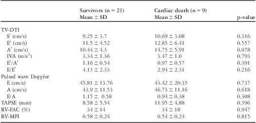
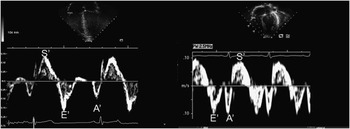
There was no significant difference between groups for right ventricular function parameters; Doppler tissue imaging, pulsed wave Doppler, tricuspid annular plane systolic excursion, or right ventricular myocardial performance index as shown in Table 4. However, an E′/A′ ratio of less than 0.95 yielded a sensitivity of 78% and specificity of 71% in favour of the primary end point. Multivariable logistic regression analysis incorporating left ventricular ejection fraction z-score, left ventricular end-diastolic dimension z-score, and tricuspid valve E′/A′ demonstrated only the left ventricular z-score to be a significant predictor of the primary end point (Fig 1).
Comparison of RV parameters by primary end point.

DTI = Doppler tissue index; FAC = fractional area change; IVA = isovolumic acceleration; MPI = myocardial performance index; RV = right ventricle; SD = standard deviation; TAPSE = tricuspid annular plane systolic excursion; TV = tricuspid valve
Left image – Doppler tissue index of tricuspid valve annulus in a normal patient: normal E′/A′ ratio and velocities. Right image – Doppler tissue index of tricuspid valve annulus in a patient with idiopathic dilated cardiomyopathy: reversal of E′/A′ ratio and low velocities.

Discussion
Our study demonstrated that right ventricular systolic and diastolic function is impaired universally in idiopathic dilated cardiomyopathy. We found that tricuspid valve Doppler tissue imaging S′, E′, right ventricle myocardial performance index, and tricuspid annular plane systolic excursion were abnormal in the study cohort when compared with controls. However, our study failed to demonstrate the utility of the right ventricular function parameters such as tricuspid valve Doppler tissue imaging S′ and E′ in isolating children with idiopathic dilated cardiomyopathy at risk of death or transplant.Reference McMahon, Nagueh and Eapen 8
Owing to the fact that right ventricular function assessment by echocardiography has significant limitations, cardiac magnetic resonance imaging is the current accepted standard measure of right ventricular function.Reference Lai, Gauvreau, Rivera, Saleeb, Powell and Geva 6 , Reference Grosse-Wortmann, Roche, Yoo, Seed and Kantor 9 The role of cardiac magnetic resonance imaging in risk stratification of idiopathic dilated cardiomyopathy is emerging and it has been shown that poor right ventricular function and fibrosis and scarring portend worse outcomes.Reference Partington, Seabra and Kwong 10 However, cardiac magnetic resonance imaging is not routinely used in patients with idiopathic dilated cardiomyopathy unless myocarditis is suspected as the aetiology. Therefore, echocardiography continues to be the preferred tool for bedside evaluation and management of idiopathic dilated cardiomyopathy.
Systolic function
Doppler tissue imaging in particular has gained wide acceptance as a surrogate marker of systolic and diastolic function.Reference Sasaki, Garcia and Lytrivi 11 – Reference Swaminathan, Ferrer, Wolff, Gómez-Marín and Rusconi 13 Our data clearly show that tricuspid valve annular S′ was abnormal as compared with controls, whereas, surprisingly, isovolumic acceleration was not.
Right ventricular systolic dysfunction as measured by decreased S′ was seen in all patients with idiopathic dilated cardiomyopathy. This finding is in agreement with previous work that showed it to be the most sensitive echocardiographic marker in children with idiopathic dilated cardiomyopathy and which correlated with right ventricular ejection fraction by cardiac magnetic resonance imaging.Reference Wang, Prakasa and Bomma 14 Isovolumic acceleration, a pre-ejection index, has shown to correlate well with ventricular contractility and has been validated against invasive measure of right ventricular elastance.Reference Wang, Prakasa and Bomma 14 , Reference Vogel, Schmidt and Kristiansen 15 In our study, tricuspid valve isovolumic acceleration in idiopathic dilated cardiomyopathy was not significantly different from the controls. It is unclear why patients with severe derangements in global left and ventricular function can have preserved pre-ejection phase contractility indices. It is possible that variability in the measurement of this parameter and the small study population impacted its statistical utility. A prior study by Pavlicek et alReference Pavlicek, Wahl and Rutz 7 showed isovolumic acceleration to be less reliable to obtain as compared with S′.
Children with idiopathic dilated cardiomyopathy have decreased tricuspid annular plane systolic excursion, suggesting the coexistence of right ventricular systolic dysfunction. The first description of tricuspid annular plane systolic excursion as a measure of right ventricular systolic function was made by Kaul et alReference Kaul, Tei, Hopkins and Shah 16 Since that time it has been primarily studied in adults, with limited studies examining its utility in children.Reference Koestenberger, Ravekes and Everett 17 Nevertheless, it has been shown to correlate well with underlying right ventricular systolic function. Although it is not completely independent of altered haemodynamics, only severely elevated mean right atrial pressure and preload dependence has been shown to alter its usefulness.Reference Rudski, Lai and Afilalo 4 Owing to the fact that cardiac catheterisation is not routinely performed on all patients with idiopathic dilated cardiomyopathy at our institution, we were not able to show a correlation between tricuspid annular plane systolic excursion and invasive measurements of right atrial pressure and pulmonary vascular resistance.
Right ventricular fractional area change is a simple but perhaps less reliable index in the assessment of right ventricular systolic function. This is especially true in idiopathic dilated cardiomyopathy where the right ventricle is dwarfed and incompletely profiled secondary to a much dilated left ventricle from the apical four-chamber view. Although right ventricular fractional area change has been found to have good correlation with cardiac magnetic resonance imaging right ventricular ejection fraction in some adult studies, its use in paediatrics is limited.Reference Wang, Prakasa and Bomma 14 , Reference Anavekar, Gerson, Skali, Kwong, Yucel and Solomon 18 In fact, a recent study by Srinivasan et alReference Srinivasan, Sachdeva, Morrow, Greenberg and Vyas 19 showed no correlation between right ventricular fractional area change and cardiac magnetic resonance imaging right ventricular ejection fraction in children.
Diastolic function
The role of diastolic dysfunction in all types of cardiomyopathy has been well documented and our finding of an abnormal E′ as compared with controls agrees with this.Reference Sasaki, Garcia and Lytrivi 11 – Reference Wang, Prakasa and Bomma 14 E′ was decreased for both the right ventricle and left ventricle, suggesting impaired biventricular relaxation. However, late diastolic inflow, the tricuspid annular A′ wave, was not aberrant as compared with controls. A′ corresponds to the atrial “kick” after early diastolic filling and subsequent diastasis. Early diastolic filling is dependent on ventricular relaxation; the reversal of diastolic waves or E′/A′ less than one indicates impaired ventricular relaxation and a relative preservation of atrial function. Contrary to findings on the right, mitral annular A′ was abnormal as compared with controls. This suggests that impaired left atrial dysfunction may be more prevalent compared with right atrial dysfunction in idiopathic dilated cardiomyopathy.
The myocardial performance index, also known as the Tei index, was first used to examine global left ventricular myocardial dysfunction in adult patients with dilated cardiomyopathy.Reference Tei, Ling and Hodge 20 Further studies showed that in children with idiopathic dilated cardiomyopathy the left ventricular myocardial performance index was abnormal.Reference Eto, Ishii, Tei, Tsutsumi, Akagi and Kato 21 Our findings of decreased left and right ventricular myocardial performance index are concordant with previously published data in this patient population subset.
Survivors versus cardiac death
On the basis of knowledge gleaned from previous studies, we had hypothesised that measures of right ventricular function would be worse in those who ultimately underwent transplantation or death.Reference McMahon, Nagueh and Eapen 8 , Reference Grosse-Wortmann, Roche, Yoo, Seed and Kantor 9 , Reference Azevedo, Albanesi Filho and Santos 22 This seemed to agree with earlier studies in adults with idiopathic dilated cardiomyopathy that right ventricular failure was predictive of long-term survival.Reference Juillière, Barbier, Feldmann, Grentzinger, Danchin and Cherrier 23 Similar studies in children have demonstrated that right ventricular myocardial performance index is significant in predicting outcome by univariate but not multivariate predictive analysis.Reference McMahon, Nagueh and Eapen 8 , Reference Azevedo, Albanesi Filho and Santos 22 In our patient cohort, right ventricular function was impaired as noted by an abnormal right ventricular myocardial performance index but did not impact our primary outcome of cardiac death. McMahon et alReference McMahon, Nagueh and Eapen 8 in their cohort observed that tricuspid valve E′ less than 8.5 centimetres per second and left ventricular ejection fraction less than 30% had a sensitivity of 44% and specificity of 100% in predicting need for hospitalisation, transplantation, or death. There was no significant difference in tricuspid valve Doppler tissue imaging E′ in our patient population as is shown in Table 4. In addition to all markers of right ventricular function, only E′/A′ less than 0.95 was predictive of cardiac death with a sensitivity of 78% and specificity of 71% (p equal to 0.05). The differences between our study and that of McMahon's may be explained by the definition of outcomes measured and by the variability in severity of disease in the two study cohorts. Decreased tricuspid valve E′ was not predictive of cardiac death in our study population.
Clinical measurements that predicted reaching the primary end point were the need for inotropic support at presentation and ventricular assist device placement. In addition, left ventricular ejection fraction z-scores were significantly different, and differences in left ventricular end-diastolic dimension z-scores were borderline significant in our patients, which met the primary end point. Both decreased left ventricular ejection fraction and increased left ventricular end-diastolic dimension have been shown to be independent predictors of poor outcome.Reference Towbin, Lowe and Colan 1 A prior extensive review of the literature by Alvarez et alReference Alvarez, Wilkinson and Lipshultz 24 demonstrated the wide disparity in the predictive power of echocardiography in patients with idiopathic dilated cardiomyopathy. A likely explanation is that idiopathic dilated cardiomyopathy is only the visible phenotype for a variety of underlying causes.Reference Towbin, Lowe and Colan 1 , Reference Jefferies and Towbin 25
Limitations
There were two primary limitations in our study. Foremost is the small study sample, which would impact statistical significance with even small changes in outliers. This was reflected in the relatively wide standard deviations for most of our measured parameters. Another limitation was the retrospective nature of this study. This restricted the nature of available clinical data that could have been evaluated, such as Ross score of functional status, measurement of biomarkers such as B-type natriuretic peptide, and a large percentage of identified patients who could not be studied because of incomplete Doppler tissue imaging data. Although our echocardiographic laboratory has a set protocol that includes Doppler tissue imaging, they were not always obtained. Patients with dilated cardiomyopathy frequently are unstable at presentation and image acquisition is limited to investigation of aetiology.
Summary
Idiopathic dilated cardiomyopathy is not purely a disease of the left ventricle. Right ventricular systolic and diastolic function is impaired almost universally in children with idiopathic dilated cardiomyopathy. The degree of dysfunction, however, is still difficult to categorise using echocardiographic measures, and hence its impact on outcomes cannot be confirmed. Previously published data of decreased tricuspid valve E′ and right ventricle fractional area change did not hold true for our patient population. The degree of left ventricular dysfunction and need for major clinical circulatory support is still the major determinant of outcomes. Certainly the right ventricle is adversely affected in idiopathic dilated cardiomyopathy, but whether any measured ventricular index can influence prediction of prognosis needs further systematic investigation.
Acknowledgements
The authors thank Dr Emilia Bagiella for her help with the statistical analysis. The authors have no financial relationships and conflicts of interest relevant to this article to disclose.