INTRODUCTION
The myxozoan parasite Tetracapsuloides bryosalmonae, is the causative agent of proliferative kidney disease (PKD) in salmonid fish (Anderson et al. Reference Anderson, Canning and Okamura1999; Canning et al. Reference Canning, Curry, Feist, Longshaw and Okamura1999, Reference Canning, Curry, Feist, Longshaw and Okamura2000; Feist et al. Reference Feist, Longshaw, Canning and Okamura2001). Mortality due to PKD in farmed fish ranges from 20 to 100%, leading to increased costs of production (Ferguson and Ball, Reference Ferguson and Ball1979; Clifton-Hadley et al. Reference Clifton-Hadley, Bucke and Richards1986; Feist and Longshaw, Reference Feist, Longshaw and Woo2006). The impacts of PKD on wild fish populations are generally poorly known although a recent outbreak in Norway (Sterud et al. Reference Sterud, Forseth, Ugedal, Poppe, Jørgensen, Bruheim, Fjeldstad and Mo2007) demonstrates the potential for mass mortality and PKD is attributed to long-term declines in Swiss brown trout populations (Borsuk et al. Reference Borsuk, Reichert, Peter, Schager and Burkhardt-Holm2006).
The life cycle of T. bryosalmonae involves exploitation of freshwater bryozoans (Phylum Bryozoa, Class Phylactolaemata) as primary hosts (see Okamura et al. Reference Okamura, Hartikainen, Schmidt-Posthaus and Wahli2011, for review). The parasite first develops as covert infections when single cells are associated with the bryozoan body wall (Morris and Adams, Reference Morris and Adams2006a ). When conditions are favourable for host growth (e.g. high temperatures and food levels for hosts; Tops et al. Reference Tops, Lockwood and Okamura2006; Hartikainen and Okamura, Reference Hartikainen and Okamura2012) these cells detach and develop within the bryozoan body cavity to form sacs filled with spores infectious to fish. Because these sacs can be observed with a stereomicroscope we refer to such infections as overt (see Okamura et al. Reference Okamura, Hartikainen, Schmidt-Posthaus and Wahli2011, for review).
Freshwater bryozoans are sessile colonial invertebrates that occur in nearly all types of freshwater environments (Massard and Geimer, Reference Massard and Geimer2008). They are suspension-feeders that capture phytoplankton and other microorganisms by creating feeding currents with ciliated tentacles (Massard and Geimer, Reference Massard and Geimer2008). Sexual reproduction is facultative, occurring briefly in early summer and resulting in the release of short-lived swimming larvae (Wood and Okamura, Reference Wood and Okamura2005). Much greater emphasis is on the asexual production of numerous dormant statoblasts, which are small (<1 mm), multicellular seed-like stages comprised of germinal tissue surrounded by resistant chitinous valves (Wood and Okamura, Reference Wood and Okamura2005). Statoblasts can tolerate poor conditions such as desiccation, freezing and lack of food (Hengherr and Schill, Reference Hengherr and Schill2011). When favourable conditions return statoblasts hatch and form a new colony (Wood and Okamura, Reference Wood and Okamura2005). Because of their resistant nature, small size (0·5–1 mm), and high production, statoblasts likely act as a means of dispersal within and between sites. Evidence in support of this includes the presence of statoblasts in waterfowl guts (Figuerola et al. Reference Figuerola, Green, Black and Okamura2004; Mouranval et al. Reference Mouranval, Guillemain, Canny and Poirier2007) and fecal material (Green et al. Reference Green, Jenkins, Bell, Morris and Kingsford2008), gene flow patterns associated with migratory waterfowl routes (Freeland et al. Reference Freeland, Noble and Okamura2000; Figuerola et al. Reference Figuerola, Green and Michot2005) and the ability of statoblasts to hatch after passing through duck digestive tracts (Charalambidou et al. Reference Charalambidou, Santamarıa and Figuerola2003). Statoblasts have also been found in the gut of many fish species, including largemouth bass, Micropterus salmoides (Lacepède), crappie, Pomoxis annularis (Rafinesque) and common carp, Cyprinus carpio (Linnaeus) (Marković et al. Reference Marković, Karin-Žnidaršić and Simonović2009). Statoblasts carried in ship ballast tanks are likely to contribute to distributions of bryozoans worldwide (Kipp et al. Reference Kipp, Bailey, MacIsaac and Ricciardi2010; Briski et al. Reference Briski, Cristescu, Bailey and MacIsaac2011).
Given the strong dispersal potential of statoblasts, it is of interest to know whether T. bryosalmonae can be vertically transmitted via statoblasts. In addition, vertical transmission may explain the presence of infected bryozoans in sites free of salmonids (Tops et al. Reference Tops, Baxa, McDowell, Hedrick and Okamura2004) as it may contribute to persistent infection in local bryozoan populations. Such vertical transmission has been demonstrated for another bryozoan–malacosporean system (Buddenbrockia allmani infecting the bryozoan, Lophopus crystallinus) whereby new colonies developed from statoblasts were shown by PCR and sequencing to carry infection (Hill and Okamura, Reference Hill and Okamura2007). The bizarre observation of the release of worm-like organisms probably representative of Buddenbrockia plumatellae from statoblasts of Plumatella fungosa also provides evidence for vertical transmission (Taticchi et al. Reference Taticchi, Gustinelli, Fioravanti, Caffara, Pieroni and Prearo2004). However, until very recently investigations have failed to find evidence for infections of T. bryosalmonae in bryozoan statoblasts (e.g. Grabner and El-Matbouli, Reference Grabner and El-Matbouli2008).
It is now clear that during overt infection T. bryosalmonae severely reduces statoblast production, causes malformation of statoblasts and thus almost entirely castrates the infected zooids of its main bryozoan host, Fredericella sultana (Hartikainen et al. Reference Hartikainen, Fontes and Okamura2013). Tetracapsuloides bryosalmonae infections have been detected by PCR in a fraction of the few statoblasts produced in such overtly infected bryozoans. However, these infections were not transmitted successfully to the mature offspring hatched from the same statoblast sources, suggesting either selective mortality of infected offspring or that the infections were subsequently lost (Hartikainen et al. Reference Hartikainen, Fontes and Okamura2013). Whether vertical transmission of infection is effective thus remains an open question. The aims of the current study were to: (1) examine the occurrence and extent of vertical transmission of T. bryosalmonae in F. sultana statoblasts, and (2) determine the effectiveness of vertical transmission by undertaking transmission studies across the T. bryosalmonae life cycle.
MATERIALS AND METHODS
The occurrence and extent of infection of statoblasts
Fredericella sultana colonies associated with submerged tree roots were collected from the River Lohr, Germany (50°01′08″N; 09°32′43″E) on 1 August 2011 and from the Rivers Avon (11°27′32″N; 41°46′29″E) and Dun (16°86′07″N; 43°42′63″E) in southern England on 29 August 2012 and 5 September 2012. Material from the Lohr was used in transmission experiments and material from the Rivers Avon and Dun were used to measure infection prevalence and vertical transmission to statoblast offspring.
Tree roots with attached colonies from the River Lohr were glued to Petri dishes, placed in an aerated 10 L bucket filled with Bryozoan Medium C (BMC) (Kumar et al. Reference Kumar, Abd-Elfattah, Soliman and El-Matbouli2013) and maintained at 18 °C (±2 °C) by feeding with cultured algae as described in Grabner and El-Matbouli (Reference Grabner and El-Matbouli2008). Briefly, 6 algal species (Cryptomonas ovata, Cryptomonas spp., Chlamydomonas spp., Chlamydomonas reinhardii, Synechococcus spp., Synechococcus leopoliensis) were cultured in sterile Guillard's WC-Medium. Cryptomonas ovata comprised 80% of food provided to F. sultana colonies and the remaining 20% was comprised of the other algae. Food was provided daily by adding 500 mL of the algal culture (concentration of 6·5 to 7·5×106 cells mL−1) to the bucket and the water was changed every week.
After 49 days, microscopic examination of the bryozoans in BMC medium revealed cells swirling in the host body cavity and the subsequent development of overt T. bryosalmonae infections in 28% of colonies observed (n = 100). Thirty statoblasts dissected from overtly infected laboratory-cultured colonies originally collected from the field were retained for PCR assays. To minimize the possibility of contamination, statoblasts were incubated in distilled water at 4 °C for 24 h (spores degrade in <24 h at 18 °C; De Kinkelin et al. Reference De Kinkelin, Gay and Forman2002) and were then washed several times with distilled water prior to preserving in RNA Later until DNA extraction.
Colonies from the Rivers Avon and Dun were collected by detaching several branches from each colony with forceps and placing these branches together in a tube specific for that colony and maintained at room temperature (20 °C) for 24 h prior to collecting all the mature statoblasts that were present in the material. Mature statoblasts were identified by the dark brown colour of valves. Statoblasts were dissected from bryozoans using a stereomicroscope (100×magnification) which also enabled determining whether T. bryosalmonae sacs were present within the bryozoan body cavity. Colonies lacking overt infections were subject to PCR to test for the presence of covert infections. Statoblasts from the same brood (i.e. from the same maternal colony) from the Rivers Avon and Dun were placed in 1·5 mL tubes with deionized water and stored at 4 °C (Rivers Avon and Dun) for 5 months. After 5 months, the statoblasts of the same brood (i.e. originating from a single maternal colony) were placed in Petri dishes filled with artificial pond water and were allowed to hatch at 20 °C over a period of 7 days. The hatched statoblasts were preserved in 100% ethanol for PCR assays for detection of infection of individual statoblasts. The tissue of maternal colonies was also preserved in ethanol to determine infection status. Associations between river, maternal colony infection, statoblast hatching status and statoblast infection were tested using Chi-square (for two-way analyses) and G tests (three-way analyses).
DNA extractions used either a QIAamp DNA mini Kit (QIAGEN) (Lohr samples) or a modified hexadecyltrimethylammonium bromide (CTAB) protocol (Tops and Okamura, Reference Tops and Okamura2003) (Avon and Dun samples). Two T. bryosalmonae-specific PCR assays were used. (1) Statoblasts from the River Lohr were diagnosed using a nested PCR with 5F (5′-CCTATTCAATTGAGTAGGAGA-3′) and 6R (5′-GGACCTTACTCGTTTCCGACC-3′) (Kent et al. Reference Kent, Khattra, Hervio and Devlin1998) in the first round, followed by a second round PCR using PKD-real F (5′-TGTCGATTGGACACTGCATG-3′) and PKD-real R (5′-ACGTCCGCAAACTTACAGCT-3′) (Grabner and El-Matbouli, Reference Grabner and El-Matbouli2009). The final 166 bp amplicon was verified to originate from T. bryosalmonae via purification from 1·5% agarose gels, cloning and sequencing. Briefly, the amplicon was purified using a MinElute gel extraction kit (QIAGEN), cloned into the pCR4-TOPO vector (TOPO TA, Invitrogen) and the recombinant plasmids purified from Escherichia coli using a QIAprep Miniprep (QIAGEN). Cloned PCR products were sequenced in a commercial sequencing laboratory (LGC Genomics GmbH, Berlin, Germany). (2) Statoblasts from the Rivers Avon and Dun were subjected to PCR with the primers 514F new (5′-ATTCAGGTCCATTCGTGAGTAACAAGC-3′) and 776R (5′-CTGCCCTTAATTGGGTGTATCAGC-3′) (diagnostic amplicon size 244 bp, PCR conditions as in Hartikainen et al. Reference Hartikainen, Fontes and Okamura2013). The PCR products were visualized on a 1·5% agarose electrophoretic gel and five positive samples were verified by direct sequencing on ABI Prism 3700×l using BigDye v1.1 chemistry.
Is vertical transmission in statoblasts effective?
We conducted a laboratory-based transmission study using bryozoan colonies originated from the River Lohr to determine whether infection of statoblasts results in effective vertical transmission to colonies that subsequently develop and whether these in turn can promote further transmission of infection to fish. Material for this study was gained by cohabiting 2-month old specific pathogen free (SPF) colonies for 8 h per day for 14 days with 5 brown trout that had been infected by exposure to naturally infected F. sultana colonies from the River Lohr 8 weeks earlier. The cohabitation experiment was conducted several times in order to obtain as many statoblasts as possible, using different SPF F. sultana colonies. A total of 19 colonies (originating from the River Lohr) were present on the Petri dishes and at the beginning of exposure each had at least three zooids. In between bouts of cohabitation fish were transferred back to their aquarium and fed with commercial fish feed. Fredericella sultana colonies were maintained in culture systems (buckets; see above) after the 8 h of cohabitation. These colonies were then allowed to grow on following exposure to fish until they produced statoblasts. Statoblasts were collected from these colonies, washed and placed in distilled water for 24 h at 4 °C.
Statoblasts produced by colonies exposed to infected fish were used in a study to address whether infection was successfully vertically transmitted to daughter colonies that hatched from these statoblasts and whether this infection could in turn be transmitted to fish. For this study we cohabited two colonies derived from these statoblasts and that were observed to have spores and sacs of T. bryosalmonae with five SPF brown trout (4 cm in length) for 8 h daily for 2 weeks. For the control, five SPF brown trout fish were cohabitated with SPF F. sultana colonies (two colonies comprising 18 zooids) from long-term laboratory stock. The aquaria were checked daily for fish mortality and extraordinary behaviour. After co-habitation, fish were placed in 20 L aquaria with flow-through water. Eight weeks post-infection, all five fish from each group were dissected and kidney, liver and spleen were sampled for DNA extraction and T. bryosalmonae-specific PCR assays using 5F and 6R primer sets.
Immunohistochemistry was performed on 30 statoblasts collected from the colonies exposed to fish (described above) and from uninfected colonies deriving from our laboratory stock. Statoblasts were washed with distilled water, incubated for 24 h at 4 °C and fixed in 10% neutral buffered formalin and paraffin-embedded 4 μm sections were labelled with T. bryosalmonae specific monoclonal antibody P01 (Aquatic Diagnostics Ltd.). Endogenous peroxidase activity in deparaffinized sections was blocked with 3% H2O2 in phosphate buffered saline (PBS) for 10 min. Following rinses with Tris-buffered saline (TBS), non-specific protein binding sites were blocked with normal sheep serum (1:10 dilution in TBS) for 30 min. The sections were then incubated for 60 min in a humid chamber at room temperature with the reconstituted P01 antibody (20 μg mL−1 in PBS). After three washes with TBS for 2 min each, the sections were incubated for 30 min with 100 μL of goat anti-mouse polyclonal antibody (IgG) conjugated with horseradish peroxidase (Dako EnVision+System-HRP AEC kit). After four washes with TBS for 2 min each, slides were incubated with the AEC substrate for 4–5 min until a pink colour appeared. The reaction was stopped by immersion of the slides in distilled water for 3–4 min. Sections were then mounted and examined microscopically.
RESULTS
The occurrence and extent of infection of statoblasts
The T. bryosalmonae-specific PCR assays demonstrated parasite presence in statoblasts produced by colonies collected from all three rivers as well as in statoblasts collected from SPF F. sultana colonies exposed to infected brown trout (see Fig. 1). Sequencing of positive samples showed 99% similarity to the 18S rRNA gene of T. bryosalmonae (GenBank accession numbers FJ981823 and AF190669).
PCR amplification of Tetracapsuloides bryosalmonae DNA from statoblasts collected from T. bryosalmonae-infected Fredericella sultana colonies under laboratory conditions. Lanes 2 to 4: first round PCR amplified by 5F-6R primers and lane 7 to 9 nested PCR amplified by PKD-real F-R primers; lanes 1 and 5: 100 bp DNA ladder (Invitrogen); lane 2: infected statoblasts DNA; lane 3: positive control; lane 4: SPF statoblasts DNA; lanes 6 and 10: 50 bp DNA ladder (Fermentas); lane 7: infected statoblasts DNA; lane 8: positive control; lane 9: SPF statoblasts DNA.

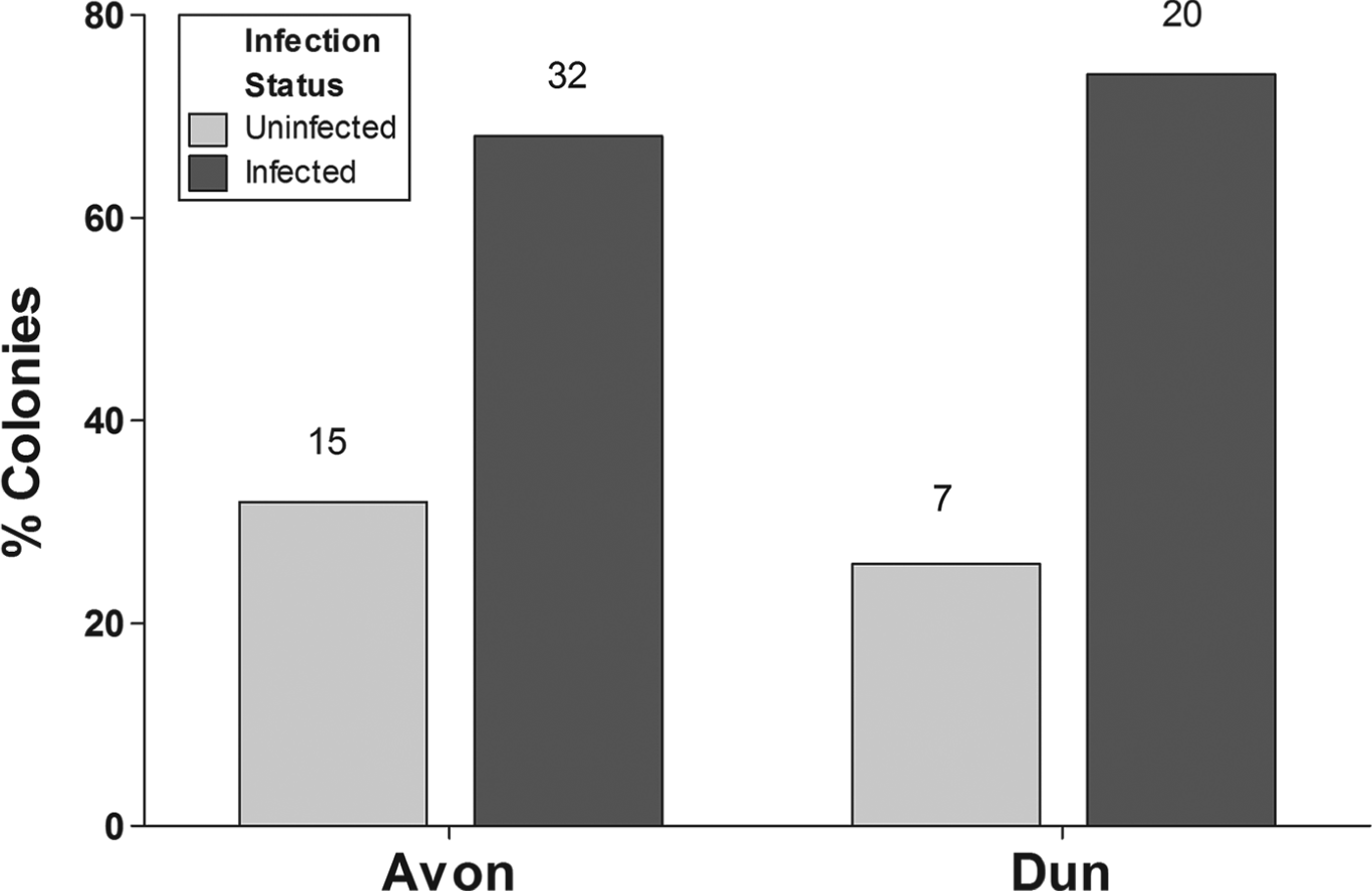
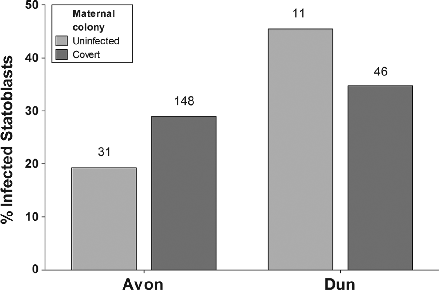
We collected 181 statoblasts from 47 F. sultana colonies from the River Avon and 57 statoblasts from 27 colonies from the River Dun. Only one overtly infected colony (with 11 T. bryosalmonae sacs) was observed (River Avon). This colony had two mature uninfected statoblasts. A high proportion of the remaining colonies were covertly infected (68% from River Avon and 74% from River Dun) (see Fig. 2) but infection status (covertly infected vs uninfected) at the time of field collection was not dependent on river (Chi-square test: χ 2 = 0·30, d.f. = 1, P = 0·59). Infected statoblasts were obtained from colonies that were both covertly infected and uninfected (presumably having lost their infection recently) at the time of collection in both sites (see Fig. 3). Higher proportions of infected statoblasts were collected from the River Dun material (35 and 45% of statoblasts collected from covertly infected and uninfected maternal colonies, respectively) than from the River Avon material (29 and 19% of statoblasts collected from covertly infected and uninfected maternal colonies, respectively) (Fig. 3) (statoblast infection status was not dependent on river: G = 1·8, d.f. = 1, P = 0·18). Notably, the highest proportions of infected statoblasts were collected from uninfected maternal colonies from the River Dun (45%) but from covertly infected maternal colonies from the River Avon (29%) (Fig. 3) (statoblast infection status was not dependent on maternal colony status: G = 0·30, d.f. = 1, P = 0·58). There was also no association between river, maternal colony infection status and statoblast infection (2×2×2 G test: G = 3·6, d.f. = 4, P = 0·463).
The percentage of maternal colonies that were covertly infected and uninfected at the time of collection from Rivers Avon and Dun. One overtly infected colony collected from the River Avon is not included. N values above bars are the numbers of colonies in each infection category per river.

The percentage of statoblasts that were infected according to the infection status of their maternal colony (covertly infected or uninfected at the time of collection from the field) from the Rivers Avon and Dun. Two uninfected statoblasts from an overtly infected colony collected from the River Avon are not included. N values above bars are the numbers of statoblasts collected from uninfected and covertly infected maternal colonies per river.

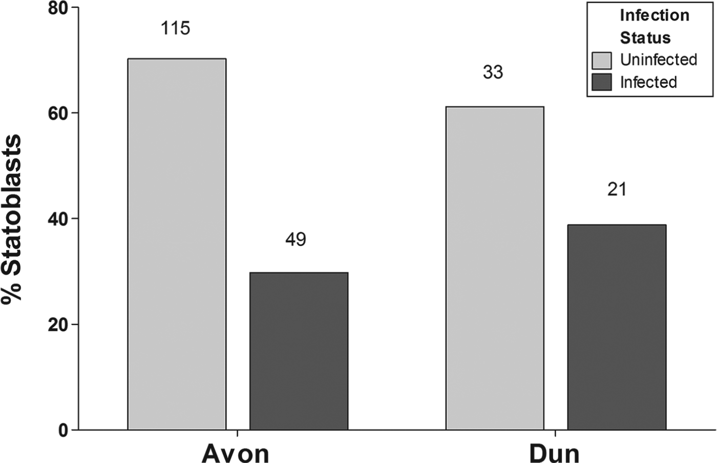
We assessed the frequency of vertical transmission by considering the infection status of pooled statoblasts produced by all infected colonies. Here we included statoblasts produced by uninfected maternal colonies that had produced at least one infected statoblast. Although there was a slightly higher proportion of infected statoblasts (as revealed by PCR) in material collected from the River Dun than in the River Avon (39 and 30%, respectively: see Fig. 4) infection status was not dependent on river (Chi-Square Test: χ 2 = 1·51, d.f. = 1, P = 0·22). The percentages of these infected statoblasts that hatched were 94% (n = 46) and 100% (n = 21) for the Rivers Avon and Dun, respectively. The uninfected colonies with no infection in any of the statoblast offspring produced 15 statoblasts in the Avon and 3 statoblasts in the Dun. The percentage of statoblasts that hatched were 87 and 100%, respectively.
The percentage of infected and uninfected statoblasts collected from infected colonies (including covertly infected maternal colonies and maternal colonies with infected statoblasts) from the Rivers Avon and Dun. Two uninfected statoblasts from an overtly infected colony collected from the River Avon are not included. N values above bars are the numbers of statoblasts according to their infection status per river.

There were notably higher proportions of hatched statoblasts that were infected (50% on the River Dun, 30% on the River Avon) relative to unhatched infected statoblasts (0% on the River Dun, 21% on the River Avon) (Fig. 5) and the effect was greatest for material from the River Dun. These patterns are reflected by the dependency of statoblast infection on hatching status (2×2 G test: G = 6·56, d.f. = 1, P = 0·0104) (uninfected statoblasts did not hatch as well as infected ones) but not on river (2×2 G test: G = 1·6, d.f. = 1, P = 0·2059), although statoblast hatching status was dependent on river (2×2 G test: G = 6·62, d.f. = 1, P = 0·0101) (Fig. 5). Overall, therefore, river, statoblast infection and hatching status were inter-dependent (2×2×2 G test: G = 22·68, d.f. = 4, P<0·0001).
The percentages of hatched and unhatched infected statoblasts collected from infected colonies (including covertly infected maternal colonies and maternal colonies with infected statoblasts) from the Rivers Avon and Dun. Numbers above bars are n-values.

Is vertical transmission in statoblasts effective?
Six of the 30 statoblasts collected for hatching from laboratory-infected F. sultana colonies (20%) successfully hatched. We confirmed that infection was successfully vertically transmitted to two of the six hatched colonies (33%) by observing free spores and mature sacs in two colonies. These infected colonies were each comprised of 2–3 zooids and were subsequently used in exposures to fish. The SPF brown trout that were cohabitated for 2 weeks with these two infected colonies did not show any abnormal behaviour or mortalities during the whole trial, and they were positive for T. bryosalmonae after 8 weeks post-infection (Fig. 6). Band intensities suggest the T. bryosalmonae stages were most abundant in the kidney but were also present in liver and spleen. No DNA was amplified from control fish.
PCR amplification of Tetracapsuloides bryosalmonae DNA from brown trout (Salmo trutta) samples cohabitated with F. sultana raised under laboratory conditions from infected statoblasts. Lane 1: 100 bp DNA ladder (Invitrogen); lanes 2–4: infected kidney, liver and spleen DNA samples of brown trout, respectively; lane 5: negative control; lane 6: positive control.

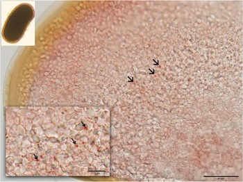
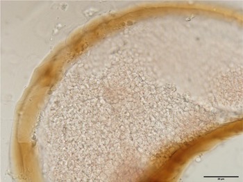
The use of T. bryosalmonae specific monoclonal antibodies revealed the presence of cryptic stages in the infected statoblasts collected from laboratory-infected F. sultana colonies (Fig. 7), which appeared as small red signals (arrows, Fig. 7). No such signal was observed in the negative control (Fig. 8).
Horizontal section of infected statoblast of Fredericella sultana with Tetracapsuloides bryosalmonae monoclonal antibody, revealed the presence of cryptic stages inside the statoblast (arrows), enlarged section (lower left) in small windows focusing on cryptic stages of T. bryosalmonae (arrows), and intact statoblast (upper left). The image shows the dark brown chitinous valve of the statoblast containing vitellogenic material and putative stained parasite stages.

Specific pathogen-free Fredericella sultana statoblast with Tetracapsuloides bryosalmonae monoclonal antibody.

DISCUSSION
Patterns of infection of maternal colonies and statoblasts
We obtained evidence for surprisingly high levels of covert infection in the maternal colonies that were collected from both the Rivers Avon and Dun (68 and 74%, respectively) relative to levels that were monitored during a previous study that examined temporal prevalences of infection in bryozoan populations in the River Cerne, also in southern England (the highest prevalence of infection being ∼50%; Tops, Reference Tops2004). There are several factors that may contribute to explaining these high levels of covert infection including: the development of new primers in this study, if bryozoans in these systems are more susceptible to infection, if a greater number of infectious spores are present or if there is a greater degree of vertical transmission of infection to new colonies produced from infected statoblasts or infected colony fragments thus promoting the spread of infection through populations (Okamura et al. Reference Okamura, Hartikainen, Schmidt-Posthaus and Wahli2011).
We found that infected statoblasts were produced in maternal colonies that were both covertly infected and uninfected during the time the statoblasts were collected. These results suggest that some colonies lose infection but nevertheless pass infection to progeny produced during a prior period of infection. This may have happened, for instance, if the colony had effectively sealed off infected regions containing statoblasts by inward pinching of the peritoneum as was observed to occur in a colony of P. fungosa infected with B. plumatellae (Canning et al. Reference Canning, Tops, Curry, Wood and Okamura2002). An alternate explanation is failure in DNA extraction from the maternal colony or that there was insufficient T. bryosalmonae DNA to form a clear band on the agarose gel. A possible explanation for apparent rescue of some statoblasts from infection is the inclusion of infected and uninfected portions of the colony in the same DNA extraction, giving a mixture of infected and uninfected statoblasts from an infected parent.
A substantial proportion of statoblasts produced by bryozoans from both the Rivers Avon and Dun were infected (30 and 39%, respectively), a finding further corroborated by detection of cryptic stages in infected statoblasts in our monoclonal antibody study. We also found that most infected statoblasts hatched and that this effect was greatest for material from the River Dun (94 and 100% in the Rivers Avon and Dun, respectively). Indeed, the hatching success was significantly associated with infection, with more infected than uninfected statoblasts successfully hatching. This may indicate complex interactions between the colony ability to support both infections and produce viable statoblasts or parasite-mediated stimulation of statoblast hatching. Our laboratory studies complemented our above findings by demonstrating that the presence of T. bryosalmonae in statoblasts can indeed result in subsequent overt infections in colonies that develop from statoblasts and that these infections, in turn, are transmitted to fish.
Implications of vertical transmission via statoblasts
Statoblasts function as dormant propagules that allow bryozoans to persist during unfavourable periods. In addition, there is good evidence that buoyant statoblasts are likely to be dispersed over great distances by waterfowl and ship ballast. Our demonstration that vertical transmission via statoblasts is effective provides evidence that T. bryosalmonae will be dispersed in space or time by hitchiking along with its host, thereby similarly evading unfavourable conditions and achieving colonization of new habitats. Fredericella sultana statoblasts are not buoyant, yet vertical transmission via statoblasts and dispersal with bryozoan hosts may explain the widespread distribution of PKD in the northern hemisphere. Indeed, long-distance dispersal achieved by infected statoblasts may explain phylogeographic patterns that suggest Europe may have been colonized by T. bryosalmonae from North America (Henderson and Okamura, Reference Henderson and Okamura2004).
The capacity for vertical transmission via statoblasts demonstrated here for T. bryosalmonae appears to be a general characteristic of malacosporeans since it has been shown in B. allmani (Hill and Okamura, Reference Hill and Okamura2007) and appears to be achieved by B. plumatellae (Taticchi et al. Reference Taticchi, Gustinelli, Fioravanti, Caffara, Pieroni and Prearo2004). This capacity should enable malacosporeans to persist indefinitely in local populations of bryozoans (Hill and Okamura, Reference Hill and Okamura2007) and, in terms of PKD, is likely to contribute to the regular annual outbreaks observed in many fish farms (El-Matbouli and Hoffmann, Reference El-Matbouli and Hoffmann2002; Okamura et al. Reference Okamura, Hartikainen, Schmidt-Posthaus and Wahli2011). Vertical transmission via colony fragmentation (Morris and Adams, Reference Morris and Adams2006b ; Hill and Okamura, Reference Hill and Okamura2007) provides another mechanism by which malacosporean infections can be amplified within local populations. Significantly, in these colonial hosts, the mechanisms of vertical transmission documented to date are achieved by infection of clonal propagules (asexually-produced statoblasts, fragments of maternal colonies). Thus, it will achieve persistent infection of clonal genotypes over time and space which, in turn, will spread the risk of loss of infection from clonal genotypes.
A body of work now demonstrates that T. bryosalmonae undergoes developmental cycling between covert and overt infections dependent on host condition. The former cause no detectable effect on bryozoan host fitness and are expressed when hosts are in poor condition while overt infections temporarily effectively castrate bryozoan hosts and are expressed when hosts are in good condition (Tops et al. Reference Tops, Lockwood and Okamura2006; Hartikainen and Okamura, Reference Hartikainen and Okamura2012). The overall driver of such developmental cycling appears to be a strategy for T. bryosalmonae to enable long-term persistence in bryozoan populations by minimizing virulence while occasionally undergoing horizontal transmission to fish. Such low virulence is anticipated when the reproductive interests of both host and parasite are aligned (Bull et al. Reference Bull, Molinieux and Rice1991) as is the case for vertical transmission. Previous to this study it was predicted that vertical transmission of T. bryosalmonae by statoblasts must occur (e.g. Tops et al. Reference Tops, Baxa, McDowell, Hedrick and Okamura2004; Grabner and El-Matbouli, Reference Grabner and El-Matbouli2008; Tops et al. Reference Tops, Hartikainen and Okamura2009). By providing the first evidence that such vertical transmission is apparently extensive and effective, our study significantly contributes to our understanding of infection dynamics in both bryozoan and fish hosts.
ACKNOWLEDGEMENTS
We are thankful to Mr Patrick Schrabauer for assistance with the images and thank Darren Butterworth and Robert Starr for permission and help in access to collecting sites.
FINANCIAL SUPPORT
This study was funded by the Austrian Science Fund (FWF), Project no. P 22770-B17, the University of Veterinary Medicine, Vienna (PL29110263), the Natural Environment Research Council (NE/019227/1), the Environment Agency and the Centre for Environment, Fisheries & Aquatic Sciences.