Introduction
Cloning by nuclear transfer of somatic cells into oocytes is currently the most efficient technique for producing copies of elite livestock and transgenic animals. Many factors influence the production of cloned animals when using the technique of nuclear transfer. One is the remodelling of the donor cell nucleus within the cytoplasm of the recipient oocyte to organize the first embryonic division. Usually, greater extents of donor cell nuclear remodelling and embryonic development can be achieved when transferring embryonic rather than somatic cell nuclei into the cytoplasm of metaphase II oocytes, although this general rule does not hold true for all species (Campbell et al., Reference Campbell, McWhir, Ritchie and Wilmut1996; Kato et al., Reference Kato, Tani and Tsunoda2000; Westhusin et al., Reference Westhusin, Long, Shin, Hill, Looney, Pryor and Piedrahita2001). In cattle, sheep, pigs and goats, fetal cells have been used to produce transgenic livestock because of their rapid growth and potential for multiple cell divisions before senescence in culture (Schnieke et al., Reference Schnieke, Kind, Ritchie, Mycock, Scott, Ritchie, Wilmut, Colman and Campbell1997; Cibelli et al., Reference Cibelli, Stice, Golueke, Kane, Jerry, Blackwell, Ponce de Leon and Robl1998; Baguisi et al., Reference Baguisi, Behboodi, Melican, Pollock, Desrempes, Cammuso, Williams, Nims, Porter, Midura, Palacios, Ayres, Denniston, Hayes, Ziomek, Meade, Godke, Gavin, Overstrom and Echelard1999; Kuhholzer et al., Reference Kuhholzer, Tao, Machaty and Hawley2000), whereas adult somatic cell cloning primarily has been used to replicate a particular female (Wilmut et al., Reference Wilmut, Schnieke, McWhir, Kind and Campbell1997; Wells et al., Reference Wells, Misica and Tervit1999) or male (Shiga et al., Reference Shiga, Fujita, Hirose, Sasae and Nagai1999; Hill et al., Reference Hill, Winger, Long, Looney, Thompson and Westhusin2000) genotype. In this context, there are several reports indicating that development rates of somatic cloned embryo remained similar regardless of donor age (Renard, Reference Renard1999; Hill et al., Reference Hill, Winger, Long, Looney, Thompson and Westhusin2000; Wakayama & Yanagimachi, Reference Wakayama and Yanagimachi2001). However, Kato et al. (Reference Kato, Tani and Tsunoda2000) noted that clones derived from adult cells frequently aborted in the later stages of pregnancy and calves developing to term showed a higher number of abnormalities than did those derived from newborn or fetal cells. Forsberg et al. (Reference Forsberg, Strelchenko, Augenstein, Betthauser, Childs, Eilertsen, Enos, Forsythe, Golueke, Koppang, Lange, Lesmeister, Mallon, Mell, Misica, Pace, Pfister-Genskow, Voelker, Watt and Bishop2002) following transfer of a large number of cloned embryos in cattle concluded that, in general, embryos cloned from fetal cells produced higher pregnancy and calving rates than those from adult cells.
Despite our general knowledge about the higher developmental competence of reconstructed embryos using fetal donor cell compared with adult ones, there are several reports indicating no difference in developmental competence of somatic cell nuclear transfer (SCNT) embryos reconstructed with fetal and adult donor cells (Hill et al., Reference Hill, Winger, Long, Looney, Thompson and Westhusin2000; Kato et al., Reference Kato, Tani and Tsunoda2000; Kasinathan et al., Reference Kasinathan, Knott, Moreira, Burnside, Jerry and Robl2001). Moreover, a clear consensus has not yet been reached as to the superior somatic cell type for nuclear transfer.
The aim of the present study was to compare the effect of fetal and adult fibroblasts as well as cumulus cells as donor cells on developmental potential of the reconstructed oocytes in ovine species. Additionally, as the majority of cumulus cells are presumed to be in the G0/G1 stage, they were chosen to investigate the development potential of NT-embryos reconstructed with nuclei from fresh cumulus cells.
Materials and Methods
Except where otherwise indicated, all chemicals were obtained from Sigma.
Establishment of fetal fibroblast cell lines
Ovine fetal cell lines were developed using a modification of a method described earlier (Freshney, Reference Freshney1994). Ovine day-40 and day-65 female fetuses were obtained from the slaughterhouse and transported to the laboratory in Dulbecco's phosphate-buffered saline (DPBS) with penicillin/streptomycin. Fetuses were rinsed in DPBS, the head, extremities and internal organs were removed and remaining tissues were finely chopped into pieces with a scalpel blade. The fibroblasts were separated from the tissue pieces by a standard trypsinization procedure described elsewhere (Freshney, Reference Freshney1994) using 0.5% trypsin and 0.25% EDTA in PBS (trypsin–EDTA) for 30–45 min at 37°C with occasional stirring. The cells were seeded into 60 mm tissue culture plates (Falcon) in a minimal essential medium (α-MEM; Gibco) supplemented with 10% fetal bovine serum (FBS; Gibco), 2 mM glutamine, 0.1 mM β-mercaptoethanol and penicillin/streptomycin. When confluence was achieved on day 6–7 of seeding, cells were trypsinized for 5 min and total cell count was determined using a haemocytometer. The recovered cells were centrifuged and the pellet was resuspended at a concentration of 1 × 10⁶ cells/ml. Aliquots were either frozen in α-MEM with 20% FBS and 10% dimethylsulfoxide (DMSO) and stored in liquid nitrogen, or 500,000 cells were transferred into new 25 mm2 tissue culture flasks. As 90% confluence was approached, the cells were passaged by trypsinization and again counted. The fetal fibroblast cell lines (40-day-old; FFC-40 and 65-day-old; FFC-65) were used for NT procedures within 4–13 subpassages.
Establishment of adult fibroblast cell lines
Ovine ear skin biopsy of a 10-month-old ewe was taken after clipping the hair and washing with disinfectant. The sample was washed three times in HBSS and finely minced with a sterile scalpel blade. The processed tissues (explants) were washed three times in hank's balanced salt solution (HBSS), then transferred to 60 × 10 mm Falcon™ plastic Petri dishes (Falcon 3004; Becton Dickinson,) and cultured in α-MEM supplemented with 40–50% fetal calf serum (FCS) and penicillin/streptomycin at 37°C and 5% CO2. After 48 h, the culture medium was replaced by medium containing α-MEM supplemented with 10% fetal bovine serum, 2 mM glutamine, 0.1 mM β-mercaptoethanol and penicillin/streptomycin. Fibroblast cells began to outgrow from the explants by the third day in culture and the explants were removed from the Petri dishes on day 6. After removal of the tissue samples, monolayers of cells were harvested at 60% confluency using trypsin–EDTA in PBS, counted and seeded, 5 × 10⁵ cells, into 25 cm2 tissue culture flasks. Cells were subpassaged (at 90% confluency) four times prior to cryopreservation in α-MEM with 20% FBS and 10% DMSO and then frozen in aliquots of 1 × 10⁶ cells per vial. The adult fibroblast cell adult lines (AFC) were used for NT procedures within 5–7 and 10 subpassages.
Preparation of cumulus cells
As the majority of cumulus cells are presumed to be in the G0/G1 stage, they are used as non-cultured cells. Fresh cumulus cells were recovered from a pool of in vitro matured oocytes harvested from abattoir ovaries. Cumulus-enclosed oocytes were transferred to HEPES-tissue culture medium (H-TCM) plus 4 mg/ml BSA supplemented with hyaluronidase (300 μg/ml) and then pipetted for 3 min to detach cumulus cells. The cumulus cells were then washed (500 g, 5 min) twice in H-SOF with 4 mg/ml BSA to remove hyaluronidase. The pellet was resuspended in H-SOF with 10% FCS until transfer (adult cumulus cell; ACC).
Donor cell preparation
Donor cells were thawed, washed and plated in 35 mm Petri dishes (Falcon 3001; Becton & Dickinson) 7–13 days prior to oocyte reconstruction and cultured in α-MEM + 10% FCS + l-glutamine + penicillin/streptomycin for the first 3–4 days. Cells were induced into quiescence by reducing the serum concentration to 0.5% for 4–9 days. Donor cells were trypsinized 1–2 h before transfer to enucleated oocytes. Briefly, cells were rinsed with two 4 ml portions of PBS to remove traces of serum. The monolayers were trypsinized with 0.5 ml of warmed trypsin–EDTA solution and incubated at 39°C for 3–4 min until cell rounding and loosening were observed. The 0.5 ml FCS was added to the cell–trypsin suspensions to inactivate the trypsin and were then transferred to 15 ml centrifuge tubes (Greiner Bio-One GmbH) and washed twice in α-MEM supplemented with 10% fetal bovine serum, 2 mM glutamine and penicillin/streptomycin. The cell pellets were resuspended and held in α-MEM supplemented with 10% fetal bovine serum, 2 mM glutamine and penicillin/streptomycin at 39°C and 5% CO2.
Oocyte maturation
Recipient oocytes were washed and selected following removal from ovine antral follicles (2–6 mm in diameter). Only oocytes that had a homogenous cytoplasm and at least three layers of cumulus cells were selected for in vitro maturation. Fifteen cumulus–oocyte complexes (COCs) were cultured per 50 μl drops of maturation medium covered with an overlay of mineral oil and incubated at 39°C in 5% CO2 for 22–24 h. The maturation medium consisted of bicarbonate-buffered tissue culture medium 199 (TCM 199) with 2 mM l-glutamine supplemented with 0.02 mg/ml cysteamine, 1 IU/ml hCG, 0.05 IU/ml FSH, 1 μg/ml E2, 100 IU/ml penicillin, 100 μg/ml streptomycin, 10% FBS and 0.2 mM Na-pyruvate.
Oocyte enucleation
At 22–24 h of maturation, cumulus-enclosed oocytes were exposed to HEPES-synthetic oviductal fluid (H-SOF) plus 4 mg/ml BSA supplemented with hyaluronidase (300 μg/ml) and then vortexed for 3 min in H-SOF plus BSA to remove cumulus cells. Denuded oocytes were washed in H-SOF and then selected for the presence of the first polar body. The enucleation process was initiated within 0.5 h of oocyte denuding. Prior to enucleation, mature oocytes were labelled with 5 μg/ml of the nuclear stain bis-benzamide (Hoechst 33342) for 5 min to visualize the metaphase plate. Groups of 20 oocytes were then transferred to an elongated droplet of micromanipulation medium (Ca2+-free H-SOF plus 4 mg/ml BSA with 7.5 μM cytochalasin B (CB)) overlaid with warmed mineral oil on the stage of an inverted microscope (IX71 Olympus) equipped with Narishige micromanipulators and epifluorescent illumination. Using the holding pipette (85 μm outside diameter) to stabilize the oocyte, a slight negative pressure was applied to the holding pipette and the enucleation pipette (15–16 μm inner diameter) was pushed through the zona pellucida until it was adjacent to the metaphase chromosomes. While exposed to UV-light (<5 s), the chromosomes and the first polar body were withdrawn from the oocyte by gentle suction applied to the enucleation pipette. Enucleated oocytes were washed and transferred into 35 μl droplets of Ca2+-free synthetic oviductal fluid (SOF) plus 4 mg/ml BSA and held for at least 30 min (39°C and 5% CO2) to await reconstruction.
Donor cell transfer and fusion
The enucleated oocytes and dispersed donor cells were manipulated in micromanipulation medium without cytochalasin. Small (<17 μm) donor cells with smooth plasma membranes was injected into the perivitelline space of each enucleated oocyte in a droplet (50 μl) of micromanipulation medium without cytochalasin using the same enucleation pipette. Close contact of the donor cell membrane with the vitelline membrane of the cytoplast was visually confirmed prior to fusion. Couplets were manually aligned between the electrodes of a 500 μm gap fusion chamber (BLS) overlaid with fusion buffer (0.3 M mannitol, 0.1 mM MgSO4.7H2O, 0.05 mM CaCl2, 0.5 mM HEPES and 0.5 mg/ml fatty acid-free BSA) and fused by two consecutive DC pulses (each 2.39 kV/cm for 40 μs) delivered by an Electrocell Manipulator (BLS). Prior to and after DC pulse, couplets were exposed to a brief exposure of AC pulse (each 6 V for 7 s). After the couplets had been exposed to the fusion pulse, they were washed in Ca2+-free H-SOF supplemented with 4 mg/ml BSA for 5 min. The post-fusion culture medium consisted of Ca2+-free SOF plus 4 mg/ml BSA with 7.5 μM CB. Fused couplets were incubated at 39°C in 5% CO2. After 0.5 h, couplets were observed for fusion. Couplets that had not fused were administered a second fusion pulse as described previously.
Activation and culture
Two hours after application of the first fusion pulse, fused couplets were activated by a 5 min exposure to 5 μM ionomycin, washed in H-SOF containing 30 mg/ml BSA to inactivate ionomycin and then incubated for 3 h in 2 mM 6-dimethylaminopurine prepared in SOFaaBSA (SOF supplemented with 2mM glutamine, essential and non-essential amino acids and 8 mg/ml BSA) medium. Following activation, five to six reconstructed embryos were washed and cultured under oil in 20 μl droplets of SOFaaBSA medium and incubated at 39°C in an atmosphere of 5% CO2, 7% O2 and 88% N2. On days 3 and 5, 10% charcoal-striped FBS was added to the culture medium. On day 2 the cleavage rate and on days 7 and 8, development to the blastocyst stage was recorded.
Statistical analysis
Data were collected over at least six replicates. All proportional data were subjected to an arc–sine transformation. Differences in overall rates of fusion, cleavage and blastocyst in fetal and adult derived reconstructed oocytes as well as the effect of passage number on aforementioned subjects within groups (fetal and adult) were analysed using one-way ANOVA. When ANOVA revealed a significant effect, the treatments were compared by the Tukey method. A p-value < 0.05 level was considered significant (SPSS version 11.5).
Results
The mean population doubling time for the first seven passages of fibroblast cell lines derived from FFC-40 (18 days in culture) and for the first 13 passages of fibroblast cell lines derived from FFC-65 (42 days in culture) were 10.1 h and 10.7 h, respectively. The corresponding value for the first 10 passages of AFC lines (30 days in culture) was 11.8 h.
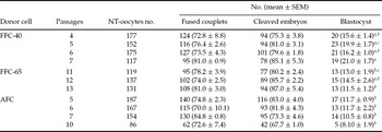
The development data of embryos reconstructed from both adult or fetal fibroblasts and cumulus cells are summarized in Tables 1 and 2. Six replicates were performed to compare developmental potential of adult and fetal cells cultured under serum-starved conditions. The ranges of cell fusion between the recipient cytoplast and fetal cell lines were 47–87% and 67–86% for 40-day and 65-day embryos, respectively. The corresponding values for adult cells were 50–87% and 82–92%, for fibroblasts and cumulus cells, respectively.
Rates of cell fusion and in vitro embryo development of reconstructed ovine oocytes using fetal and adult donor cells.

a –cNumbers with different superscripts in the same column differ significantly (p < 0.001).
ACC: adult cumulus cells; AFC: adult fibroblast cell; FFC-40: fetal fibroblast cell-40 (40-day-old); FFC-65: fetal fibroblast cell-65 (65-day-old).
Influence of the number of donor cell passages on fusion and in vitro embryo development rates of reconstructed ovine oocytes using fetal and adult fibroblasts.

a–cNumbers with different superscripts in the same column differ significantly (p < 0.001).
AFC: adult fibroblast cell; FFC-40: fetal fibroblast cell-40 (40-day-old); FFC-65: fetal fibroblast cell-65 (65-day-old).
The rates of electro-fusion was not affected by either the age (fetal vs. adult) or type (fibroblast vs. cumulus) of donor cells and passage numbers in fetal (4–7 and 11–13) and adult (5–7 and 10) fibroblast-derived NT embryos (Table 2).
Cleavage rate of the reconstructed oocytes 48 h after cell fusion was not significantly different between oocytes reconstructed with fetal and adult donor cells. When the effect of passage number was analysed within groups (fetal vs. adult), there were no significant differences in cleavage rate of reconstructed oocytes (Table 2). The range of cleavage rates was 64–100% and 71–94% for NT embryos reconstructed with FFC-40 and FFC-65, respectively. The corresponding values for NT embryos reconstructed with AFC and ACC were 64–94% and 68–77%, respectively.
Blastocyst production ranges were 8–25% and 6–19% of successfully fused oocytes for fetal and adult cell-derived NT embryos, respectively. The rate of blastocyst formation was significantly higher in FFC-40 and ACC-derived reconstructed embryos compared with AFC-derived NT embryos (Table 1).
Discussion
In this study, there was no significant difference in cell cycle length between fibroblasts derived from fetuses and a young adult lamb. The cell cycle length, however, was slightly longer in the young adult donor lamb than in the fetuses. The cell lines from both fetuses and young adult lamb continued to grow to at least 60 population doublings in culture without becoming senescent, which is higher than what has been demonstrated in other studies (Cibelli et al., Reference Cibelli, Stice, Golueke, Kane, Jerry, Blackwell, Ponce de Leon and Robl1998; Hill et al., Reference Hill, Winger, Long, Looney, Thompson and Westhusin2000; Lanza et al., Reference Lanza, Cibelli, Blackwell, Cristofalo, Francis, Baerlocher, Mak, Schertzer, Chavez, Sawyer, Lansdorp and West2000; Kasinathan et al., Reference Kasinathan, Knott, Moreira, Burnside, Jerry and Robl2001). The higher life span of donor cells in this study may be due to the culture conditions or species-specific differences.
We compared the developmental rates of reconstructed ovine oocytes using adult and fetal fibroblasts under serum-starved (Freshney, Reference Freshney1994) culture conditions as well as fresh cumulus cells as donor cells to determine which cell type has higher capability to develop to the blastocyst stage.
Despite the perceived differences in the time needed for dissolution of the plasma membrane of various donor cell types, no difference was found in the average fusion rate among couplets reconstructed with FFC-40, FFC-65, AFC, or ACC donor cells. These findings are in agreement with those for other domestic species (Reggio et al., Reference Reggio, James, Green, Gavin, Behboodi, Echelard and Godke2001).
There is, however, evidence indicating the difference in cell fusion between fibroblasts and cumulus cells as well as between adult and fetal fibroblasts (Hill et al., Reference Hill, Winger, Long, Looney, Thompson and Westhusin2000; Lee et al., Reference Lee, Wu, Tian, Barber, Hoagland, Riesen, Lee, Tu, Cheng and Yang2003). The discrepancy may be attributable to the differences in the membrane properties of these cells such as their protein and/or lipid compositions (Lee et al., Reference Lee, Wu, Tian, Barber, Hoagland, Riesen, Lee, Tu, Cheng and Yang2003).
In the current study, 48 h after cell fusion, the average rates of embryonic cleavage to the 2-cell stage were not different among oocytes reconstructed with different cell types. This finding confirms what has been previously reported, indicating that nuclear formation and normal division of SCNT embryos was not markedly affected by the donor cell type (Tian et al., Reference Tian, Kubota, Enright and Yang2003; Ng et al., Reference Ng, Chen, Yip, Liow, Tong, Martelli, Tan and Martelli2004). First embryonic division in reconstructed ovine oocytes was identical to the rate achieved routinely in cattle (Kato et al., Reference Kato, Tani and Tsunoda2000), sheep (Campbell et al., Reference Campbell, McWhir, Ritchie and Wilmut1996; Wilmut et al., Reference Wilmut, Schnieke, McWhir, Kind and Campbell1997), goats (Baguisi et al., Reference Baguisi, Behboodi, Melican, Pollock, Desrempes, Cammuso, Williams, Nims, Porter, Midura, Palacios, Ayres, Denniston, Hayes, Ziomek, Meade, Godke, Gavin, Overstrom and Echelard1999; Keefer et al., Reference Keefer, Baldassarre, Keyston, Wang, Bhatia, Bilodeau, Zhou, Leduc, Downey, Lazaris and Karatzas2001) and pigs (Lai et al., Reference Lai, Tao, Machaty, Kuhholzer, Sun, Park, Day and Prather2001; Park et al., Reference Park, Lai, Cheong, Im, Sun, Wu, Day and Prather2001). This finding indicates that the nuclei of both fetal and adult ovine fibroblasts as well as adult cumulus cells have similar remodelling potential in term of cleavage rate.
As shown in Tables 1 and 2, although no differences were detected in the cleavage rates of embryos from the three different cell types, FFC-40 produced significantly higher rate of blastocyst development compared with the FFC-65 and AFC. This finding is in agreement with our general knowledge indicating the higher pregnancy and calving rates of cloned embryos derived from fetal cells than those from adult cells. However, there are several reports in other species indicating the lack of significant difference in development of fused oocyte–fibroblast couplets to blastocysts between fibroblasts derived from fetal and from adult cell (Hill et al., Reference Hill, Winger, Long, Looney, Thompson and Westhusin2000; Kato et al., Reference Kato, Tani and Tsunoda2000; Kasinathan et al., Reference Kasinathan, Knott, Moreira, Burnside, Jerry and Robl2001). Similarly, Renard et al. (Reference Renard1999), Hills et al. (Reference Hill, Winger, Long, Looney, Thompson and Westhusin2000) and Wakayama & Yanagimachi (Reference Wakayama and Yanagimachi2001) also reported that developmental rates of somatic cloned embryos remained similar regardless of the donor age.
In the current study, it is not clear why the blastocyst rate in FFC-65 reconstructed embryos is significantly lower than FFC-40. As the culture condition and all procedures were identical, one possibility for the lower rate might be the higher number of passages in FFC-65 compared with FFC-40. However, this is in contrast to our general knowledge indicating that cells derived from higher passages contain less epigenetic modifications and are more receptive to nuclear reprogramming (Enright et al., Reference Enright, Jeong, Yang and Tian2003).
Nevertheless, one explanation for the higher potential of fetal cells in producing healthy live births might be the fact that somatic cells of adult animals have accumulated more genetic aberrations and are more terminally differentiated than fetal cells and are thus more likely to fail at full-term development (Tian et al., Reference Tian, Kubota, Enright and Yang2003). Whether this concept could explain the higher remodelling potential of FFC-40 in reconstructed oocytes compared with AFC remains to be elucidated.
The higher rate of blastocyst formation derived form ACC-reconstructed embryos compared with AFC was in agreement with several reports indicating that among the somatic cell types tested, the consensus from numerous laboratories is that cumulus cells give the highest cloning efficiency and result in the least number of abnormalities in cloned animals (Tian et al., Reference Tian, Kubota, Enright and Yang2003). It has been documented that DNA from cumulus cells is more prone to be reprogrammed following nuclear transfer (Tian et al., Reference Tian, Kubota, Enright and Yang2003). Furthermore, it was reported that cumulus cell-derived cloned mice do not have widespread imprinting dysregulations (Rideout et al., Reference Rideout, Eggan and Jaenisch2001). Another report supporting the superiority of cumulus cells for nuclear transfer comes from the study by Forsberg et al. (Reference Forsberg, Strelchenko, Augenstein, Betthauser, Childs, Eilertsen, Enos, Forsythe, Golueke, Koppang, Lange, Lesmeister, Mallon, Mell, Misica, Pace, Pfister-Genskow, Voelker, Watt and Bishop2002) and it has also been reported that cumulus and oviduct epithelial cells are the most suitable nuclear donors compared with cells from the liver, testis, skin, or ear (Kato et al., Reference Kato, Tetsuya, Sotomaru, Kurokawa, Kato, Doguchi, Yasue and Tsunoda1998, Reference Kato, Tani and Tsunoda2000).
The number of donor cell passages is another contributing factor in cloning by nuclear transfer. Most reports of successful cloning in domestic animal species have used cells of limited passages (3–9) as sources of donor nuclei (Wilmut et al., Reference Wilmut, Schnieke, McWhir, Kind and Campbell1997; Kato et al., Reference Kato, Tetsuya, Sotomaru, Kurokawa, Kato, Doguchi, Yasue and Tsunoda1998; Wells et al., Reference Wells, Misica and Tervit1999; Hill et al., Reference Hill, Winger, Long, Looney, Thompson and Westhusin2000; Reggio et al., Reference Reggio, James, Green, Gavin, Behboodi, Echelard and Godke2001). It has been reported that nuclei from both early passage (8–16) and late passage (17–32) donor cells were capable of supporting in vitro development after nuclear transfer in cattle, although the rate of blastocyst formation was lower when using the late passage cells (Roh et al., Reference Roh, Shim, Hwang and Yoon2000). In the present study, the number of donor cell passages ranged between 4 and 13 and the rates of fusion and embryo development were not influenced by the number of passages in each cell type groups (fetal vs. adult). This finding is in agreement with what has been reported by Kubota et al. (Reference Kubota, Yamakuchi, Todoroki, Mizoshita, Tabara, Barber and Yang2000) indicating no differences in the development of NT-embryos when using adult fibroblasts that ranged in passage numbers from 5–15.
During cell passage, both genetic and epigenetic alterations that might affect nuclear remodelling would be expected to accumulate in the cultured cells. Other possible disruptions to the regulation of the imprinted genes could also be induced by repeated culture, thereby leading to perturbations in embryonic and fetal development (Walker et al., Reference Walker, Hartwich and Seamark1996; Wakayama et al., Reference Wakayama, Rodriguez, Perry, Yanagimachi and Mombaerts1999). In the present study, because of the limited number of passages at least in FFC-40 (4–7 passages) and in AFC (5–7 and 10 passages), such genetic and epigenetic alterations or other aberrations would be expected to be limited or exerted equally for both cell types. Although, it seems the lower developmental potential in oocytes reconstructed with FFC-65 is, at least, partially attributed to their slightly higher passage number (11–13).
Previous studies have shown that blastocyst development of NT embryos derived from fetal fibroblasts is ranged between 18 and 43% (Wilmut et al., Reference Wilmut, Schnieke, McWhir, Kind and Campbell1997; Cibelli et al., Reference Cibelli, Stice, Golueke, Kane, Jerry, Blackwell, Ponce de Leon and Robl1998; Wells et al., Reference Wells, Misica, McMillan and Tervit1998) and that of adult cells ranged between 9 and 49% (Kato et al., Reference Kato, Tetsuya, Sotomaru, Kurokawa, Kato, Doguchi, Yasue and Tsunoda1998; Vignon et al., Reference Vignon, Lebourhis, Chesne, Marchal, Heyman and Renard1999; Wells et al., Reference Wells, Misica and Tervit1999). Although the rates of blastocyst development in the present study is comparable with previous reports, the reason for the relatively low rates of blastocyst formation for both fetal and adult NT derived embryos (13–18% and 10.9–17.5%, respectively) could be attributed to the age of the slaughtered ewe lambs (3–6-month-old) as a source of recipient oocytes. As documented, pre-pubertal oocytes show some structural and functional limitations compared to those from adult donors including small size, defective coupling between cumulus cells and oocytes, decreased amino acid uptake and reduced protein synthesis and energy metabolism (Ledda et al., Reference Ledda, Bogliolo, Leoni, Calvia and Naitana1996, Reference Ledda, Bogliolo, Calvia, Leoni and Naitana1997; O'Brien et al., Reference O'Brien, Dwarte, Ryan, Maxwell and Evans1996). Therefore, immature pre-pubertal oocytes used in the present study as recipient cytoplasts could be a contributing factor for the lower developmental rate of the fused couplets to blastocyst. In this context, comparing embryos produced in vitro from both immature pre-pubertal and adult oocytes, Majerus et al. (Reference Majerus, Lequarrè, Ferguson, Kaidi, Massip, Dessy and Donnay2000) observed in the former a significant delay in development to morula but not to blastocyst stage.
In summary, the present experiment demonstrates that ovine fetal and adult fibroblasts that have undergone few passages (4–13) as well as fresh cumulus cells are comparable in their ability to achieve cell fusion and embryonic cleavage to the 2-cell stage in reconstructed oocytes. Moreover, the age of the donor animal has a pronounced impact on the development of fused oocyte–fibroblast couplets to blastocyst and that the fresh cumulus cells have similar remodelling potential to that of fetal fibroblasts in oocytes matured in vitro in term of blastocyst formation rate.
Acknowledgements
The authors would like to thank the Research Institute of Animal Embryo Technology for technical and financial supports, Shahrekord University and Shahrekord's slaughterhouse staff for their cooperation.