Highlights
-
• Peripheral neuropathies are increasingly recognized in spinal muscular atrophy (SMA), most commonly motor axonal, with occasional sensory or demyelinating involvement.
-
• SMN deficiency affects peripheral nerves beyond motor neurons, consistent with experimental and developmental evidence.
-
• Nerve conduction studies may complement clinical scales in selected patients for phenotyping and longitudinal monitoring.
Introduction
Spinal muscular atrophy (SMA) is an autosomal recessive motor neuron disorder caused by deletion or mutation of SMN1. The classic form of SMA is characterized by progressive degeneration of α-motor neurons in the spinal cord. Reference Mercuri, Pera, Scoto, Finkel and Muntoni1 Although a higher SMN2 copy number is generally associated with a milder clinical phenotype, substantial variability remains within patient subgroups defined by SMN2 copy number, including clear discordance between the expected phenotype and the actual clinical presentation. Reference Cuscó, Bernal and Blasco-Pérez2 This variability is particularly evident among individuals with three SMN2 copies. Reference Dangouloff, Boemer, Dideberg, Caberg and Servais3,Reference Dosi and Masson4 The classic phenotype of SMA therefore reflects a neuromuscular condition marked by progressive loss of anterior horn motor neurons. Reference Kolb and Kissel5
The compound motor action potential (CMAP) represents the overall electrical output generated by motor units innervating a muscle or group of muscles when subjected to supramaximal nerve stimulation. Reference Pino, Rich and Kolb6 CMAP is obtained during routine motor nerve conduction studies (NCSs) and constitutes a rapid and accessible measurement. Reference Bjelica, Wohnrade and Osmanovic7 In children with SMA type I, the reduction in CMAP amplitude observed in both upper and lower limbs may indicate widespread degeneration of spinal motor neurons, Reference Ogata, Natarajan and Sripadrao8 resulting in a neurophysiological pattern compatible with motor axonal neuropathy. CMAP assessment may also serve as a valuable biomarker for monitoring the effectiveness of currently available therapies, including nusinersen, risdiplam and onasemnogene abeparvovec. Reference Ogata, Natarajan and Sripadrao8–Reference Yonekawa, Komaki, Saito, Sugai and Sasaki11 Nevertheless, the literature also reports cases involving sensory nerve impairment, showing both axonal and demyelinating features. Reference Pro, Tozzi and D’Amico12,Reference Anagnostou, Miller and Fehlings13
The aim of this systematic review is to summarize the different neuropathic patterns described in the literature among patients with SMA, highlighting the neurophysiological characteristics in both treated and untreated individuals. In addition, we seek to determine whether neurophysiology, based on currently available data, can serve as a reliable biomarker for evaluating the effectiveness of modern SMA therapies.
Materials and Methods
Study registration and reporting
This systematic review was conducted in accordance with the PRISMA 2020 guidelines Reference Page, McKenzie and Bossuyt14 and was prospectively registered in the International Prospective Register of Systematic Reviews (PROSPERO, ID: CRD1231714). All methodological steps – including search, study selection, data extraction and synthesis – were defined a priori.
Eligibility criteria
Inclusion criteria
Studies were eligible if they met the following criteria:
Population: patients of any age diagnosed with 5q-SMA of any subtype (0–4) confirmed by biallelic SMN1 deletion and/or pathogenic SMN1 variants.
Condition of interest: NCSs in genetically confirmed SMA patients.
Outcomes: qualitative or quantitative data on:
-
Presence and classification of neuropathy (axonal vs demyelinating; motor vs sensory vs mixed);
-
Motor and/or sensory amplitudes, conduction velocities or latencies;
-
Longitudinal neurophysiological evolution (stable, improved and worsened);
-
Associations with SMN2 copy number or disease-modifying therapies (nusinersen, risdiplam and onasemnogene abeparvovec).
Study design: original research reporting patient-level or cohort-level data: case reports, case series, cohort studies and clinical trials; retrospective or prospective studies.
Language and setting: articles published in English, without restrictions on publication year or geographical setting.
Publication type: letters to the editor were included only if they provided original clinical, genetic or electrophysiological data.
Exclusion criteria
Studies were excluded if they met any of the following:
-
SMA not genetically confirmed (no SMN1 testing; clinical diagnosis only; linkage studies; non-5q-SMA);
-
Neuropathies unrelated to SMA;
-
Animal or in vitro studies without extractable human data;
-
Studies focusing exclusively on central nervous system pathology without peripheral nerve, root or dorsal root ganglia (DRG) assessment;
-
Articles reporting only muscle or anterior horn cell pathology without peripheral nerve data;
-
Insufficient data to characterize neuropathy (no NCS);
-
Reviews, meta-analyses, commentaries, editorials or expert opinions without original data;
-
Conference abstracts, posters or oral presentations without a full peer-reviewed manuscript;
-
Non-English publications.
Information sources and search strategy
A comprehensive search was performed in the following electronic databases from inception to 4 November 2025:
-
PubMed/MEDLINE
-
Embase
-
Scopus
The core search string was: (“SMA” OR “Spinal muscular atrophy”) AND (“Neuropathy” OR “Neuropathies”). Reference lists of included studies and relevant reviews were screened manually to identify additional eligible articles.
Study selection
All retrieved records were imported into a reference management system, and duplicates were removed. A total of 11,886 unique records were screened. Six independent reviewers assessed titles and abstracts using the predefined eligibility criteria. Full texts of potentially eligible studies were then retrieved and reviewed in duplicate by the same six reviewers. Disagreements were resolved through discussion; when consensus was not reached, a third reviewer adjudicated.
Data extraction
Clinical, genetic and electrophysiological data were extracted into a standardized Microsoft Excel spreadsheet. Variables collected included:
-
Study characteristics (author, year, country and design).
-
Number of SMA patients; sex; age at diagnosis; age at first NCS.
-
SMA subtype (0–4).
-
SMN1 genotype and SMN2 copy number.
-
Type of neuropathy (motor/sensory/mixed; axonal/demyelinating/mixed).
-
NCS parameters at baseline and follow-up.
-
Exposure to nusinersen, risdiplam, onasemnogene abeparvovec or combinations; drug-naïve status.
-
Longitudinal evolution (stable, improved and worsened).
Data extraction was performed independently and in duplicate by six reviewers (G.P., L.C., M.G., B.V., G.A. and A.I.) using the predefined spreadsheet. Discrepancies were resolved by discussion and, when necessary, by consultation with a seventh reviewer (C.A.C.). After initial extraction, a random 20% sample of entries was cross-checked for consistency, and no major errors or missing data were identified.
Quality assessment
Because the included studies consisted mainly of case reports, case series and small observational cohorts, methodological quality was assessed using Joanna Briggs Institute (JBI) critical appraisal tools, applied according to study design.
The assessment was performed independently by two reviewers, focusing on:
-
Clarity of SMA genetic diagnosis.
-
Adequacy and replicability of neurophysiological or neuropathological methods.
-
Completeness and transparency of outcome reporting.
-
Risk of selection and reporting bias.
Risk-of-bias judgments were subsequently summarized qualitatively across studies, focusing on the most frequent domains of concern.
Data synthesis and statistical analysis
Given the clinical heterogeneity across studies (age, SMA subtype, SMN2 copy number, neurophysiological methods and therapy exposure), a descriptive synthesis was planned. NCS findings, neuropathy classifications and follow-up changes were summarized narratively and in structured tables. When comparable quantitative data were available across ≥2 studies, summary statistics were calculated.
All analyses and visualizations were performed using R (R Foundation for Statistical Computing) within RStudio (2025.09.2).
A large language model-based assistant (OpenAI) was used to support English language editing. The tool was not used to generate, select or screen studies, nor to extract, code or analyze data. All data extraction, statistical analyses and clinical interpretations were performed by the authors, who carefully checked and approved the final manuscript.
Results
Study selection and characteristics
After full-text screening, 23 studies published between 1999 and 2025 met the eligibility criteria and were included in the quantitative synthesis. Reference Pro, Tozzi and D’Amico12,Reference Anagnostou, Miller and Fehlings13,Reference Axente, Sporea, Mirea, Burcea and Ion15–Reference Rudnik-Schöneborn, Barisić and Eggermann35 Study designs were heterogeneous and predominantly observational. There were given as follows:
-
8 single-patient case reports; Reference Anagnostou, Miller and Fehlings13,Reference Fernández, Peciña, Muñoz-Cabello, Antínolo and Borrego19–Reference García-Cabezas, García-Alix and Martín21,Reference Koul, Abdelrahim, Al-Nabhani and Al-Futaisi25,Reference Li, Li, Wang and Wu27,Reference Li, Perera and Varghese28,Reference Reid, Arends and Wirtz32
-
5 case series with 2–5 patients; Reference Harding, Kariya and Monani22,Reference MacLeod, Taylor, Lunt, Mathew and Robb29–Reference Ravid, Topper and Eviatar31,Reference Rudnik-Schöneborn, Barisić and Eggermann35
-
10 larger series/cohorts with ≥10 patients. Reference Pro, Tozzi and D’Amico12,Reference Axente, Sporea, Mirea, Burcea and Ion15–Reference Farrar, Vucic and Lin18,Reference Kariyawasam, D’Silva and Howells23,Reference Koszewicz, Ubysz, Dziadkowiak, Wieczorek and Budrewicz24,Reference Li, Sun and Xiang26,Reference Ros, Sleutjes and Goedee33,Reference Rudnik-Schöneborn, Goebel and Schlote34
Geographically, studies originated from Europe, North America, Asia and Australia, with no obvious regional clustering of neurophysiological patterns.
The study selection process is shown in Figure 1.
PRISMA flow chart.

Quality assessment
Using the appropriate JBI appraisal tools, overall methodological quality was low-to-moderate across studies. The most frequent sources of bias were incomplete reporting of patient selection, limited description of NCS protocols and absence of blinded outcome assessment. Case reports and small case series were particularly affected by restricted sample size and lack of systematic follow-up, whereas larger cohorts showed more consistent methodology but still variable detail in electrophysiological reporting. No study preregistered a protocol or reported sample-size calculations.
Demographic and clinical characteristics of population
Across the 23 included studies, a total of 395 patients with clinically and genetically confirmed 5q-SMA underwent NCSs. Sex was specified for most participants: 183 males (46.3%) and 188 females (47.6%), whereas sex was not reported for 24 patients (6.1%). Thus, the overall sex distribution in the available data was approximately balanced.
Clinical SMA subtype (0–4) was reported for 338 out of 395 patients (85.6%). Among these, 2 patients had SMA0 (0.5%), 107 SMA1 (27.1%), 160 SMA2 (40.5%) and 69 SMA3 (17.5%); no cases of SMA4 were identified. In 57 patients (14.4%), the SMA type was not specified in the original reports or was provided only in aggregated form (e.g., “SMA II–III” or “type unclear”), preventing precise classification. Overall, the cohort spans the full phenotypic spectrum of 5q-SMA, from severe infantile forms to adolescent and adult-onset types. Demographic and clinical characteristics are summarized in Table 1.
Study-level demographic characteristics, SMA clinical spectrum, age at first NCS and therapeutic context at baseline (n = 23 studies)

CMT1A = Charcot-Marie-Tooth disease type 1A; NCS = nerve conduction study; SMA = spinal muscular atrophy.
Genetic background and SMN2 copy number
SMN1 status
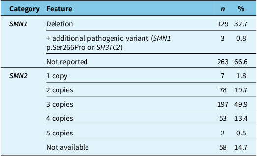
SMN1 genotype was explicitly reported in 132/395 patients (33.4%). Among these:
-
129 patients (32.7%) had biallelic SMN1 deletions;
-
3 patients had SMN1 deletion combined with an additional pathogenic variant: 1 patient on SMN1 gene (p.Ser266Pro - Li L) and 2 patients on SH3TC2 gene. Reference Rudnik-Schöneborn, Barisić and Eggermann35
For 263 patients (66.6%), SMN1 genotype was not specified beyond confirmation of 5q-SMA.
Across studies, no evidence suggested that SMN1 point mutations or missense variants produced a distinct peripheral neuropathy pattern compared with classic deletion-mediated SMA.
SMN2 copy number
SMN2 copy number was reported in most cohorts, although not always at the individual-patient level. Aggregated across studies:
-
1 copy: 7 patients (1.8%)
-
2 copies: 78 patients (19.7%)
-
3 copies: 197 patients (49.9%)
-
4 copies: 53 patients (13.4%)
-
5 copies: 2 patients (0.5%)
For 58 patients (14.7%), SMN2 copy number was not reported.
Aggregated genetic data are reported in Table 2.
Aggregated genetic characteristics (SMN1 genotype and SMN2 copy number) across all included SMA patients

SMA = spinal muscular atrophy.
NCS methodology
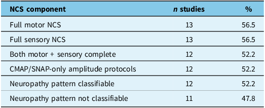
NCS methodology varied substantially across the 23 included studies. Full motor NCSs – defined as assessment of CMAP amplitude, distal latency and motor conduction velocity in at least one proximal and one distal nerve – were performed in 13 studies (56.5%). Full sensory NCS, including sensory nerve action potential (SNAP) amplitude and sensory conduction velocity in at least one upper- and one lower-limb nerve, were also reported in 13 studies (56.5%). Both components were fully assessed in 12 studies (52.2%).
Twelve studies (52.2%) used simplified amplitude-only protocols (CMAP and/or SNAP without conduction velocities or distal latencies), predominantly in biomarker- or treatment-focused cohorts. These designs enabled quantification of motor unit integrity but did not allow differentiation between axonal and demyelinating mechanisms.
Overall, neuropathy pattern (motor, sensory, sensorimotor; axonal vs demyelinating) could be classified in 12/23 studies (52.2%), while 11/23 studies (47.8%) lacked sufficient electrophysiological detail.
Percentages refer to the number of studies rather than to the number of patients, because reporting of NCS parameters was heterogeneous and often incomplete at the individual-patient level.
Completeness of NCS methodology across studies is summarized in Table 3.
Completeness of NCS methodology across the 23 included studies. Definitions: full motor = CMAP amplitude + distal latency + MCV in ≥1 proximal and ≥1 distal nerve. Full sensory = SNAP amplitude + SCV in ≥1 upper and ≥1 lower limb nerve. CMAP/SNAP-only = amplitude-only protocols without velocities or latencies

CMAP = compound motor action potential; NCS = nerve conduction study; SNAP = sensory nerve action potential; MCV = motor conduction velocity; SCV = sensory conduction velocity.
Neurophysiological findings
Neurophysiological patterns could be evaluated in 12 of 23 studies (52.2%), which reported sufficient NCS detail to classify neuropathy type (axonal vs demyelinating; motor, sensory or sensorimotor).
The remaining 11 studies (47.8%) provided CMAP and/or SNAP amplitude data only, without conduction velocities or distal latencies, preventing formal neuropathy classification.
Axonal abnormalities
Axonal involvement represented the most consistent neurophysiological finding across the included literature.
-
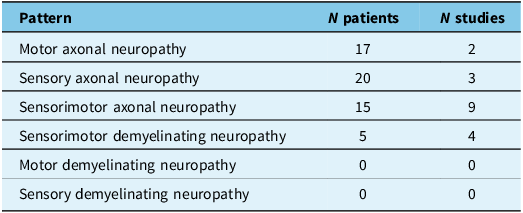
Sensorimotor axonal neuropathy was the most frequent pattern, identified in 15 patients across 9 studies. Reference Anagnostou, Miller and Fehlings13,Reference Duman, Uysal, Skjei, Kizilay, Karauzum and Haspolat17,Reference Fernández-Torre, Teja and Castellanos20,Reference Li, Li, Wang and Wu27–Reference MacLeod, Taylor, Lunt, Mathew and Robb29,Reference Ravid, Topper and Eviatar31,Reference Reid, Arends and Wirtz32,Reference Rudnik-Schöneborn, Goebel and Schlote34 This distributed pattern suggests widespread axonal dysfunction rather than isolated motor or sensory fiber involvement.
-
Sensory axonal neuropathy was reported in 20 patients across 3 studies. Reference Pro, Tozzi and D’Amico12,Reference Duman, Uysal, Skjei, Kizilay, Karauzum and Haspolat17,Reference Rudnik-Schöneborn, Barisić and Eggermann35
-
Motor axonal neuropathy was documented in 17 patients across 2 studies. Reference Duman, Uysal, Skjei, Kizilay, Karauzum and Haspolat17,Reference MacLeod, Taylor, Lunt, Mathew and Robb29
Demyelinating abnormalities
-
Demyelinating features were distinctly uncommon.
-
Sensorimotor demyelinating neuropathy was reported in five patients across four studies (Duman; Fernández; Ravid; Rudnik-Schöneborn).
-
No study reported isolated motor demyelinating neuropathy.
-
No study reported isolated sensory demyelinating neuropathy.
Extractable neurophysiological patterns are summarized in Table 4.
Extractable neurophysiological patterns from studies with classifiable NCS (n = 12)

NCS = nerve conduction study.
Longitudinal NCS data
Longitudinal electrophysiological follow-up was reported only in a subset of cohorts and was methodologically heterogeneous. As shown by the aggregated dataset, almost all follow-up data were derived from CMAP/SNAP-only amplitude protocols, whereas complete motor and sensory NCS were rarely repeated over time.
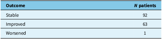
Among the 12 studies that performed full motor and sensory NCS at baseline, only three patients underwent repeat assessment with complete NCS parameters (2 stable, 1 worsened and 0 improved).
In contrast, amplitude-only CMAP/SNAP protocols provided the vast majority of longitudinal observations: across 153 patients with CMAP/SNAP follow-up, 90 (58.8%) were classified as stable and 63 (41.2%) as improved, with no patient reported as worsened.
Longitudinal electrophysiological outcomes are summarized in Table 5.
Longitudinal nerve conduction study outcomes extracted from complete and CMAP/SNAP-only follow-up assessments

CMAP = compound motor action potential; SNAP = sensory nerve action potential.
Electrophysiological follow-up by treatment category
Longitudinal electrophysiological data were available in a subset of studies and are summarized by treatment category.
Given the heterogeneity of study designs, patient populations and electrophysiological protocols, Figure 2 is intended to provide a descriptive overview of reported electrophysiological outcomes by treatment category and should not be interpreted as a comparative analysis between treatments.
Reported electrophysiological outcomes at follow-up by treatment category (descriptive overview).

Electrophysiological outcomes by treatment category are shown in Figure 2.
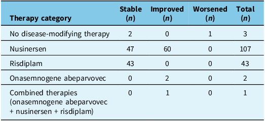
Electrophysiological outcomes differed substantially across treatment groups (Figure 2). Among untreated patients (n = 3), two remained stable and one worsened.
Nusinersen-treated patients (n = 107) showed the most balanced pattern, with 60 patients (56.1%) demonstrating improvement in CMAP/SNAP amplitudes and 47 (43.9%) remaining stable; no deterioration was reported.
Risdiplam-treated patients (n = 43) exhibited uniform stability, with no documented improvements or worsening.
All three patients assessed after onasemnogene abeparvovec treatment showed improvement, while the single patient receiving combined therapies also improved.
Notes
-
1. The majority of follow-up data derive from amplitude-only CMAP/SNAP evaluations, particularly in treatment-focused cohorts.
-
2. Nusinersen and gene-replacement therapy (onasemnogene abeparvovec) show the highest proportion of improvement.
-
3. Risdiplam-treated patients display uniformly stable electrophysiological trajectories in available data.
-
4. Untreated patients show mixed outcomes, but the sample size is small (n = 3).
-
5. Combined therapy data are limited to a single patient.
Electrophysiological outcomes by treatment category are reported in Table 6 and illustrated in Figure 2.
Electrophysiological follow-up outcomes stratified by treatment category

Bars indicate the number of patients classified as improved, stable or worsened at longitudinal electrophysiological follow-up within each treatment category (n shown above each bar). This figure provides a descriptive synthesis of heterogeneous studies and does not support comparative inference. Follow-up evaluations were predominantly based on CMAP/SNAP amplitude-only protocols.
Discussion
This systematic review synthesizes NCS findings in genetically confirmed 5q-SMA across 23 studies published between 1999 and 2025, comprising 395 patients spanning the clinical spectrum from SMA0 to SMA3. Three overarching observations emerge. First, despite the traditional view of 5q-SMA as a predominantly motor neuron disorder, peripheral nerve involvement is repeatedly described, particularly in severe infantile phenotypes. Reference Anagnostou, Miller and Fehlings13,Reference Duman, Uysal, Skjei, Kizilay, Karauzum and Haspolat17,Reference Rudnik-Schöneborn, Goebel and Schlote34 Second, interpretation of the neurophysiological literature is substantially constrained by heterogeneous NCS methodology and incomplete reporting, with formal neuropathy classification feasible in only approximately half of the included studies. Third, longitudinal electrophysiological data – although limited and largely based on amplitude-only protocols – suggest that treated patients are more often reported as stable or improved, whereas deterioration is rarely documented and appears mainly in the small untreated sample. Reference Axente, Sporea, Mirea, Burcea and Ion15,Reference Kariyawasam, D’Silva and Howells23,Reference Li, Sun and Xiang26,Reference Ros, Sleutjes and Goedee33
A key finding of this review is that the available evidence is not methodologically uniform enough to support robust prevalence estimates of specific neuropathy subtypes across the full pooled cohort. While slightly more than half of studies reported complete motor and sensory NCS, only 12 of 23 studies provided sufficient detail to classify neuropathy type (axonal vs demyelinating; motor vs sensory vs sensorimotor). Conversely, nearly half relied on simplified amplitude-only protocols, particularly in treatment-focused or biomarker-oriented cohorts. Reference Axente, Sporea, Mirea, Burcea and Ion15,Reference Boulay, Delmont, Audic, Chabrol and Attarian16,Reference Kariyawasam, D’Silva and Howells23,Reference Li, Sun and Xiang26,Reference Pant and Verma30 These designs are well suited for quantifying motor unit integrity and longitudinal change but intrinsically preclude mechanistic classification. As a result, apparent differences in reported neuropathy patterns across studies likely reflect differences in ascertainment and reporting rather than true biological divergence.
These methodological limitations are compounded by the overall low-to-moderate quality of the literature. Common sources of bias included incomplete reporting of patient selection, limited description of NCS acquisition parameters and lack of blinded outcome assessment, particularly in single-patient reports and small case series. Reference Anagnostou, Miller and Fehlings13,Reference Fernández-Torre, Teja and Castellanos20,Reference Reid, Arends and Wirtz32 None of the included studies preregistered a protocol or reported sample-size calculations, further limiting inferential strength.
Across studies, two partially overlapping neurophysiological scenarios emerge that help reconcile seemingly contradictory findings.
Several case reports, small series and pathology-informed studies describe severe infantile SMA presenting with neurophysiological patterns resembling a peripheral sensorimotor axonal neuropathy or sensory neuronopathy, including reduced or absent SNAPs. Reference Anagnostou, Miller and Fehlings13,Reference Duman, Uysal, Skjei, Kizilay, Karauzum and Haspolat17,Reference García-Cabezas, García-Alix and Martín21,Reference MacLeod, Taylor, Lunt, Mathew and Robb29,Reference Rudnik-Schöneborn, Goebel and Schlote34 In some cases, these findings led to initial misdiagnoses such as congenital neuropathy, Guillain–Barré syndrome or critical illness neuropathy. Reference Pro, Tozzi and D’Amico12,Reference Fernández-Torre, Teja and Castellanos20 Collectively, these studies demonstrate that sensory abnormalities and mixed axonal patterns do not exclude SMA, particularly in severe infantile phenotypes.
In contrast, cohorts focusing on adolescents and adults, or on upper-limb sensory nerves, frequently report preserved SNAPs and a predominantly motor neurogenic profile. Reference Farrar, Vucic and Lin18,Reference Li, Li, Wang and Wu27,Reference Ros, Sleutjes and Goedee33 In these studies, electrophysiology supports the concept of SMA as a “pure motor” disorder at the peripheral nerve level, despite marked motor neuron dysfunction. Importantly, this pattern does not negate sensory vulnerability in SMA but suggests that sensory involvement is phenotype-, age- and nerve-dependent.
This dual-scenario framework places negative findings (preserved SNAPs in older patients) and positive findings (sensory abnormalities in severe infantile phenotypes) on a biological continuum rather than treating them as contradictory observations.
Among the subset of studies with sufficient electrophysiological detail to classify neuropathy patterns, axonal involvement clearly predominates. Motor axonal neuropathy, sensory axonal neuropathy and sensorimotor axonal neuropathy were repeatedly reported across multiple studies. Reference Duman, Uysal, Skjei, Kizilay, Karauzum and Haspolat17,Reference MacLeod, Taylor, Lunt, Mathew and Robb29,Reference Ravid, Topper and Eviatar31,Reference Rudnik-Schöneborn, Goebel and Schlote34 Sensorimotor axonal neuropathy was the most frequently reported mixed pattern, particularly in severe infantile SMA. Reference Anagnostou, Miller and Fehlings13,Reference Fernández-Torre, Teja and Castellanos20,Reference Reid, Arends and Wirtz32 In contrast, demyelinating abnormalities were distinctly uncommon and never reported as isolated motor or isolated sensory demyelinating neuropathy.
Demyelinating features were reported in only a small number of patients and always in a sensorimotor distribution. Reference Pro, Tozzi and D’Amico12,Reference Duman, Uysal, Skjei, Kizilay, Karauzum and Haspolat17,Reference Fernández, Peciña, Muñoz-Cabello, Antínolo and Borrego19,Reference Rudnik-Schöneborn, Barisić and Eggermann35 Importantly, several of these cases had plausible alternative explanations. Fernández et al. Reference Fernández, Peciña, Muñoz-Cabello, Antínolo and Borrego19 described a patient with SMA II and a PMP22 duplication consistent with Charcot-Marie-Tooth disease type 1A, while Rudnik-Schöneborn et al. Reference Rudnik-Schöneborn, Barisić and Eggermann35 reported compound genetic findings including variants in SH3TC2, a gene associated with hereditary demyelinating neuropathy. These cases highlight the importance of considering coexisting hereditary neuropathies or genetic modifiers when demyelinating features are observed. Taken together, the available clinical and neurophysiological evidence does not support primary demyelination as a characteristic or defining feature of 5q-SMA.
Experimental data provide biological plausibility for myelin involvement in SMA: Hunter et al. Reference Hunter, Aghamaleky Sarvestany, Roche, Symes and Gillingwater36 demonstrated intrinsic Schwann cell defects in SMA mouse models, including reduced SMN expression and impaired myelin protein production reversible by SMN restoration. However, the rarity of demyelinating patterns in the clinical literature suggests that, while possible, demyelination is not a common or defining feature of 5q-SMA.
Sensory involvement represents a central and nuanced finding in this review. Developmental studies have shown that SMN expression, while highest in anterior horn motor neurons, is also present at lower levels in posterior horn neurons and DRG from fetal life into adulthood. Reference Tizzano, Cabot and Baiget37 This distribution provides a plausible biological substrate for sensory involvement in SMA.
Clinically, sensory abnormalities were reported both as isolated sensory axonal neuropathy and as part of a broader sensorimotor axonal pattern. Reference Duman, Uysal, Skjei, Kizilay, Karauzum and Haspolat17,Reference Ravid, Topper and Eviatar31,Reference Rudnik-Schöneborn, Goebel and Schlote34 Pathology-based studies further support genuine sensory neuron involvement in severe infantile SMA. Reference MacLeod, Taylor, Lunt, Mathew and Robb29,Reference Rudnik-Schöneborn, Goebel and Schlote34 In contrast, large cohorts of adolescents and adults demonstrated preserved median SNAPs in the vast majority of patients, with abnormalities more often attributable to age or comorbidities such as diabetes or chemotherapy exposure. Reference Ros, Sleutjes and Goedee33 These observations suggest that sensory involvement in SMA is phenotype-dependent, nerve-dependent, and modulated by disease severity and systemic factors.
Longitudinal NCS data were available only in a subset of studies and were predominantly based on amplitude-only protocols. In nusinersen-treated cohorts, CMAP/SNAP amplitudes were reported as stable or improved in most patients, with no reported deterioration. Reference Axente, Sporea, Mirea, Burcea and Ion15,Reference Kariyawasam, D’Silva and Howells23,Reference Li, Sun and Xiang26 Patients treated with risdiplam showed uniformly stable electrophysiological trajectories in available reports, Reference Ros, Sleutjes and Goedee33 while all patients assessed after onasemnogene abeparvovec showed improvement. Reference Pant and Verma30 In contrast, deterioration was observed only in the small untreated sample. Reference Pro, Tozzi and D’Amico12
These findings should be interpreted cautiously: follow-up intervals, baseline severity and electrophysiological endpoints varied widely across studies, limiting direct comparability. In this context, reported longitudinal “improvements” in CMAP or SNAP amplitudes should be interpreted as stabilization or relative improvement within substantial methodological constraints, rather than as unequivocal evidence of axonal regeneration or true neurophysiological recovery; such changes may reflect developmental maturation, improved survival of motor units, technical variability or disease stabilization under treatment. Nonetheless, the overall pattern is consistent with stabilization or improvement of peripheral motor unit measures in treated patients.
Taken together, the findings of this review indicate that peripheral nerve involvement is a relevant but heterogeneous component of the 5q-SMA spectrum. Axonal abnormalities predominate when neuropathy patterns can be classified, sensory involvement is biologically plausible and most evident in severe infantile phenotypes, and demyelinating features are rare and frequently confounded by alternative explanations. From a diagnostic standpoint, the presence of sensory abnormalities on NCSs should therefore not exclude 5q-SMA from the differential diagnosis, particularly in infants and neonates with severe phenotypes, in whom mixed axonal or sensory neuronopathic patterns have been repeatedly reported. Reference Anagnostou, Miller and Fehlings13,Reference Duman, Uysal, Skjei, Kizilay, Karauzum and Haspolat17,Reference García-Cabezas, García-Alix and Martín21,Reference MacLeod, Taylor, Lunt, Mathew and Robb29,Reference Rudnik-Schöneborn, Goebel and Schlote34 Instead, such findings should prompt careful molecular evaluation for SMN1-related SMA, alongside systematic exclusion of coexisting hereditary neuropathies or genetic modifiers, especially when demyelinating features are observed. Reference Fernández, Peciña, Muñoz-Cabello, Antínolo and Borrego19,Reference Hunter, Aghamaleky Sarvestany, Roche, Symes and Gillingwater36
Future studies would benefit from standardized NCS reporting, including both motor and sensory studies with distal latencies and conduction velocities, assessment of both upper- and lower-limb nerves, and explicit documentation of comorbidities and coexisting genetic neuropathies. Such standardization is essential to clarify the true frequency, mechanisms and clinical relevance of peripheral neuropathy in 5q-SMA, particularly in the era of disease-modifying therapies.
Supplementary material
The supplementary material for this article can be found at https://doi.org/10.1017/cjn.2026.10582.
Acknowledgments
We thank the nurses from the Child Neurology and Psychiatry Unit for their cooperation. We would like to thank the association Progetto Pulcino for its ongoing support to the Child Neurology and Psychiatry Unit, AUSL-IRCCS of Reggio Emilia.
Author contributions
Conceptualization: C.A.C. and C.F.; methodology: M.G., G.P., L.C., A.P., S.R., C.A.C., D.F. and C.F.; data curation: C.A.C., M.G., G.P. L.C., A.I., G.A., B.V. and C.F.; writing – original draft preparation: C.A.C., M.G. and G.P.; writing – review and editing: C.A.C. and C.F.; supervision: C.F. All authors have read and agreed to the published version of the manuscript.
Funding statement
This research received no specific grant from any funding agency in the public, commercial or not-for-profit sectors.
Competing interests
The authors declare that they have no conflicts of interest related to this work.
Use of AI tools
A large language model-based assistant (OpenAI) was used solely to support English-language editing and clarity. The tool was not used to generate scientific content, determine study eligibility, extract or code data, perform statistical analyses or interpret results. All decisions and interpretations were made by the authors, who reviewed and approved the final manuscript.