




No CrossRef data available.
Article contents
Study on the ecological distribution of alveolar Echinococcus in Hulunbeier Pasture of Inner Mongolia, China
Published online by Cambridge University Press: 01 March 2004
Abstract
A study on the ecological distribution of alveolar Echinococcus was carried out in the Hulunbeier Pasture of Inner Mongolia, China during 1998 and 1999. Animals examined included wolves (Canis lupus), red foxes (Vulpes vulpes), sand foxes (Vulpes corsac), domestic dogs (Canis familiaris), Microtus brandti, Meriones unguiculatus, Citellus dauricus, Allactaga sibirica, Phodopus sungorus and Ochotona daurica. Three wolves were found to be infected with E. granulosus. Two sand foxes were infected with E. multilocularis. The majority of infections of alveolar echinococcus was found in M. brandti. Based on the structure of metacestodes found in the livers of naturally infected M. brandti, 3 main variants were observed. Type I had small alveolar cysts with thin cyst walls. Type II had a larger cyst with a thick cyst wall. Infection of laboratory mice with the gravid segments isolated from the naturally infected sand foxes led to the formation of mature Type I alveolar metacestodes in the lungs and Type II metacestodes in the livers of infected animals, respectively.
Information
- Type
- Research Article
- Information
- Copyright
- 2004 Cambridge University Press
INTRODUCTION
Alveolar echinococcosis is a well-described zoonosis caused by the cestode Echinoccocus multilocularis. The disease is a threat to human health in many areas around the world. In the last decade, cases of alveolar hydatid disease have been reported in many countries, including China (Lang & Chen, 1997; Bai & Ma, 1998), India (Subeder et al. 1996), Japan (Sasaki, Wakisaka & Kurisu, 1994), Taiwan (Young et al. 1996), Australia (Bresson-Hadni et al. 1996), Germany (Merkle et al. 1997), Switzerland and France (Piotin et al. 1997), Spain (Barbero et al. 1998), Poland (Bobrowska et al. 1996), Turkey (Turgut, Benli & Eryilmaz, 1997), Morocco (Lahlou et al. 1998) and Brazil (Ferreira et al. 1995). In 1863, Leuckart described the multicystic nature of the alveolar form of hydatid disease, which he designated as a variant of cystic hydatid disease caused by Echinococcus granulosus. Based on the morphology of the parasite and host occurrence, Rausch & Schiller (1954) described E. sibiricensis, recovered from arctic fox (Alopex lagopus) and sled dogs from St Lawrence Island, Alaska. E. sibiricensis is considered to be conspecific with E. multilocularis that was reported in southern Germany (Vogel, 1957). However, due to certain biological differences between E. multilocularis and E. sibiricensis, the former has been designated as E. multilocularis multilocularis and the latter E. multilocularis sibiricensis occurring in northwestern North America (Vogel, 1957).
In 1985, we initiated an investigation on the ecology of rodents and wild carnivores in Hulunbeier Pasture in Inner Mongolia, China. During the investigation we found alveolar Echinococcus in rodents (Microtus brandti) and adult Echinococcus in the sand-fox (Vulpes corsac). We noticed that the morphology, especially the shorter hook structure, was very similar to that reported for E. m. sibiricensis. The structure of the scolex found in Inner Mongolia also differed from that of E. m. multilocularis found in the northwest area of China (Li et al. 1985; Tang et al. 1988). Our studies in the Hulunbeier Pasture were undertaken from April to September during 1998 and 1999. We observed variation in growth-form of metacestodes of E. multilocularis, resembling either the described subspecies multilocularis or sibiricensis. A third variant of a metacestode of E. multilocularis was also found. In this communication we report results of our observations on the distribution of these 3 variants in rodents.
MATERIALS AND METHODS
Field study
The Hulunbeier Pasture of Inner Mongolia, China, is situated at lat. 47–53°N, and long. 115–126°E (Fig. 1). The winter season is from September to April. Severe snowstorms are common in the region; temperatures in winter can drop to less than −30 °C. Capturing rodents in Hulunbeier Pasture is feasible only from April to September. In 1998 and 1999, studies were carried out during those months. Rodents were collected in 4 counties at 8 different locations: Hao-li-bao, West Lan-tun, Xi-ni River, Bao-ge-de, Hulurn, Erl-dun, Xiwu-zue and Yingdamuji.

Fig. 1. Geological location and the ecosystem of the Hulunbeier Pasture of Inner Mongolia. Map of China, arrow shows the site of the Hulunbeier Pasture.
Mousetraps were randomly set around mouse holes in the pastures. The captured rodents were labelled for locations and times of capture and preserved in 10% formalin for further studies. The species of rodents were identified in the laboratory. The rodents were dissected individually for evidence of lesions in the internal organs, such as liver and lungs. The organs displaying lesions were embedded in wax, sectioned serially and stained by the Periodic acid-Schiff (PAS) method.
Wild carnivores examined included 9 wolves (Canis lupus), 6 red foxes (Vulpes vulpes), and 36 sand foxes (Vulpes corsac). Twenty-eight domestic dogs were examined in the study.
Infection of experimental animals in the laboratory
Experimental infections were established in laboratory white mice (QM strain, Xiamen University, China). The animals were divided into 2 groups and orally infected with gravid segments of adult parasites isolated from 2 naturally infected sand foxes, respectively. Mice were examined on day 40 or 9 months post-infection for the presence of aveolar cysts in different organs. The organs displaying lesions were fixed in 10% formalin, embedded in wax. Serial sections were stained by the PAS method.
RESULTS
Investigation of the final hosts from the Hulunbeier Pasture
No specimens of E. multilocularis were found in dogs and red foxes. Three wolves were infected with E. granulosus (Fig. 2A). Two sand foxes were infected with E. multilocularis (Fig. 2B and C).

Fig. 2. Adults and mature protoscoleces of Echinococcus from the Hulunbeier Pasture. (A) Adult Echinococcus granulosus. (B) Adult Echinoccocus multilocularis sibiricensis that was found in the Hulunbeier Pasture. (C) Adult Echinoccocus multilocularis multilocularis. (D) Mature protoscolex of E. m. multilocularis from naturally infected M. brandti and experimentally infected white mice. (E) Mature protoscolex of E. m. sibiricensis from naturally infected M. brandti and experimentally infected white mice.
About 300 adult cestodes were recovered from one of the sand foxes. All were morphologically similar to E. m. sibiricensis (Fig. 2B), as reported by Tang et al. (1988). The total length of the strobila was 1·78–2·26 mm. The mean number of segments was 3–4, the mean length of the large hooks was 26–27 μm; the mean length of the small hooks was 20–22 μm; the position of the genital pore was anterior to middle; the mean number of testes was 14–20 and all were located posterior in relation to the genital pore. Experimental infection of white mice with the adult cestodes of E. m. sibiricensis led to the death of many infected animals at the early stage of infection and the formation of mature alveolar metacestodes in the lungs. The alveolar metacestodes were similar to those for E. m. sibiricensis (Type I metacestodes) found in the liver of naturally infected voles (Figs 2E and 3A). About 2000 E. multilocularis were found in the second infected sand fox. Morphological variations were observed in adult worms. Some were morphologically similar to E. m. sibiricensis from the first sand fox (Fig. 2B); others resembled the cestodes described by Skrjabin & Abuladse (1964), in that the uterus had a different shape and position (Fig. 2C) in the gravid segment. Infection of white mice with eggs from mixed adult worms isolated from the second infected fox led to the formation of mature alveolar metacestodes either in the liver or lungs. The majority of the infected mice had only 1 type of mature metacestode. The structure of alveolar metacestodes in the lungs was similar to that found for E. m. sibiricensis. However, the structure of alveolar metacestodes in the livers was similar to that for E. m. multilocularis (Type II metacestodes). Only small numbers of infected mice were found to have both Type I metacestodes in the lungs and Type II metacestodes in the livers. The structures of mature protoscoleces that were found in the livers (Figs 2D and 3C) of the infected mice were different from those found in the lungs (Figs 2E and 3B) of infected mice.

Fig. 3. PAS staining of tissue sections containing Echinococcus m. sibiricensis (A and B) and E. m. multilocularis (C, D and E) from naturally infected M. brandti or experimentally infected white mice. (A) An alveolar vesicle of E. m. sibiricensis found in the liver of naturally infected M. brandti from the Hulunbeier Pasture. (B) Mature protoscoleces in brood capsule cavity of E. m. sibiricensis found in the lung of experimentally infected white mouse 9 months post-infection. Arrow indicates the membrane surrounding the mature type I protoscoleces, which differs from the structure of mature type II protoscolices seen in (C) and (E). (C) Mature protoscolex of E. m. multilocularis found in the liver of experimentally infected white mouse. Arrow indicates the connection of the mature protoscolex to the inner wall of type II cyst. (D) Alveolar cysts of E. m. multilocularis found in the liver of experimentally infected white mouse 3 months post-infection. Arrows indicate the area of type II protoscolex formation. (E) Alveolar cysts with protoscoleces of E. m. multilocularis found in the liver of experimentally infected white mouse 6 months post-infection.
Infection of Echinococcus in rodents from the Hulunbeier Pasture
In the investigation, 3717 animals belonging to 6 species were examined (Table 1) as follows: 3249 Microtus brandti (87·4%), 403 Meriones unguiculatus (10·8%), 27 Citellus dauricus (0·7%), 16 Allactaga sibirica (0·4%), 21 Phodopus sungorus (0·6%). One lagomorph, Ochotona daurica, also was examined. Similar to our investigation in 1985, Microtus brandti was found to be the dominant rodent in that area (Tang et al. 1988). From these 6 animal species, we only found Aveolar echinococcus in Microtus brandti.

In total, 792 rodents from Xi-ni River, Xiwu-zue and Yingdamuji were negative for Echinococcus infection (Table 2). Many rodents from the remaining 5 pastures (Hao-li-bao, West Lan-tun, Bao-ge-de, Hulurn Su-mu and Erl-dun) were found to be positive for aveolar Echinococcus infections (Table 2). Out of 3249 M. brandti examined, 198 were found to be infected with alveolar Echinococcus with an infection rate of 6·09%. The infection of alveolar echinococcus in M. brandti was not evenly distributed among different counties in Inner Mongolia. The infection rates varied from 0% (in 3 counties) to as high as 19·0% in Hulurn Su-mu of West County (Table 2). Even with the high infection rate, there were no mature protoscoleces found in the studies carried out in 1998 and 1999.

Of the 2457 voles collected from the 5 locations in 2 counties where infected rodents were found, 1294 were females and 1163 males (Table 3). The infection rate of female voles was slightly higher than that of males. Out of 198 Echinococcus-positive voles, 106 were females (53·5%) and 92 males (46·5%). Body lengths of voles examined ranged from about 78–173 mm for females and 73–174 mm for males; the infected voles were found to be the larger individuals. Lengths of infected female voles ranged from about 112 to 167 mm, and from 106 to 174 mm for males.

Based on the structures of metacestodes found in the livers of naturally infected M. brandti, 3 variants were observed in voles of the Hulunbeier Pasture. Type I had small alveolar cysts with thin cyst walls (Fig. 3A). Our observations indicated that the brood capsules of type I metacestodes were derived from the layer of germinal tissue. Type II metacestodes had larger cysts, with thick cyst walls (Fig. 3D and E), and brood capsules were thought to arise through budding of germinal cells. The third variant had a solid cyst (data not shown), evidently an alveolar metacestode of this type has not been described in the literature.
Our studies indicated that the type I metacestode was the dominant form in Hulunbeier Pasture. In 188 infected M. brandti, 148 were considered to have the type I alveolar metacestode (78·7% of total infected). The prevalence of type I was 6·0% in M. brandti. Thirty M. brandti (16%) were found to be infected by type II. The overall infection rate for type II in M. brandti was 1·2%. Only 10 M. brandti (5·3%) from 188 infected voles were identified to be type III metacestode. The overall infection rate for type III in M. brandti was 0·32%.
Seasonal variation of alveolar infections in M. brandti from the Hulunbeier Pasture
A total of 2457 M. brandti was captured from 5 sites in 2 counties (Table 3). During the spring season (April) 546 voles were examined. Thirty-five of them were found to be infected with alveolar metacestodes (6·4%). In the summer (July) 1248 voles were examined for the alveolar infection. Sixty-two voles were found to be positive with alveolar infection (infection rate at 5%). In September, 663 voles were examined, 101 of them were found to be infected with alveolar metacestodes (infection rate at 15·2%).
The sex-ratio of voles examined in different seasons varied slightly (Table 3). In April and September, more males were captured, and more females were found in July. However, infection rates of alveolar metacestodes were found to be higher in female voles in all seasons. The female vole infection rate in April was 7·5% (20/267) in comparison with 5·4% for male voles (15/279). In July, the infection rate for females was 5·3% (38/719), and 4·5% (24/529) for males. The infection rate in September was 15·6% (48/308) for females and 14·9% (53/355) for males. Overall, the infection rate for females was 8·2% (106/1294), and 7·9 for males (92/1163).
DISCUSSION
In 1988, we reported the identification of E. m. sibiricensis in the Hulunbeier Pasture, Inner Mongolia, China (Tang et al. 1988). The classification was based on the structures, including the scolex, of adult worms which were similar to the reported E. m. sibiricensis found in Russia (Wardle et al. 1974) and differed from that reported for E. m. multilocularis in Northwest China (Li et al. 1985). Further investigations on the epidemiology of Echinococcus in the Hulunbeier Parsture carried out in 1998 and 1999, indicated that there were 3 variants of alveolar metacestodes in the voles examined based on the structure of early unilocular cysts and formation of brood capsules in the livers of infected voles, though the true nature of type III variant of alveolar metacestodes was not clear.
Although differences between E. m. multilocularis and E. m. sibiricensis have been reported (Rausch & Schiller, 1954; Vogel, 1957), for many years these 2 parasites were considered to be the different strains of the same species. E. m. multilocularis is mainly found in Europe and E. m. sibiricensis in Alaska, Siberia. However, it is interesting to note that these 2 types of adult worms were identified from the same infected sand fox in the Hulunbeier Pasture. Infection of the same strain of white mice with these 2 types of adult worms lead to the formation of 2 types of mature protoscoleces either in the livers or in the lungs of infected animal, respectively. It is noteworthy that E. m. multilocularis (collected from Hulunbeier) only formed protoscoleces in the livers of experimentally infected mice. The structure of protoscoleces of E. m. multilocularis in the infected mice was similar to that reported for E. m. multilocularis in Northwest China (Li et al. 1985). In comparison with the infection of E. m. multilocularis, there was a strong host response to the infection of E. m. sibiricensis in mice. Although there were parasite capsules detected in the livers of infected mice at an early stage of infection, the mid-stage of alveolar vesicles and mature protoscoleces were detected only in the lungs of mice, and many mice died during the early stage of infection. The structure of protoscoleces of E. m. sibiricensis found in the lungs of infected mice was similar to that reported for E. m. sibiricensis from Alaska (Rausch, 1954; Tang et al. 1988). Our findings raise some questions regarding the true nature of these two Echinococcus parasites. We are now working on further investigation on the developments of these two parasites in different animal models.
In comparison with the investigation carried out in 1985 in 4 counties in the Hulunbeier Pasture, which had average infection rate of voles (M. brandti) at 3·4% (Tang et al. 1988), results from 1998 and 1999 investigations revealed an infection rate at 6·1% in voles from 5 locations within the Hulunbeier Pasture. An increase in the infection rate of alveolar Echinococcus in voles may represent the trend of echinococcosis within wild animals in the Hulunbeier Pasture. However, the nature of human infection with alveolar Echinococcus is not clear at this stage, and needs further investigation.
In 1998 and 1999, the number of voles captured in East and West counties was dramatically reduced in comparison to that in 1985. The infection rate of E. multilocularis in the sand fox was also reduced, being 5·56% (2/36) for E. m. sibiricensis and 2·78% (1/36) for E. m. multilocularis in 1998 and 1999, in contrast to 33·3% (2/6) for E. m. sibiricensis and 16·6% (1/6) for E. m. multilocularis in 1985 (Tang et al. 1988). The reduction in vole number and the infection of E. multilocularis in sand fox might be due to the fact that, during 1997 and 1998, there were efforts in that area to eliminate rodents in the Hulunbeier Pasture.
In the investigations of 1998 and 1999, type I alveolar metacestodes with an infection rate of 6·02% (148/2457) were found to be the dominant alveolar variant in M. brandti. This result was consistent with the finding that E. m. sibiricensis was the main type of adult worm in the infected sand foxes. Although only 30 voles (1·22%) from a total of 2457 were found to be infected with type II alveolar metacestode, the parasite was found to have wide distribution in all positive sites examined. The distribution of E. m. multilocularis in Northwest China has been confirmed by studies carried out on rodents infected naturally or experimentally, and also on histochemistry specimens of infected human samples (Li et al. 1985; Hong et al. 1987).
In 1954, Rausch & Schiller designated the Echinococcus found in Siberia and Alaska as E. sibiricensisRausch and Schiller, 1954. The main differences between E. sibiricensis and European E. multilocularis included the size of the rostellum hooks of adult worms and the intermediate hosts of the parasites. The size of the large rostellum hooks for E. sibiricensis was approximately 23–29 μm, and 19–26 μm for the small rostellum hooks. However, the size of the large hook of E. multilocularis was reported to be 28–34 μm, and 22·7–31 μm for the small hooks (Rausch, 1953; Vogel, 1957). The intermediate host for E. sibiricensis is the vole (Microtus). For many years E. sibiricensis and E. multilocularis were designated as different subspecies of the same species (Vogel, 1957).
The specimens of E. m. sibiricensis that we collected from the Hulunbeier Pasture in 1985 had a rostellum hook size of 26–27 μm and small hook size of 20–22 μm, which were much smaller than that reported for E. m. multilocularis in Northwest China (Li et al. 1985; Tang et al. 1988). Differences were also found in the species of intermediate hosts, the size of protoscoleces and the hooks of scoleces between these two parasites (Li et al. 1985; Tang et al. 1988). During the investigations carried out in 1998 and 1999, we further confirmed differences in the structure of early alveolar cysts and the formation of brood capsules between E. m. sibiricensis and E. m. multilocularis. The results from naturally infected voles in the Hulunbeier Pasture and from experimentally infected mice indicate that E. m. sibiricensis and E. m. multilocularis likely co-exist in Northeastern China. These results indicate that these two parasites are genetically segregated, even though they were found in the same location and in the same animal. Our studies strongly suggest that E. m. sibiricensis and E. m. multilocularis might be different species of parasite with similar adult morphology.
The authors would like to thank Mrs V. Rausch and Dr R Rausch for scientific discussions and many great suggestions regarding the research, and for comments on the manuscript. This research was supported by the Nature Science Foundation of China. Grant number: 39730400.
References
REFERENCES
BAI, G. &
MA, Y. G.
(1998).
A case of alveococcosis.
Chinese Journal of Parasitology and Parasitic Disease 16, 20.Google Scholar
BARBERO, D. M. J.,
CASCON, P. T. E.,
GORDILLO, G. I. &
LAFUENTE, M. J.
(1998).
Hepatomegaly and asthenia.
Revista Clinica Espanola 198, 43–44.Google Scholar
BOBROWSKA, E.,
GRZESZCZUK, A.,
BARWIJUK-MACHALA, M.,
WIERZBICKA, I.,
FLISIAK, R. &
PROKOPOWIEZ, D.
(1996).
Family cases of alveolar echinococcosis.
Przeglad Epidemiologiczny 50, 287–293.Google Scholar
BRESSON-HADNI, S.,
HUMBERT, P.,
PAINTAUD, C.,
AUER, H.,
LENYS, D.,
LAURENT, R.,
VUITTON, D. A. &
MIGUET, J. P.
(1996).
Skin localization of alveolar echinococcosis of the liver.
Journal of the American Academy of Dermatology 34, 873–875.CrossRefGoogle Scholar
FERREIRA, M. S.,
NISHIOKA, S. D. E. A.,
ROCHA, A. &
D'ALESSANDRO, A.
(1995).
Echinococcus vogeli polycystic hydatid disease report of two Brazilian cases outside the Amazon region.
Transactions of the Royal Society of Tropical Medicine and Hygiene 89, 286–287.CrossRefGoogle Scholar
HONG, L. X. &
LIN, Y. G.
(1987).
Studies on the development and histopathology of alveolar cestode of Echinococcus multilocularis in human and animal hosts.
Endemic Diseases Bulletin 2,
51–61.Google Scholar
LAHLOU, M. K.,
MAJDANE, M.,
ESSADEL, A.,
BENAMER, S.,
MOHAMMADINE, E.,
TAGHY, A.,
CHAD, B.,
ZIZI, A. &
BELMAHI, A.
(1998).
A primitive retroperitoneal hydatid cyst.
Annales de Chirurgie 52, 102.Google Scholar
LANG, Z. &
CHEN, Z. X.
(1997).
Pathological observation of 6 cases of liver alveolar echinococossis from Sichuan province.
Chinese Journal of Parasitology and Parasitic Diseases 15, 296–297.Google Scholar
LI, W. X.,
ZHANG, G. C.,
LIN, Y. G. &
HONG, L. X.
(1985).
The occurrence of Echinococcus multilocularis Leuckart, 1863 the natural animal host in China and its morphological study.
Acta Zoologica Sinica 31, 365–371.Google Scholar
MERKLE, E. M.,
KRAMME, E.,
VOGEL, J.,
KRAMER, S.,
SCHULTE, M.,
USADEL, S.,
KERN, P. &
BRAMBS, H. J.
(1997).
Bone and soft tissue manifestation of alveolar echinococcosis.
Skeletal Radiology 26, 289–292.CrossRefGoogle Scholar
PIOTIN, M.,
CATTIN, F.,
KANTELIP, B.,
MIRALBES, S.,
GODARD, J. &
BONNEVILLE, J. F. (1997).
Disseminated intracerebral alveolar echinococcosis: CT and MRI.
Neuroradiology 39, 431–433.CrossRefGoogle Scholar
RAUSCH, R.
(1953).
The taxonomic value and variability of certain structures in the cestode Genus Echinococcus (Rud., 1801), and a review of recognized species.
Commemoration Issue for Dr G. S. Thapar. Lucknow, India.
RAUSCH, R.
(1954).
Studies on the helminth fauna of Alaska. XX. The histogenesis of the alveolar larva of Echinococcus species.
Journal of Infectious Diseases 94, 178–186.Google Scholar
RAUSCH, R.
(1967).
On the ecology and distribution of Echinococcus spp. (Cestoda: Taeniidae) and characteristics of their development in the intermediate host.
Annales de Parasitologie Humaine et Comparée 42, 19–63.Google Scholar
RAUSCH, R. &
SCHILLER, E. L.
(1954).
Studies on the helminth fauna of Alaska. 24. Echinococcus sibiricensis n. sp. From St Lawrence Island.
Journal of Parasitology 40, 659–662.Google Scholar
SASAKI, Y.,
WAKISAKA, S. &
KURISU, K.
(1994).
Effects of peripheral axotomy of the inferior alveolar nerve on the levels of neuropeptide Y in rat trigeminal primary afferent neurons.
Brain Research 21,
108–114.CrossRefGoogle Scholar
SUBEDAR, S.,
GRIWAN, M. S.,
UMESH, K.,
REKESH, D. &
SINGH, S.
(1996).
Retrovesical hydatid cyst.
Journal of the Indian Medical Association 94, 178.Google Scholar
SKRJABIN, K. E. &
ABULADSE, V. P.
(1964).
Fundamental Cestology (IV). Science Publisher, Muscow.
TANG, C. T.,
CUI, G. W.,
QIAN, Y. C.,
LU, S. M. &
LU, H. C.
(1988).
On the occurrence of Echinococcosis Multilocularis in Hulunbeier Pasture, Nei Mongolian Autonomous Region.
Acta Zoologica Sinica 34, 172–179.Google Scholar
TURGUT, M.,
BENLI, K. &
ERYILMAZ, M.
(1997).
Secondary multiple intracranial hydatid cysts caused by intracerebral embolism of cardiac echinococcosis: an exceptional case of hydatidosis. Case report.
Journal of Neurosurgery 86, 714–718.CrossRefGoogle Scholar
VOGEL, H.
(1957).
Über den Echinococcus multilocularis Suddeutschlands. I. Das Bandwurmstadium von Stammen menschlicher und tierischer Herkunft.
Zeitschrift für Tropenmedizin und Parasitologie 8, 404–454.Google Scholar
WARDLE, E. N.
(1974).
Letter: Endotoxaemia in liver disease.
Lancet 11, 930–931.CrossRefGoogle Scholar
YOUNG, T. H.,
HSIEN, T. Y.,
LIU, Y. C.,
CHAO, Y. C.,
HSU, C. T.,
LEE, H. S. &
TANG, H. S.
(1996).
Hydatid cysts in the liver.
Journal of the Formosan Medical Association 95, 176–179.Google Scholar

Fig. 1. Geological location and the ecosystem of the Hulunbeier Pasture of Inner Mongolia. Map of China, arrow shows the site of the Hulunbeier Pasture.
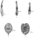
Fig. 2. Adults and mature protoscoleces of Echinococcus from the Hulunbeier Pasture. (A) Adult Echinococcus granulosus. (B) Adult Echinoccocus multilocularis sibiricensis that was found in the Hulunbeier Pasture. (C) Adult Echinoccocus multilocularis multilocularis. (D) Mature protoscolex of E. m. multilocularis from naturally infected M. brandti and experimentally infected white mice. (E) Mature protoscolex of E. m. sibiricensis from naturally infected M. brandti and experimentally infected white mice.
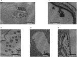
Fig. 3. PAS staining of tissue sections containing Echinococcus m. sibiricensis (A and B) and E. m. multilocularis (C, D and E) from naturally infected M. brandti or experimentally infected white mice. (A) An alveolar vesicle of E. m. sibiricensis found in the liver of naturally infected M. brandti from the Hulunbeier Pasture. (B) Mature protoscoleces in brood capsule cavity of E. m. sibiricensis found in the lung of experimentally infected white mouse 9 months post-infection. Arrow indicates the membrane surrounding the mature type I protoscoleces, which differs from the structure of mature type II protoscolices seen in (C) and (E). (C) Mature protoscolex of E. m. multilocularis found in the liver of experimentally infected white mouse. Arrow indicates the connection of the mature protoscolex to the inner wall of type II cyst. (D) Alveolar cysts of E. m. multilocularis found in the liver of experimentally infected white mouse 3 months post-infection. Arrows indicate the area of type II protoscolex formation. (E) Alveolar cysts with protoscoleces of E. m. multilocularis found in the liver of experimentally infected white mouse 6 months post-infection.

Table 1. Prevalence of alveolar echinococcosis in wild animals from Inner Mongonia, China (1998–1999)

Table 2. Prevalence of three types of alveolar echinococcosis in Microtus brandti from eight pastures in four counties

Table 3. Prevalence of three types of alveolar echinicoccosis in Microtus brandti from two counties during different seasons
 You have
Access
- 35
- Cited by