Introduction
Nitrogen oxide (NO) is produced in the tissues of humans and animals from the amino acid L-arginine. NO mediates the tone of smooth muscles (Ghimire et al., Reference GHIMIRE, ALTMANN, STRAUB and ISENBERG2017; Moncada et al., Reference MONCADA, PALMER and HIGGS1991; Vanhoutte, Reference VANHOUTTE2018), activity of the autonomic and central neural systems (Moncada et al., Reference MONCADA, PALMER and HIGGS1991; Malyshev et al., Reference MALYSHEV, ZENINA, GOLUBEVA, SALTYKOVA, MANUKHINA, MIKOYAN, KUBRINA and VANIN1999), cell differentiation and apoptosis (Rossig et al., Reference ROSSIG, FICHTLSCHERER, BREITSCHOPF, HAENDELER, ZEIHER, MULSCH and DIMMELER1999; Li et al., Reference LI, BILLIAR, TALANIAN and KIM1997; Kim et al., Reference KIM, CHUNG, SIMMONS and BILLIAR2000), immune responses (Moncada et al., Reference MONCADA, PALMER and HIGGS1991; Deroee et al., Reference DEROEE, NARAGHI, SONTOU, EBRAHIMKHANI and DEHPOUR2009), expression of different genes (Vasil'eva et al., Reference VASIL'EVA, STUPAKOVA, LOBYSHEVA, MIKOYAN and VANIN2001; Zhou and Brune, Reference ZHOU and BRUNE2005) and epigenetic effects (Socco et al., Reference SOCCO, BOVEE, PALCZEWSKI, HICKOK and THOMAS2017; Vasudevan et al., Reference VASUDEVAN, BOVEE and THOMAS2016). In living tissues, NO is a short-living substance and readily transformed to nitrite and nitrate. S- nitrosothiols (RSNO) and dinitrosyl-iron complexes (DNIC) (Rossig et al., Reference ROSSIG, FICHTLSCHERER, BREITSCHOPF, HAENDELER, ZEIHER, MULSCH and DIMMELER1999; Vanin, Reference VANIN2009; Vanin et al., Reference VANIN, MULLER, ALENCAR, LOBYSHEVA, NEPVEU and STOCLET2002; Severina et al., Reference SEVERINA, BUSSYGINA, PYATAKOVA, MALENKOVA and VANIN2003) may also be formed.
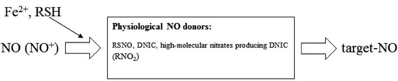
RSNO, DNIC and some high molecular nitro-compounds capable of producing DNIC (RNO2) are considered a physiological depot of NO (NO donors) that mediate its physiological effects (Rassaf et al., Reference RASSAF, PREIK, KLEINBONGARD, LAUER, HEI, STRAUER, FEELISCH and KELM2002; Vanin, Reference VANIN2009) (Figure 1). Because of these donors, physiological effects can last many times longer than the lifetime of molecular NO (Moncada et al., Reference MONCADA, PALMER and HIGGS1991; Rassaf et al., Reference RASSAF, PREIK, KLEINBONGARD, LAUER, HEI, STRAUER, FEELISCH and KELM2002). Nitrite and nitrate are considered the final products of NO metabolism (Moncada et al., Reference MONCADA, PALMER and HIGGS1991; Rossig et al., Reference ROSSIG, FICHTLSCHERER, BREITSCHOPF, HAENDELER, ZEIHER, MULSCH and DIMMELER1999).
Proposed mechanism of NO physiological activity (according to Vanin Reference VANIN2009; Vanin et al., Reference VANIN, MULLER, ALENCAR, LOBYSHEVA, NEPVEU and STOCLET2002; Severina et al., Reference SEVERINA, BUSSYGINA, PYATAKOVA, MALENKOVA and VANIN2003).

All NO effects on smooth muscle tone are realised via the interaction with the enzyme guanylate cyclase (GC) producing cyclic guanosine monophosphate (cGMP) (Moncada et al., Reference MONCADA, PALMER and HIGGS1991). NO activates GC via a reversible interaction with this enzyme (Moncada et al., Reference MONCADA, PALMER and HIGGS1991; Stalmer et al., Reference STALMER, SINGEL and LOSCALZO1992).
Apoptosis refers to genetically scheduled cell death. A group of cysteine proteases (caspases) play the key role in this process. It has been shown that NO can influence apoptosis via caspase inhibition (Rossig et al., Reference ROSSIG, FICHTLSCHERER, BREITSCHOPF, HAENDELER, ZEIHER, MULSCH and DIMMELER1999; Li et al., Reference LI, BILLIAR, TALANIAN and KIM1997; Kim et al., Reference KIM, CHUNG, SIMMONS and BILLIAR2000).
NO has been shown to be involved in the regulation of some expression of different genes (Vasil'eva et al., Reference VASIL'EVA, STUPAKOVA, LOBYSHEVA, MIKOYAN and VANIN2001; Zhou and Brune, Reference ZHOU and BRUNE2005), supposedly by nitrosation of SH-groups of definite proteins (Vasil'eva et al., Reference VASIL'EVA, STUPAKOVA, LOBYSHEVA, MIKOYAN and VANIN2001). The role of NO in immune response is related to activity of inducible NO-synthase (iNOS) in neutrophils and macrophages (Moncada et al., Reference MONCADA, PALMER and HIGGS1991; Carreras et al., Reference CARRERAS, PARGAMENT, CATZ, PODEROSO and BOVERIS1994).
The role of NO in embryogenesis
It is well known that maturation of the egg cell (Tiwari et al., Reference TIWARI, PRASAD, PANDEY, PREMKUMAR, TRIPATHI, GUPTA, CHETAN, YADAV, SHRIVASTAV and CHAUBE2017) and embryogenesis is accompanied by NO production. Accumulation of NO metabolites in the incubation medium of the fertilised egg (Battaglia et al., Reference BATTAGLIA, CIOTTII, NOTARANGELO, FRATTO, FACCHINETTI and DE ALOYSIO2003; von Mandach et al., Reference VON MANDACH, LAUTH and HUCH2003), in amniotic fluid and in blood of new born animals has been established (Blum et al., Reference BLUM, MOREL, HAMMON, BRUCKMAIER, JAGGY, ZURBRIGGEN and JUNGI2001; Ribeiro et al., Reference RIBEIRO, OGANDO, FARINA and FRANCHI2004; Vignini et al., Reference VIGNINI, TURI, GIANNUBILO, PESCOSOLIDO, SCOGNAMIGLIO, ZANCONI, SILVI, MAZZANTI and TRANQUILLI2008; Titov et al., Reference TITOV, IVANOVA, PETROV, SEREZHENKOV, MIKOYAN, VANIN and OSIPOV2012a; Reference TITOV, VINNIKOVA, AKIMOVA and FISININ2012b; Reference TITOV, AKIMOVA, IVANOVA and FISININ2012c; Reference TITOV, KONDRATOV and IVANOVA2015).
Embryogenesis includes differentiation of tissues, formation of blood vessels and regulation of their tone, gene expression and other processes potentially influenced by NO. There is still no unambiguous opinion whether NO plays any specific role in embryogenesis. It has been shown that NO influences embryonic development on the earliest stages of embryogenesis before tissue differentiation (Sengoku et al., Reference SENGOKU, TAKUMA, HORIKAWA, TSUCHIYA, KOMORI, SHARIFA, TAMATE and ISHIKAWA2001; Tirone et al., Reference TIRONE, CONTI, MANENTI, NICOLOSI, D'ORLANDO, AZZONI and BRUNELLI2016; Tomankova et al., Reference TOMANKOVA, ABAFFY and SINDELKA2017). In all these hypotheses the supposition was made on the basis of the effects of exogenous input of blockers of NO synthase and NO donors. It does not give quantitative representation regarding the real participation of NO in various processes. Such representation requires data on the content of NO metabolites in different parts of an embryo, possible relationships between these concentrations and specific features of embryonic and post-natal development. Avian embryos are suitable for such research because they develop outside the mother's body and have a relatively large size.
Basic patterns of NO metabolism in developing avian embryo
In the earliest studies the intensity of NO production was assessed by measuring nitrate and nitrite concentration as final products of NO metabolism (Blum et al., Reference BLUM, DOSOGNE, HOEBEN, VANGROENWEGHE, HAMMON, BRUCKMAIER and BURVENICH2000; Gladwin et al., Reference GLADWIN, SHELHAMER, SCHECHTER, PEASE-FYE, WACLAWIW, PANZA, OGNIBENE and CANNON2000). Now there is more knowledge on NO donors and their physiological role. According to certain researchers (Hickok et al., Reference HICKOK, SAHNI, SHEN, ARVIND, ANTONIOU, FUNG and THOMAS2011) concentrations of NO donors in cells can reach several tens of micromoles, which is comparable to nitrate concentrations. Based on the schema
NO → NO donors → NO targets → nitrate
the intensity of NO synthesis can be estimated as total concentration of nitro- and nitroso-compounds if it is assumed that these compounds do not enter the developing avian embryo exogenously. An indication of the intensity of metabolism can be gained from the ratio of nitrate to NO donors.
Titov et al. (Reference TITOV, AKIMOVA, VINNIKOVA and FISININ2009; Reference TITOV, VINNIKOVA, AKIMOVA and FISININ2012b; Reference TITOV, KOSENKO, AKIMOVA and FISININ2013; Reference TITOV, KONDRATOV and IVANOVA2015) showed that, by controlling the content of NO donors and nitrate in various parts of the embryo by specific sensors, the original avian embryo contains nitro - and nitroso-compounds at a concentration several micromol. The development of the bird embryo is accompanied by intense production of different nitro- and nitroso-compounds that accumulate inside the embryo. In the first three days of embryo development it predominantly accumulates the NO donors nitrosothiols, dinitrosyl-iron complexes and high molecular nitro-compounds. This process has the same intensity in embryos of all breeds of the same species (Titov et al., Reference TITOV, KONDRATOV and IVANOVA2015). The source of these compounds is still unknown but is probably the amniotic membrane where the NO-synthase enzyme has been found (Khan et al., Reference KHAN, KUSAKABE, WAKITANI, HIYAMA, TAKESHITA and KISO2012).
In amniotic fluid, which accumulates NO donors, their content on the third day of embryo development can reach several tens of millimole (Titov et al., Reference TITOV, KONDRATOV and IVANOVA2015). From the second to the fifth day of development NO donors accumulated in the amnion are transformed to nitrate which is removed to outside of the amnion. This process in the embryo of meat breeds is many times higher than in egg breeds (Titov et al., Reference TITOV, VINNIKOVA, AKIMOVA and FISININ2012b;
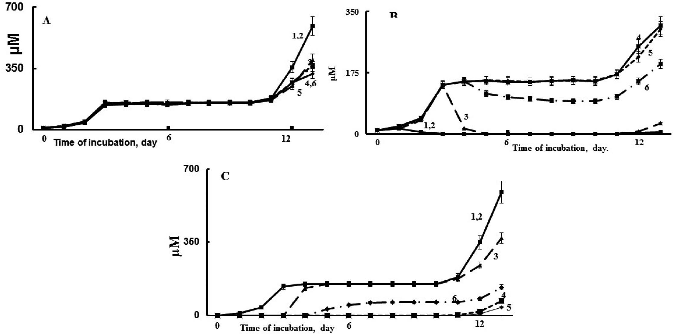
2015). The dynamics of accumulation and transformation of NO donors and nitrate in embryo of meat- and egg-type breeds is presented in Figure 2.
Dynamics of accumulation of total NO metabolites (A), NO donors (B), and nitrate (C) in developing chicken embryo of cross ‘Cobb 500’ (1), cross ‘Smena-8’ (2) and its lines B56 (Cornish) (3), and B79 (Plymouth Rock) (4), cross ‘Hisex White’ (5) and its line X1 (6) (Titov et al., Reference TITOV, KONDRATOV and IVANOVA2015).

The dynamics of the accumulation of NO metabolites in the embryos of meat- and egg-type breeds have not been significantly distinguished (Figure 2A). In the embryos of egg-type poultry (a cross of ‘Hisex White’ with maternal line B79 of ‘Smena-8’) the NO donors accumulated during the first three days of development and any decrease in subsequent days was negligible. In the eggs of meat-type poultry (crosses of ‘Cobb 500’ and ‘Smena-8’ line B56 with the egg-meat type line Hisex X1) NO donors accumulated during the first three days and then concentration greatly decreased (Figure 2B). Nitrate concentration increased with decreasing NO donors’ concentrations (Figure 2C). However, the total concentration of nitro- and nitroso-compounds within the eggs remained constant until 11 days of incubation. It should be noted that the decrease in concentration of NO donors started non-simultaneously; viz in line B56 on the third day of incubation; in the cross ‘Cobb 500’ and ‘Smena-8’ on second day. Therefore, a structure mediating NO oxidation appears or manifests itself not immediately but sometime after the beginning of embryo development. In case of line X1 the NO donor's transformation was not complete (Figure 2B). As long as the amniotic sac is impermeable for nitro- and nitroso-compounds (Titov et al., Reference TITOV, AKIMOVA, VINNIKOVA and FISININ2009, Reference TITOV, VINNIKOVA, AKIMOVA and FISININ2012b) the metabolites of NO can be transported from the amnion to the allantois only via the germ. Consequently, the structure mediating NO oxidation should be located in the germ.
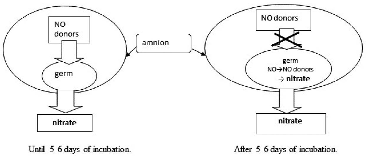
NO donors accumulated in the amnion can be consumed by germ tissues until five to six days of incubation. After this time, the amniotic NO donors seem to be unavailable for the germ, as long as the amniotic concentration of NO donors remains the same until the destruction of the amniotic membrane (Titov et al., Reference TITOV, VINNIKOVA, FISININ, BLIZNETSOVA and RETSKY2008, Reference TITOV, AKIMOVA, VINNIKOVA and FISININ2009, Reference TITOV, VINNIKOVA, AKIMOVA and FISININ2012b). After this period the germ continues the consumption of NO donors now produced by its own tissues (Figure 3).
Embryonic NO consumption before and after 5-6 days of incubation (Titov et al., Reference TITOV, KONDRATOV and IVANOVA2015).

Until 11 days of incubation synthesis is of low intensity and cannot influence total concentration of nitro- and nitroso-compounds in embryo. After 11 days of incubation, however, this process become more intense, and the divergence between meat- and egg-type poultry continues. The embryos of meat-type poultry accumulate mainly nitrate, whereas embryos of egg-type poultry accumulate mostly NO donors. The resulting nitrate is transporting to the allantois (Titov et al., Reference TITOV, AKIMOVA, VINNIKOVA and FISININ2009; Reference TITOV, VINNIKOVA, AKIMOVA and FISININ2012b).
As result of intensive consumption of NO donors, the amnion of meat-type poultry embryos contains substantially less NO donors than those from egg-type birds and forms from 3-5 days of incubation. The allantois in meat-type embryo contains substantially more nitrate than in egg-type embryos.
It has been previously shown in more than 40 breeds, crosses and lines of five avian species that at the end of the second trimester of embryonic development (day 13 in chickens) all meat-type eggs are characterised by low concentrations of NO donors in the amnion (only a few or tens of micromoles) with high concentrations of nitrate in the allantois (hundreds of micromoles). Other species are characterised by high amniotic concentrations of NO donors (millimoles) and low concentrations of nitrate in allantois (tens of micromoles) (Titov et al., Reference TITOV, AKIMOVA, VINNIKOVA and FISININ2009; Reference TITOV, VINNIKOVA, AKIMOVA and FISININ2012b; Reference TITOV, KOSENKO, AKIMOVA and FISININ2013; Reference TITOV, KONDRATOV and IVANOVA2015). The difference between this parameter in egg- and meat-type birds could attain hundreds of times in difference.
Intense accumulation of nitro- and nitroso-compounds in the embryo after 11 days of incubation is the result of the accumulation of these substances in the tissues of the germ. At the end of embryogenesis, the concentrations of nitro- and nitroso-compounds in tissues can reach 1000 nanomoles/g while, at 10-20 days post-hatch this parameter decreases to few tens of nanomoles/g (Titov et al., Reference TITOV, KOSENKO, AKIMOVA and FISININ2013; Reference TITOV, KONDRATOV and IVANOVA2015). The process of transformation of NO donors into nitrate continues during the entire period of embryogenesis.
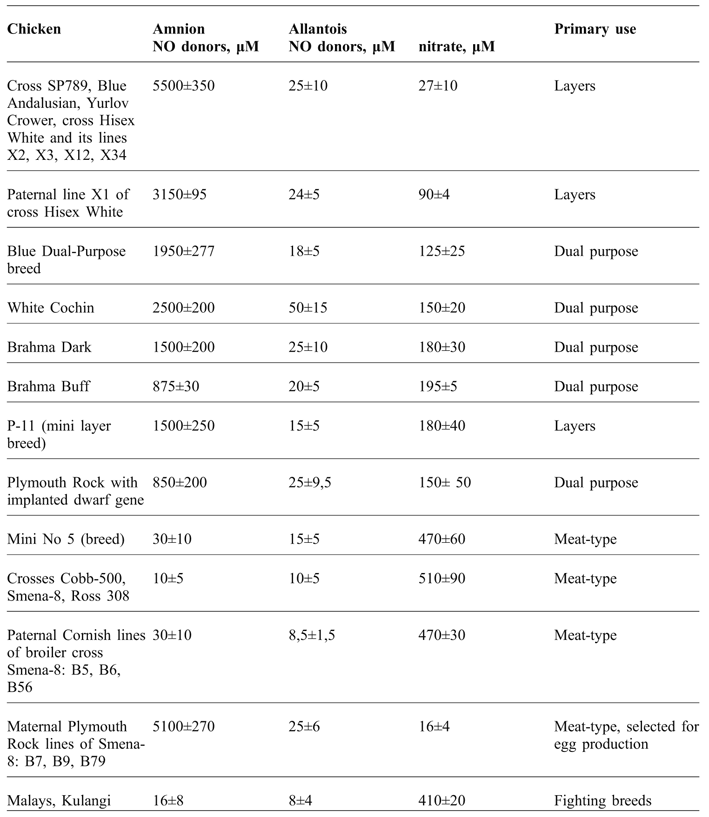
Earlier authors have proposed the ratio of concentrations of NO donors in the amnion and nitrate in the allantois at the end of second trimester of incubation as a suitable criterion for estimating the intensity of embryonic NO metabolism (Titov et al., Reference TITOV, VINNIKOVA, AKIMOVA and FISININ2012b; Reference TITOV, KOSENKO, AKIMOVA and FISININ2013; Reference TITOV, KONDRATOV and IVANOVA2015; Vinnikova et al., 2008). This parameter has been shown to remain relatively constant within a single cross or breed (with variation of no more than 10%) but substantially varies (at a magnitude of hundreds of times) between meat- and egg-type breeds of poultry (Titov et al., Reference TITOV, AKIMOVA, VINNIKOVA and FISININ2009; Reference TITOV, VINNIKOVA, AKIMOVA and FISININ2012b; Table 1).
Not only do the processes that are associated with NO oxidation determine body weight, but selection of chicken for higher rates of postembryonic growth has always been accompanied by the increase in the intensity of embryonic NO oxidation.
Concentrations of NO metabolites in the amnion and allantois at day 14 of incubation in different chicken breeds, crosses, and lines* (Titov et al., Reference TITOV, AKIMOVA, VINNIKOVA and FISININ2009; Reference TITOV, VINNIKOVA, AKIMOVA and FISININ2012b; Reference TITOV, KOSENKO, AKIMOVA and FISININ2013).

*In the amnion, in all cases, the nitrate and nitrite concentration <100 nM.
In the allantois of all layer breeds low concentrations of nitrate is found, while NO donors in the amnion is high (ca. 5 mM). The paternal line X1 of the layer cross Hisex White is characterised by higher body weight and skeletal strength than the final hybrid (Royter et al., Reference ROYTER, YEGOROVA, VARAKINA and SHAKHNOVA2005). NO oxidation in the X1 embryo is more intense than in the embryo of final hybrid (Table 1). In the embryos of broiler crosses, Cobb-500, Ross 308 and Smena-8, highly intense NO metabolism was found (in the order of hundreds of μM nitrate in the allantois and few μM NO donors in the amnion). However, the lines of these crosses (paternal Cornish and maternal Plymouth Rock lines) substantially differed from each other. All Cornish lines were characterised by high intensity of NO oxidation and Plymouth Rock lines had a low index of intensity. Both Cornish and Plymouth Rock are meat-type breeds. But Plymouth Rock exceeded the Cornish breed in egg production by 30-40%. Fighting breeds (selected for higher bodyweight and aggressiveness) have high intensity of NO metabolism.
The intensity of NO metabolism in dual purpose breeds was intermediate between the egg and meat breeds. The similar indices were found for the mini breeds. These strains are the result of selection for meat productivity, relatively high egg production, and high feed conversion ratio (Royter et al., Reference ROYTER, YEGOROVA, VARAKINA and SHAKHNOVA2005). Embryonic NO metabolism in meat-type mini breeds was similar to that in ‘standard’ meat-type breeds.
Data on the intensity of embryonic NO metabolism in quail, Guinea fowl and ostrich were also similar. In the embryo of all meat breeds, NO metabolism is substantially higher than in embryo of egg breeds (Titov et al., Reference TITOV, AKIMOVA, VINNIKOVA and FISININ2009; Reference TITOV, VINNIKOVA, AKIMOVA and FISININ2012b).
The role of NO in embryonic myogenesis
Myogenesis occurs in all embryos while intensive oxidation NO donors to nitrate mostly take place in meat-type embryos. Therefore, the question arises: what process and what structure is responsible for the transformation of NO donors?
According to Cazzato et al. (Reference CAZZATO, ASSI, MOSCHENI, BRUNELLI, DE PALMA, CERVIA, PERROTTA and CLEMENTI2014), injection of NO-synthase inhibitor (Nώ-propyl-L-arginine (NPLA) into the fertilised chicken embryo on the first day of incubation (500 ng/g of embryonic tissue) led to significant (over 30%) decrease of embryonic weight at the third day of incubation. Concurrent injection of NO donors (DETA-NO, 2 mg/g of embryonic tissue) was shown to cancel this effect. But if these substances were injected at days three and four of incubation then at day five there were no differences between injected and intact embryos. The authors suggested that the NO source inhibited in this experiment could influence embryonic development only at its earliest stage.
The data from ELISA analysis showed that the injection of NPLA at days one or two led to a significant decrease of myosin and myocyte-specific factor 2a content on the third day. The injection with NO-inhibitor was found to decrease concentration of myogenin. Concurrent injections of a NO donor cancelled these effects. It could be suggested that the synthesis of these substances is anyhow related to NO.
The effects of the NO-synthase inhibitor and NO donor injected at day one and the absence of these effects for injections at day three could be related to the formation of the amniotic sac at day three. Its membrane is impermeable to nitro- and nitroso-compounds (Titov et al., Reference TITOV, VINNIKOVA, AKIMOVA and FISININ2012b; Reference TITOV, KOSENKO, AKIMOVA and FISININ2013). However, this does not mean that NO produced in the embryo can influence embryonic development only at the earliest stages (Figure 3).
It is known that the myotome of the chicken embryo is formed from 45-50 to 90-96 hours of incubation, i.e. at days two to four. During this time the number of cells in the myotome increases 15-fold. The proliferation of muscle cells stops on day 12 of development. Some authors have reported the presence of NO-synthase in embryonic muscle cells and the participation of NO in development and differentiation of myoblasts (Lee et al., Reference LEE, BAEK, MOON, SONG, CHUNG, HA and KANG1994; Stamler et al., 2001; Tirone et al., Reference TIRONE, CONTI, MANENTI, NICOLOSI, D'ORLANDO, AZZONI and BRUNELLI2016).
It has been shown that the activity of Ca2+-dependent NO-synthase in chicken embryonic myoblasts is maximal during the development of myofibrils from mononuclear myoblasts (Stamler et al., 2001). NO donors activate this process (Lee et al., Reference LEE, BAEK, MOON, SONG, CHUNG, HA and KANG1994; Stamler et al., 2001).
It should be noted, however, that 1) the intense embryonic NO metabolism does not stop at day 12 of incubation; and 2) in embryos from egg-type chickens the proliferation of myoblasts and the formation of myofibrils occurs as for those from meat-type birds, but the intensity of NO metabolism in embryos of egg-type strains is tremendously lower than in meat-type ones (Borkhunova et al., 2014).
According to published data (Anderson, Reference ANDERSON2000) the injection of NO-synthase inhibitors inhibited the activation of satellite cells after muscle tissue damage. It should be noted that the activation of satellite cells can occur after 12 days of incubation as well. According to Borkhunova et al. (2014), muscle development in all breeds of the same poultry species has the similar speed. Morphological differences between birds of different breeds become evident after hatching. However, after hatching, concentrations of NO metabolites in chick tissues of different poultry breeds equalise (Titov et al., Reference TITOV, KONDRATOV and IVANOVA2015). Therefore, NO oxidation in embryo may be associated with processes that manifest after hatching, apparently via a chain of interactions of some mediators.
Transformation of NO donors into nitrate in embryos of some breeds starts as early as at day two of incubation. The question arises: what structure and process can promote NO oxidation on this stage of incubation? Is this process related to myogenesis directly, or the interdependence between myogenesis and NO metabolism in different breeds as described above, or is it just a coincidence? The effect of growth enhancers on NO synthesis and oxidation would be interesting to explore.
The influence of green light as an exogenous factor stimulating myogenesis on the rate of embryonic NO metabolism
Green light is an exogenous factor stimulating post-natal muscle growth when applied at the embryonic stage of development. However, there is no consensus on the possible mechanism of this stimulating action. Some authors (Rozenboim et al., Reference ROZENBOIM, HUISINGA, HALEVY and EL HALAWANI2003; Reference ROZENBOIM, PIESTUN, MOBARKEY, BARAK, HOYZMAN and HALEVY2004; Reference ROZENBOIM, EL HALAWANI, KASHASH, PIESTUN and HALEVY2013; Sobolewska et al., Reference SOBOLEWSKA, ELMINOWSKA-WENDA, BOGUCKA, SZPINDA, WALASIK, BEDNARCZYK and PARUSZEWSKA-ACHTEL2011; Halevy et al., Reference HALEVY, PIESTUN, ALLOUH, ROSSER, RINKEVICH, RESHEF, ROZENBOIM, WLEKLINSKI-LEE and YABLONKA-REUVENI2004; Reference HALEVY, PIESTUN, ROZENBOIM and YABLONKA-REUVENI2006) have suggested that green light enhances proliferation and differentiation of myoblasts and increases the amount of myosatellocytes in developing embryos. All these effects enhance postnatal muscle growth.
Halevy et al. (Reference HALEVY, PIESTUN, ROZENBOIM and YABLONKA-REUVENI2006) reported that exposure of avian embryos (Cobb broilers) to green light during incubation increased the counts and accelerated maturation of embryonic myoblasts. The stimulating effect results from the acceleration of myoblastic proliferation and differentiation. The breast muscle weight in broilers from eggs exposed to green light was higher compared to unexposed eggs. The count of cells capable for the proliferation reached maximum at day three post-hatch and was higher by 20% in the exposed birds. It has been proposed that irradiation increased the amount of the cells capable of proliferation and differentiation, but not the speed of these processes. It is important to note that green light did not produce any effect in the culture of isolated myoblasts. Therefore, the effect of green light is indirect.
In trials with green light exposure of embryos during incubation a modest increase (by 5-11%) in live bodyweight of hatched chicks at three weeks post-hatch was seen in all studied breeds and crosses (Titov et al., Reference TITOV, AKIMOVA, IVANOVA and FISININ2012c). These data were in agreement with the results of other studies (Rozenboim et al., Reference ROZENBOIM, HUISINGA, HALEVY and EL HALAWANI2003; Reference ROZENBOIM, PIESTUN, MOBARKEY, BARAK, HOYZMAN and HALEVY2004). Exposure of embryos to green light decreased concentrations of nitro- and nitroso-compounds in the amnion by up to 40% and increased the concentration of nitrate in allantois (Titov et al., Reference TITOV, AKIMOVA, IVANOVA and FISININ2012c).
Analysis of nitro- and nitroso-compounds concentration in the whole embryo showed that the exposure to green light did not increase total concentration of these compounds, but promoted the transformation of NO donors to nitrate (Titov et al., Reference TITOV, KONDRATOV and IVANOVA2015).
Exposure of eggs to green light from one to 10, one to 14 and two to 14 days of incubation induced a maximal effect on the concentrations of nitro- and nitroso-compounds in the embryo. The exposure from six to 14 days of incubation, however, produced no effect. The exposure from one to six days of incubation led to less marked effect, with 70% of maximal effect recorded (Titov et al., Reference TITOV, AKIMOVA, IVANOVA and FISININ2012c).
It may be supposed from the available data that green light does not affect amniotic nitro- and nitroso-compounds directly. The light supposedly affects certain factors determining oxidation of NO donors to nitrate. This proposition is further evidenced by the absence of any effect when the light was applied after six days of incubation, despite the presence of NO donors in the embryo.
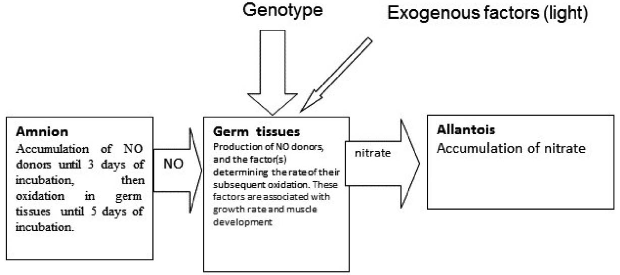
It can be assumed that the intensification of meat tissue growth may be related to genetically conditioned activation of an unknown factor within the embryonic tissues. This process can be, to a certain extent, conditioned by exogenous factors (i.e. green light). Activity of the factor is accompanied by the oxidation of NO to nitrate (Figure 4).
NO synthesis and oxidation during avian embryogenesis.

Conclusions
In avian embryogenesis the metabolism of nitric oxide (NO) is closely related to speed of growth and meat productivity. A strong correlation between meat productivity in adult chickens and intensity of NO oxidation during embryogenesis has been determined. NO oxidation occurs in the germ tissues as a product of oxidation accumulated in the allantois. Green light, an exogenous factor enhancing meat productivity, has been found to intensify embryonic NO metabolism. Thus, intensity of NO oxidation is directly connected with the intensification of the rate of muscle growth. The difference between amounts of NO oxidised to nitrate in the embryo of egg and meat breeds have two orders of magnitude, although the amount of produced NO in all embryos is about the same. This allows to use the indices of the NO metabolites concentration in the embryo as a suitably sensitive parameter to control the breeding process already at the embryo stage, and for the testing of exogenous factors that influence meat productivity which have an impact at the embryonic stage.
There is evidence to suggest that NO exerts no active influence on the rate of body growth and muscle tissue development. Embryonic oxidation of nitric oxide, apparently due to the presence of structures in the germ tissues, is coupled with the intensification of growth and muscle tissue development. This structure appears at the beginning of embryo development. The mechanism of relationship between embryonic NO oxidation with the velocity of growth and muscle development are not yet clear.