A number of diseases are characterised by the accumulation of excessive or inappropriate scar tissue, resulting in organ dysfunction. Notable examples include idiopathic pulmonary fibrosis (IPF), liver fibrosis, kidney fibrosis and systemic sclerosis (SSc)/scleroderma. Many of these occur in tissues with epithelial, mesenchymal or vascular structures in close opposition. In a broader context, many other disorders with prominent tissue remodelling, such as cardiac failure, atherosclerosis and asthma, have a significant fibrotic component. Although almost every cell type has been implicated in the pathogenesis of fibrosis, fibroblasts, as mediators of fibrillar collagen deposition, are the cellular sine qua non. Fibroblasts are somewhat ill-defined. They are found in most tissues, especially those with prominent epithelial and microvascular components (e.g. skin, lung, liver and kidney). Fibroblasts are identified primarily by their location and morphology. In tissue culture, they are often characterised by their appearance and the absence of specific markers. Fibroblasts are important in normal development, tissue homeostasis and wound healing. Injury and repair of most tissues begin with epithelial or endothelial damage, resulting in exudation of blood or platelet-rich plasma into extravascular spaces, leading to the formation of a fibrinous clot (provisional matrix) (Ref. Reference Clark and Clark1). Fibroblasts migrate into the provisional matrix, proliferate and produce de novo extracellular matrix (ECM) components, such as fibronectin and collagen, creating a fibroblast-populated granulation tissue. Simultaneously, fibroblasts acquire a myofibroblastic phenotype. Because myofibroblasts actively elaborate and remodel abundant ECM, and because persistence and activation of myofibroblasts are seen in abnormal scar tissue, myofibroblasts are thought to be the end effector cell and ‘final common pathway’ for most fibrotic diseases. In the first part of this review, factors promoting myofibroblastic differentiation will be considered. The principal mediators that affect myofibroblast differentiation are listed in Table 1. In the second half of this review, factors affecting the prolonged survival of myofibroblasts in scar tissue will be discussed.
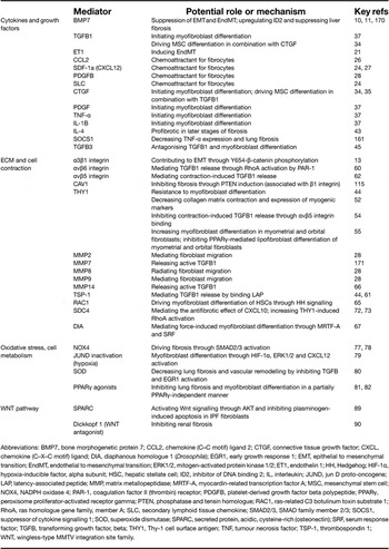
Key mediators of myofibroblast differentiation

Abbreviations: BMP7, bone morphogenetic protein 7; CCL2, chemokine (C–C motif) ligand 2; CTGF, connective tissue growth factor; CXCL, chemokine (C–X–C motif) ligand; DIA, diaphanous homologue 1 (Drosophila); EGR1, early growth response 1; EMT, epithelial to mesenchymal transition; EndMT, endothelial to mesenchymal transition; ERK1/2, mitogen-activated protein kinase 1/2; ET1, endothelin 1; HH, Hedgehog; HIF-1α, hypoxia-inducible factor, alpha subunit; HSC, hepatic stellate cell; ID2, inhibitor of DNA binding 2; IL, interleukin; JUND, jun D proto-oncogene; LAP, latency-associated peptide; MMP, matrix metallopeptidase; MRTF-A, myocardin-related transcription factor A; MSC, mesenchymal stem cell; NOX4, NADPH oxidase 4; PAR-1, coagulation factor II (thrombin) receptor; PDGFB, platelet-derived growth factor beta polypeptide; PPARγ, peroxisome proliferator-activated receptor gamma; PTEN, phosphatase and tensin homologue; RAC1, ras-related C3 botulinum toxin substrate 1; RhoA, ras homologue gene family, member A; SLC, secondary lymphoid tissue chemokine; SMAD2/3, SMAD family member 2/3; SOCS1, suppressor of cytokine signalling 1; SOD, superoxide dismutase; SPARC, secreted protein, acidic, cysteine-rich (osteonectin); SRF, serum response factor; TGFB, transforming growth factor, beta; THY1, Thy-1 cell surface antigen; TNF, tumour necrosis factor; TSP-1, thrombospondin 1; WNT, wingless-type MMTV integration site family.
Origin of myofibroblasts
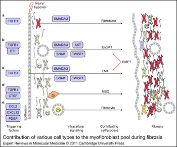
Specialised contractile fibroblasts called myofibroblasts emerge following tissue injury and their role in normal wound healing has been well described (Ref. Reference Gabbiani2). Fibroblasts are the principal effector cells during fibrosis (scarring) in most tissues, as shown by their persistence and excessive matrix deposition (mostly collagen I, collagen III and fibronectin) (Ref. Reference Hardie, Glasser and Hagood3). Although tissue fibrosis (e.g. IPF, a chronic disease with progressive scarring of the lung parenchyma) was originally thought to be mediated exclusively by resident mesenchymal cells that proliferated and synthesised excessive amounts of ECM (Ref. Reference Gabbiani2, Reference Hinz4), it has recently been demonstrated in many tissues that myofibroblasts can also be derived from epithelial to mesenchymal transition (EMT), endothelial to mesenchymal transition (EndMT) and bone-marrow-derived mesenchymal precursors [fibrocytes or mesenchymal stem cells (MSCs)]. Whether myofibroblasts derived from different cellular origins have distinct roles in the injury–repair process and whether they take part in the normal repair response as opposed to fibrosis have yet to be elucidated. The possible cellular origins of myofibroblasts are shown in Figure 1.
Contribution of various cell types to the myofibroblast pool during fibrosis. Factors released from cells upon tissue injury trigger the formation of myofibroblasts predominantly from fibroblasts (a); however, endothelial cells (EndMT) (b), epithelial cells (EMT) (c), MSCs (d) and fibrocytes (e) can also contribute to fibrosis. Abbreviations: AKT, v-akt murine thymoma viral oncogene homologue; BMP7, bone morphogenetic protein 7; CCL2, chemokine (C–C motif) ligand 2; CTGF, connective tissue growth factor; CXCL12, chemokine (C–X–C motif) ligand 12; EndMT, endothelial to mesenchymal transition; EMT, epithelial to mesenchymal transition; ET1, endothelin 1; MSC, mesenchymal stem cell; PDGF, platelet-derived growth factor; SMAD2/3, SMAD family member 2/3; SNAI1, Snail homologue 1 (Drosophila); TGFB1, transforming growth factor, beta1; TWIST1, twist homologue 1 (Drosophila).

Epithelial to mesenchymal transition
EMT has a crucial role in the differentiation of multiple tissues and organs, and in tissue repair, and can also contribute to organ fibrosis and promote carcinoma progression through the local formation of interstitial myofibroblasts from organ epithelium (Ref. Reference Thiery5). Mediators triggering EMT include different members of the transforming growth factor β (TGFB) superfamily, wingless-type MMTV integration site family members (WNTs), Notch, epidermal growth factor (EGF), hepatocyte growth factor, fibroblast growth factor (FGF), hypoxia-inducible factor (HIF) and many others (Refs Reference Thiery5, Reference Kalluri and Neilson6). A wide array of transcription factors such as SNAI1 and TWIST1 are involved in regulating EMT (Ref. Reference Zavadil and Böttinger7). Fibroblasts expressing fibroblast-specific protein-1 (FSP1, also called S100A4) can arise in large numbers by local EMT during renal fibrogenesis (Ref. Reference Iwano8). Although FSP1 is often used as a fibroblast marker, it has also been shown to be a marker of a specific subset of inflammatory macrophages in liver injury, fibrosis and cancer (Ref. Reference Osterreicher9). Bone morphogenic protein 7 (BMP7), a member of the TGFB superfamily that is known to antagonise TGFB signalling, has been demonstrated to inhibit progression of fibrosis and to abolish EMT- or EndMT-derived fibroblasts in liver (Ref. Reference Zeisberg10), cardiac (Ref. Reference Zeisberg11) and kidney fibrosis (Ref. Reference Zeisberg12). In the presence of active TGFB1, α3β1 integrin mediates tyrosine phosphorylation of β-catenin at Y654 and formation of pY–β-catenin/pSMAD2 transcriptional complexes in alveolar epithelial cells (AECs), promoting EMT and contributing to lung fibrosis (Ref. Reference Kim13). Human bronchial epithelial cells are also able to undergo EMT in vitro upon TGFB stimulation through a primarily SMAD2/3-dependent mechanism (Ref. Reference Câmara and Jarai14). However, other groups have found no evidence of EMT in liver (Ref. Reference Scholten15) or kidney (Ref. Reference Humphreys16) fibrosis. Despite many recent studies defining mechanisms of EMT, its exact role in vivo in many diseases and models remains unclear (Ref. Reference Tanjore17).
Endothelial to mesenchymal transition
EndMT has an important role during cardiovascular development and in various vascular pathologies. There are many similarities between EMT and EndMT regarding the loss of expression of cell-specific markers and increased expression of mesenchymal markers such as alpha 2 smooth muscle actin (ACTA2), fibronectin, FSP1, and collagens I and III (reviewed in Refs Reference Li and Bertram18, Reference Nataraj, Ernst and Kalluri19, Reference Potenta, Zeisberg and Kalluri20). EndMT is also induced by TGFB1 in a SMAD2/3-dependent manner and can be reversed by BMP7 during cardiac fibrosis (Ref. Reference Zeisberg11). FSP1 expression induced by high glucose levels in human umbilical vein endothelial cells is inhibited by siRNA against endothelin 1 (ET1, EDN1), along with decreased phosphorylation of SMAD3 and v-akt murine thymoma viral oncogene homologue (AKT), decreased expression of TGFB and SNAI1, and decreased motility of these cells (Ref. Reference Widyantoro21), suggesting a role of ET1 in EndMT. EndMT has also been demonstrated in bleomycin-induced lung fibrosis in lacZ-expressing TIE2-cre transgenic mice (Ref. Reference Hashimoto22). The contribution of EndMT to vascular remodelling (Ref. Reference Arciniegas23), cancer progression (Ref. Reference Potenta, Zeisberg and Kalluri20) and diabetic renal fibrosis (Ref. Reference Li and Bertram18) has been reviewed elsewhere.
Fibrocytes
A population of circulating cells with fibroblast properties, called fibrocytes, was first described in 1994. These cells specifically enter sites of tissue injury and express collagen I, vimentin, fibronectin, CD11b, β2 integrin (ITGB2), CD34 and protein tyrosine phosphatase, receptor type, C (PTPRC), but are negative for esterase, CD14, CD25 and CD54 expression. Although the fibrocytes present in wound chambers of a sex-mismatched bone marrow chimeric mouse model did not arise from the transferred bone marrow, others have demonstrated bone marrow origin of lesional cells during lung fibrosis (Ref. Reference Hashimoto24) and renal fibrosis (Ref. Reference Iwano8), and their numbers in blood have been demonstrated to correlate with disease activity of IPF (Ref. Reference Moeller25). Several chemoattractants, such as CCL2 (Ref. Reference Moore26), stromal-cell-derived factor 1 (SDF-1a, also called CXCL12) (Refs Reference Hashimoto24, Reference Andersson-Sjöland27, Reference García-de-Alba28, Reference Phillips29), platelet-derived growth factor beta (PDGFB) (Ref. Reference García-de-Alba28) and secondary lymphoid tissue chemokine (SLC, also called CCL21) (Refs Reference Hashimoto24, Reference Abe30), have been shown to have a role in fibrocyte migration in vitro or in vivo during lung fibrosis, indicating that circulating fibrocytes might contribute to the expansion of the fibroblast or myofibroblast population in IPF (Ref. Reference Strieter31). An elegant lineage-tracing study in the bleomycin model has demonstrated that in the active fibrotic stage 2 weeks after injury, one-third of FSP1+ cells were of epithelial origin and one-fifth were of bone marrow origin, but neither contributed significantly to lesional myofibroblasts (Ref. Reference Tanjore17). Thus it is possible that EMT, EndMT and fibrocytes or other bone-marrow-derived precursors might have more important roles in normal repair than in fibrosis.
Mesenchymal stem cells
Although exogenous MSCs can ameliorate fibrosis (Ref. Reference Asanuma32), MSCs from tracheal aspirates of premature infants, but not bone-marrow-derived MSCs, also have the capacity to differentiate into myofibroblasts in response to TGFB1 (Ref. Reference Popova33). Bone-marrow-derived MSCs require an additional profibrotic signal such as connective tissue growth factor (CTGF) to complete their differentiation from a fibroblastic to a myofibroblastic phenotype (Ref. Reference Lee34).
Cytokines and growth factors
Sources for profibrotic cytokines in the lungs include macrophages and other inflammatory cells, epithelial cells and mesenchymal cells. TGFB1, tumour necrosis factor-alpha (TNF-α), PDGF, CTGF, interleukin 1 beta (IL-1B), IL-6 and IL-4 have well-established roles in myofibroblast differentiation (Refs Reference Atamas and White35, Reference Knight36, Reference Lasky and Brody37). Regulators of these key players and the roles of other cytokines are under ongoing investigation. Understanding the networks of interaction of cytokines and growth factors is probably more important than determining the roles of individual mediators. TGFB1 has a central role not only in inducing myofibroblastic differentiation and fibrosis, but also in the differentiation of regulatory T cells that suppress lymphocyte activation. However, in the presence of IL-6 (for mouse cells) or IL-21 (for human cells), TGFB1 facilitates the generation of proinflammatory, IL-17-producing TH17T helper lymphocytes (Ref. Reference Yang38). Bleomycin- and IL-1B-induced IL-17A production and lung fibrosis are TGFB dependent, suggesting cooperative roles for IL-17A and TGFB in the development of fibrosis (Ref. Reference Wilson39). IL-17A and IL-17F stimulate inflammatory responses in human colonic subepithelial myofibroblasts, which contribute to the pathogenesis of inflammatory bowel disease (Ref. Reference Yagi40). IL-6 is produced by IL-1A-stimulated human cardiac myofibroblasts (Ref. Reference Turner41) and during acute exacerbations of IPF (Ref. Reference Collard42). The effect of IL-4 is somewhat contradictory. Based on intratracheal bleomycin administration to Il4 −/– or Il4 +/+ mice, IL-4 appears to have an early anti-inflammatory or immunosuppressive role; however, during later stages of fibrosis, IL-4 seems to have a profibrotic role (Ref. Reference Huaux43). THY1−, but not THY1+, rat lung fibroblasts respond to PDGFBB, IL-1B, IL-4 or bleomycin treatment with increased TGFB activity, SMAD3 phosphorylation, and expression of ACTA2 and fibronectin (Ref. Reference Zhou, Hagood and Murphy-Ullrich44). TGFB activation by IL-4 and bleomycin is mediated by thrombospondin 1 (TSP-1), whereas the effects of IL-1B require the activation of plasmin and matrix metallopeptidases (MMPs) (Ref. Reference Zhou, Hagood and Murphy-Ullrich44). The mutant form of TGFB3, which has improved bioavailability and activity, reduces fibroblast or myofibroblast transdifferentiation and re-epithelialisation density in a mouse skin-wounding model (Ref. Reference Waddington45). TNF-α antagonises TGFB1-mediated human pulmonary myofibroblast differentiation through ERK1/2-dependent induction of early growth response 1 (EGR1) (Ref. Reference Liu, Kelm and Strauch46).
Myogenic transcription factors
Myogenesis, which is required for the development and regeneration of skeletal muscle, is regulated by myogenic regulatory factors including MYOD1 and myogenin. Mitogen-activated protein kinase kinase kinase 7 (MAP3K7) has been shown to induce myogenic differentiation through the activation of p38 MAPK and AKT kinase in mouse myoblasts and embryonic fibroblasts, whereas the RAS–RAF–MEK–ERK pathway was inhibitory (Ref. Reference Bhatnagar47). FIZZ1 (found in the inflammatory zone 1; also known as resistin-like alpha) is increased markedly in fibrotic lungs and induces myofibroblast differentiation (Ref. Reference Liu48). Similarly, Yin Yang 1 is found in fibrotic lung and potently induces fibroblast ACTA2 expression (Ref. Reference Lin49).
THY1
A distinct feature of IPF is the development of fibroblastic foci, which represent areas of active fibrosis and correlate clearly with poor outcome. Fibroblasts within fibroblastic foci of usual interstitial pneumonia (UIP)/IPF patients express ACTA2, but show no expression of THY1 (Refs Reference Hagood50, Reference Sanders51). Intratracheal bleomycin induces more severe lung fibrosis in THY1-knockout mice as demonstrated by histological evaluation and TGFB activation (Ref. Reference Hagood50), indicating a lung-fibrosis-suppressing function of THY1. Confirming the in vivo results in lung fibrosis, THY1− rat lung fibroblasts have significantly higher expression of myofibroblast and myogenic regulatory factor genes and proteins, cause significantly greater collagen contraction and are more resistant to apoptosis compared with THY1+ cells (Ref. Reference Sanders, Kumbla and Hagood52). Most of the myofibroblastic features of THY1− lung fibroblasts are reversible upon re-expression of THY1 (Refs Reference Sanders, Kumbla and Hagood52, Reference Sanders53), indicating an important mechanistic role. Indeed, this has been confirmed by demonstrating that THY1 inhibits activation of latent TGFB1 and myofibroblastic differentiation in lung fibroblasts (Refs Reference Zhou, Hagood and Murphy-Ullrich44, Reference Zhou54). Unlike lung fibroblasts, only THY1+ TGFB-treated human myometrial or orbital fibroblasts expressed ACTA2, whereas only THY1− orbital fibroblasts differentiated into lipofibroblasts (Ref. Reference Koumas55), possibly as a result of paracrine antiadipogenic factors secreted by THY1-expressing cells (Ref. Reference Lehmann56). This seemingly contradictary modulation of fibrosis by THY1 might be due to differences in the local tissue environment (e.g. ECM and integrin expression). The role of TGFB1 and other fibrogenic factors in myogenic differentiation of fibroblasts (i.e. expression of MYOD and myogenin together with ACTA2, sarcomeric myosin, myocardin and desmin) was confirmed in THY1− fibroblasts (Ref. Reference Sanders, Kumbla and Hagood52).
ECM, physical force and cell contractility
In early granulation tissue, the mechanical tension of the open wound gives the initiation signal for the differentiation of protomyofibroblasts (Refs Reference Eckes57, Reference Tomasek58), which show the morphological characteristics of myofibroblasts, such as stress fibres, but do not express ACTA2. Lack of a mechanical load on fibroblasts in a collagen matrix (similar to the environment of an early wound) produces an ‘inflammatory’ phenotype that is characterised by high expression of inflammatory mediators and proteases, along with low proliferation (Ref. Reference Eckes57). Further differentiation into mature, ACTA2-expressing myofibroblasts is driven by high tissue stiffness, the presence of TGFB1 and the ED-A splice variant of fibronectin in the later stages of wound healing. Mechanical load induces an ‘activated’ proliferating fibroblast phenotype that synthesises abundant ECM and protease inhibitors, as well as fibrogenic mediators. Integrins connect the cytoskeleton to the ECM and are able to transmit mechanical signals in both directions across the plasma membrane (Ref. Reference Katsumi59). Release of active TGFB from the latent TGFB complex has been shown to be mediated by RhoA activation by the protease-activated receptor 1 (PAR-1) in an αvβ6-integrin-dependent manner in epithelial cells (Ref. Reference Jenkins60), probably as a result of cytoskeletal changes, in addition to TSP-1- and CD36-mediated protease activity (Ref. Reference Wang61). This observation is supported by results showing that myofibroblast contraction directly releases active TGFB1 from the mechanoresistant ECM through integrins (Ref. Reference Wipff62).
THY1 has been shown to bind integrin αvβ5 of rat lung fibroblasts and to inhibit contraction-induced latent TGFB1 activation and TGFB1-dependent lung myofibroblast differentiation, presumably by interfering with the binding of latent TGFB with integrin αvβ5 (Ref. Reference Zhou54). The functional integrin-binding motif (RLD) of THY1 is necessary for contraction-induced lung myofibroblast differentiation (Ref. Reference Zhou54). Furthermore, THY1-knockout mice with exacerbated lung fibrosis and increased active TGFB have increased LTBP4 expression compared with wild-type mice (Ref. Reference Zhou63), suggesting another mechanism by which THY1 is able to reduce the release of active TGFB.
Integrins regulate cell spreading and migration through activation of the Rho family of small GTPases such as RhoA, CDC42 and RAC (Ref. Reference Giancotti and Ruoslahti64). RAC1 becomes activated during differentiation of quiescent hepatic stellate cells (HSCs) into myofibroblastic cells (Ref. Reference Choi65). A constitutively active form of RAC1 was found to generate enhanced Hedgehog (HH) signalling, with downregulation of HH-interacting protein and increased expression of SHH (HH ligand) and GLI2, both in vitro in myofibroblastic HSCs and in vivo, in a bile-duct-ligation-induced mouse liver fibrosis model (Ref. Reference Choi65). The crosstalk between specific integrins and TGFB signalling is reviewed elsewhere (Ref. Reference Margadant and Sonnenberg66). Actin assembly is regulated by formin proteins, such as the mammalian diaphanous-regulated formin (DIA), which becomes activated after interaction with the Rho family of small GTPases. DIA has been shown to mediate force-induced myofibroblast differentiation by increasing translocation of myocardin-related transcription factor-A (MRTF-A, also called MKL1) and activation of serum response factor (SRF), which are activators of ACTA2 transcription (Ref. Reference Chan67). Changes in intracellular free cytoplasmic Ca2+ concentration contribute to myofibroblast differentiation by influencing cell contractility and mechanical communication between cells (reviewed in Ref. Reference Follonier Castella68).
Most matrix proteins (fibronectin, vitronectin, TSP-1, laminin and collagens) contain binding domains for members of the integrin family, as well as heparin-binding domains that mediate interactions with cell surface proteoglycans such as syndecans (Refs Reference Couchman and Woods69, Reference Yoneda and Couchman70). Syndecans can regulate microfilament assembly and cell shape and influence small GTPase activation. CTGF (CCN2) is required for syndecan 4 (SDC4) and TSP-1 expression, and loss of SDC4 expression is responsible for at least part of the Ctgf −/− phenotype (Ref. Reference Kennedy71). Ctgf −/− mouse embryonic fibroblasts show a disorganised actin network, impaired migration and less contractile force (Ref. Reference Kennedy71). The role of SDC4 in lung fibrosis was shown by administration of recombinant CXCL10 protein, an antifibrotic chemokine that directly interacts with SDC4, which inhibited fibrosis in wild-type mice, but not in Sdc4 −/− mice (Ref. Reference Jiang72). Besides the THY1-integrin-binding motif (RLD), the heparin-binding domain of THY1, by interaction with SDC4, is also required to stimulate focal adhesion formation by stimulating RhoA through protein kinase C alpha (PRKCA) activation (Ref. Reference Avalos73). Therefore, the ECM has an important role in the process of integrin- and THY1-mediated TGFB release and influences myofibroblast differentiation through signalling pathways such as HH signalling and small GTPase activation by means of integrins, cell surface proteoglycans and THY1.
Oxidative stress and cell metabolism
NADPH oxidases (NOX) are multisubunit enzymes that generate superoxide by transferring electrons from NADPH to molecular oxygen. The production of reactive oxygen species (ROS) by NOX enzymes is important during phagocytic burst to kill pathogens and also for a variety of essential biological processes, including developmental and differentiation processes, ECM crosslinking, hormone biosynthesis, cellular senescence, apoptosis, responses to oxygenation (oxygen sensing), and cellular signalling responses to growth factors, hormones and cytokines (Ref. Reference Leto74). The presence of an oxidant–antioxidant imbalance in IPF is indicated by increased oxidative processes and altered levels of antioxidants in the lungs of affected patients (Ref. Reference Kinnula75). NOX4 mRNA and protein expression has been found to be increased in TGFB1-treated human lung fibroblasts (Ref. Reference Hecker76), in cardiac fibroblasts (Ref. Reference Cucoranu77) and in IPF lungs compared with controls (Ref. Reference Amara78). ROS inhibitors decrease TGFB1-induced ACTA2 expression; furthermore, siRNA against NOX4 attenuates lung fibrosis (Ref. Reference Hecker76) and decreases TGFB1-induced SMAD2/3 phosphorylation in human cardiac fibroblasts (Ref. Reference Cucoranu77) and in fibroblasts from control and IPF lungs (Ref. Reference Amara78), supporting the importance of NOX4 in myofibroblast differentiation. Oxidative stress can be stimulated by JUND inactivation or by treatment with desferrioxamine, which mimics hypoxia. The resulting accumulation of HIF-1α and phosphorylation of ERK1/2 leads to the activation of CXCL12/CXCR4, which converts fibroblasts into myofibroblasts in a carcinogenesis model (Ref. Reference Toullec79). Furthermore, overexpression of extracellular superoxide dismutase is able to attenuate bleomycin-induced lung fibrosis and vascular remodelling, and blocks the early upregulation of TGFB1 and EGR1 (Ref. Reference Van Rheen80).
Fibroblasts can also be differentiated into ‘lipofibroblasts’ or even adipocytes. Adipogenesis is initiated by CCAAT/enhancer-binding protein, beta (C/EBP-β) and C/EBP-γ and driven by peroxisome proliferator-activated receptor gamma (PPARγ), and has an important role in conditions such as thyroid eye disease in which an autoimmune inflammatory response specifically targeted to the orbit results in remodelling and expansion of connective and adipose tissues, including proliferation and differentiation of fibroblasts to adipocytes (Ref. Reference Lehmann56). THY1 can also influence lipofibroblast differentiation: only THY1− orbital and myometrial fibroblasts differentiate into lipofibroblasts after treatment with PPARγ ligands (Ref. Reference Koumas55). The nuclear hormone receptor PPARs heterodimerise with retinoid X receptors (RXRs), thus forming a complex that regulates gene expression. PPARγ controls lipid and glucose metabolism and is also implicated in inflammation regulation. Primary human pulmonary fibroblasts express PPARγ and RXR proteins and are capable of PPARγ-dependent gene transcription (Refs Reference Burgess81, Reference Milam82). The PPARγ agonist rosiglitazone decreases serum-induced proliferation, migration and ACTA2 expression as well as serum-, PDGFBB- or TGFB1-induced ERK phosphorylation of human fetal lung fibroblasts (Ref. Reference Lin83). PPARγ agonists also inhibit TGFB-driven myofibroblast differentiation in vitro (Refs Reference Burgess81, Reference Milam82) in an AKT-dependent (Ref. Reference Kulkarni84) and SMAD2/3-, AP-1- and partially PPARγ-independent manner (Ref. Reference Ferguson85), and inhibit in vivo bleomycin-induced lung fibrosis (Ref. Reference Milam82). Haem oxygenase-1 (HO-1) is the inducible form of haem oxygenase that participates in the conversion of haem to equimolar concentrations of biliverdin, iron and carbon monoxide, and has antioxidant and anti-inflammatory properties. Increased HO-1 expression by PPARγ ligands is also independent of PPARγ and can be reversed by N-acetyl cysteine, a thiol antioxidant, but does not influence TGFB-induced ACTA2 expression (Ref. Reference Ferguson86). TGFB can activate ERK1/2 and FAK–PI3K–AKT, and inhibits PTEN in a SMAD-independent manner, leading to myofibroblast differentiation and fibrosis (Refs Reference Kulkarni84, Reference Fagone87). These effects can be partially reversed by resveratrol (Ref. Reference Fagone87) or PPARγ ligands bearing an electrophilic centre (Ref. Reference Kulkarni84).
Other critical signalling pathways
The WNT family proteins have been shown to have a role in organogenesis (e.g. kidney development), tissue homeostasis and tumour formation (Ref. Reference Kikuchi, Yamamoto and Kishida88). Activation of the WNT–β-catenin signalling pathway leads to the nuclear translocation of β-catenin and complex formation with members of the T-cell factor (TCF)/lymphoid enhancer factor-1 (LEF1) family of transcription factors. IPF fibroblasts constitutively express significantly more SPARC, a matricellular protein that modulates cell–matrix interactions, and nuclear β-catenin than control fibroblasts. SPARC mediates activation of β-catenin through activation of AKT and inhibition of glycogen synthase kinase 3 beta (GSK3B) (Ref. Reference Chang89). In addition, SPARC regulates expression of plasminogen activator inhibitor, type 1 (PAI-1, also called SERPINE1) in IPF fibroblasts (Ref. Reference Chang89). After obstructive kidney injury, the expression of the majority of WNT family members is upregulated upon accumulation of β-catenin in renal tubular epithelial cells of fibrotic kidney. Gene therapy with the WNT antagonist Dickkopf 1 inhibits myofibroblast activation and renal fibrosis (Ref. Reference He90). ERK phosphorylation can also contribute to WNT pathway activation by stabilising Myc protein, as shown in a murine neoplasia model (Ref. Reference Lee91). TGFB can influence the WNT pathway in the presence of α3β1 integrin by mediating the phosphorylation of β-catenin at position Y654 (in contrast to Ser33, Ser37 and Thr41 phosphorylation by GSK3B), leading to EMT of AECs and lung fibrosis (Ref. Reference Kim13).
Epigenetics, microRNA and noncoding RNAs
Epigenetic alterations result in heritable changes in gene function without changes in the DNA sequence; thus, they offer an extra layer of transcriptional control regarding how, when and where genes are expressed (Ref. Reference Egger92). Epigenetic regulation is important for the diversity of cell types arising during development, and it is also critical to maintain the stability and integrity of expressed gene profiles. DNA methylation and chromatin modifications have been extensively studied in cancer research. DNA methylation and histone modifications are closely related, although the critical initiating events in silencing remain to be clarified. DNA methylation tends to result in longer-term transcriptional silencing, whereas histone modifications might have shorter-term effects (Ref. Reference Reik93). The post-translational modifications of histone proteins together with DNA methylation comprise the epigenome of a cell, which controls access to the genes and changes throughout development (Ref. Reference Laurent94). Other epigenetic mechanisms, such as microRNAs and chromatin structural alterations, are also important. We have shown that although the THY1 promoter region is usually demethylated in fibroblasts, hypermethylation can cause loss of THY1 expression in IPF (Ref. Reference Sanders53).
There is growing evidence for epigenetic alterations in fibrotic diseases. Methylation of Friend leukaemia virus integration 1 (FLI1) is associated with increased collagen expression in scleroderma fibroblasts (Ref. Reference Wang, Fan and Kahaleh95) and histone deacetylase HDAC4 is required for TGFB-induced myofibroblastic differentiation of skin fibroblasts (Ref. Reference Glenisson, Castronovo and Waltregny96). Treatment of human lung fibroblasts with the pan-histone deacetylase inhibitor trichostatin A blocks TGFB1-induced expression of ACTA2 and type-1 collagen through an HDAC4-dependent mechanism (Ref. Reference Guo97). Epigenetic silencing of THY1 by DNA hypermethylation occurs specifically within fibroblastic foci in IPF (Ref. Reference Sanders53). Inhibition of DNA methyltransferase activity induces myofibroblastic differentiation of lung fibroblasts (Ref. Reference Hu98). MicroRNAs are single-stranded RNA molecules of 21–23 nucleotides in length, which can be complementary to multiple mRNAs and induce silencing of multiple transcripts. They have been found to regulate reprogramming of gene expression in several types of cancer and in fibrosis in other organs, such as the heart (Ref. Reference van Rooij99). miR-21, which is found in lesional fibroblasts in lung fibrosis and increases expression of ACTA2 and fibronectin (Ref. Reference Liu100), also mediates TGFB1-induced myofibroblastic differentiation in cancer stroma by targeting programmed cell death 4 (PDCD4), a negative regulator of several myofibroblastic genes (Ref. Reference Yao101). The relationships between different epigenetic mechanisms can be complicated. Myofibroblastic differentiation of hepatic stellate cells involves an ‘epigenetic relay’ system involving miR-132, methyl-CpG-binding protein 2 (MECP2), histone methylation and PPARγ (Ref. Reference Mann102).
In summary, tissue injury triggers the formation of myofibroblasts predominantly from fibroblasts; however, endothelial cells, epithelial cells, MSCs and fibrocytes can also contribute to the myofibroblast pool (Fig. 1). In addition to the well-known functions of TGFB1, TNF-α, PDGF, CTGF, IL-1B, IL-6 and IL-4 in fibrosis, a cooperative role of IL-17A and TGFB1 as well as an antifibrotic effect of mutated TGFB3 have been suggested by recent studies. The ECM has an important role in the process of integrin- and THY1-mediated TGFB activation and influences myofibroblast differentiation through signalling pathways such as HH signalling, small GTPase activation by means of integrins, cell surface proteoglycans and THY1. PPARγ ligands can antagonise several effects of TGFB1, whereas antioxidant strategies such as knockdown of NOX4 decrease fibrosis by restoring the oxidant–antioxidant balance. Activation of the WNT pathway and epigenetic changes, such as methylation of THY1 and FLI1 promoters, also contribute significantly to the fibrotic process.
Myofibroblast survival or death by apoptosis
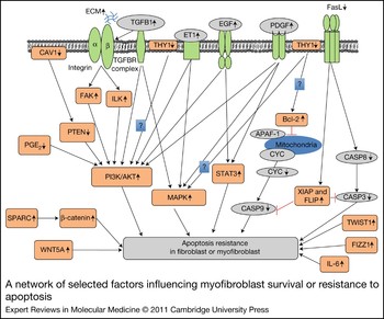
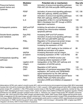
There is much support for an important role for dysregulation of apoptotic cell death in the development of IPF, as well as fibrosis, in most tissues (Ref. Reference Chabaud103). Myofibroblasts, regardless of their origins (see above), are the key effector cells in fibrogenesis. In the process of normal wound healing, myofibroblasts undergo apoptosis and rapidly disappear; failure of apoptosis leads to myofibroblast expansion, excessive ECM production, persistent tissue contraction and pathological scar formation. In the lungs of IPF patients, a series of consistent findings supports the correlation of dysregulated apoptosis with the pathogenesis of fibrosis (Refs Reference Fattman104, Reference Maher105, Reference Jinta106). By contrast, widespread epithelial apoptosis is observed, and epithelial apoptosis might be important in the initiation of fibrotic remodelling. The contrast of increased epithelial cell apoptosis with fibroblast resistance to apoptosis in the IPF lung has been termed the ‘apoptosis paradox’ (Refs Reference Fattman104, Reference Maher105, Reference Kuhn and McDonald107, Reference Drakopanagiotakis108, Reference Beon109). Myofibroblast survival is a key determinant of whether fibrosis will progress or resolve (Ref. Reference Uhal110), and is due in part to enhanced responsiveness to growth factors and fibrotic cytokines, in addition to resistance to apoptosis (Fig. 2). Myofibroblast proliferation and survival require signals from the cellular and extracellular microenvironment, especially cell–cell and cell–matrix contacts, which provide key signals for proliferation and survival. Modification of prosurvival signalling in fibroblasts by a protein kinase inhibitor (AG1879) protects against fibrotic tissue injury in the bleomycin fibrosis model, indicating that targeting of prosurvival signalling pathways in fibroblasts or myofibroblasts might provide a novel and effective antifibrotic therapy (Refs Reference Vittal111, Reference Horowitz112, Reference Garneau-Tsodikova and Thannickal113). Recent studies have greatly improved our understanding of cellular and molecular factors regulating myofibroblast survival (Refs Reference Vuga114, Reference Xia115, Reference Huang116, Reference Wynes, Frankel and Riches117). The principal mediators affecting myofibroblast survival or apoptosis are listed in Table 2, and the main signalling pathways involved are depicted in Figure 3.
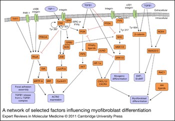
A network of selected factors influencing myofibroblast differentiation. Several integrin-activated signalling pathways (e.g. FAK–JNK–AP-1, PI3K–AKT or MEK1/2–ERK1/2) are involved in myofibroblast differentiation. The potent profibrotic effect of TGFB1 is demonstrated by its influence on several pathways. Recent results showing the role of HH ligands, DIA, PPARγ ligands, HIF-1α and NOX4 are also illustrated. Abbreviations: ACTA2, alpha 2 smooth muscle actin; AKT, v-akt murine thymoma viral oncogene homologue; c-fos, FBJ murine osteosarcoma viral oncogene homologue; c-Jun, jun proto-oncogene; CTGF, connective tissue growth factor; CXCL12, chemokine (C–X–C motif) ligand 12; CXCR4, chemokine (C–X–C motif) receptor 4; DIA, diaphanous homologue 1 (Drosophila); EMT, epithelial to mesenchymal transition; EndMT, endothelial to mesenchymal transition; ERK1/2, mitogen-activated protein kinase 1/2; FAK, PTK2 protein tyrosine kinase 2; FYN, FYN oncogene related to SRC, FGR, YES; GRB2, growth factor receptor-bound protein 2; HH ligand, Hedgehog ligand; HIF-1α, hypoxia-inducible factor 1, alpha subunit; JNK, JUN N-terminal kinase; JUND, jun D proto-oncogene; L-TGFB1, latent transforming growth factor, beta1; MEK1/2, mitogen-activated protein kinase kinase 1/2; MRTF-A, myocardin-related transcription factor A; NOX4, NADPH oxidase 4; PAR-1, coagulation factor II (thrombin) receptor; PI3K, phosphoinositide-3-kinase; PIP3, phosphatidylinositol-(3,4,5)-trisphosphate; PRKCA, protein kinase C, alpha; PPARγ, peroxisome proliferator-activated receptor gamma; PTEN, phosphatase and tensin homologue; RAC1, ras-related C3 botulinum toxin substrate 1; RhoA, ras homologue gene family, member A; SMAD2/3, SMAD family member 2/3; SRC, v-src sarcoma viral oncogene homologue; SFK, SRC family kinase; SHC, SHC (Src homology 2 domain containing) transforming protein; SOS, son of sevenless homologue (Drosophila); SRF, serum response factor; TGFB1, transforming growth factor, beta1; THY1, Thy-1 cell surface antigen; TSP-1, thrombospondin 1; Tyr, tyrosine.

A network of selected factors influencing myofibroblast survival or resistance to apoptosis. Several integrin-activated signalling pathways are involved in myofibroblast survival. The prominent role of the AKT survival pathway is demonstrated by the convergence of several pathways. Recent results showing the role of WNT/β-catenin, TWIST1 and FIZZ1 pathways are also illustrated. (The question marks indicate where literature or our own studies suggest an effect, but the mechanism is unknown. See main text of the article.) Abbreviations: AKT, v-akt murine thymoma viral oncogene homologue; APAF-1, apoptotic peptidase activating factor 1; Bcl-2, B-cell CLL/lymphoma 2; CASP, caspase; CYC, cytochrome c; ECM, extracellular matrix; EGF, epidermal growth factor; ET1, endothelin 1; FAK, PTK2 protein tyrosine kinase 2; FasL, FAS (TNF receptor superfamily, member 6) ligand; FIZZ1, resistin-like alpha; FLIP, CASP8 and FADD-like apoptosis regulator; FOXO3, forkhead box O3; IL-6, interleukin 6; ILK, integrin-linked kinase; MAP, mitogen-activated protein; PDGF, platelet-derived growth factor; PGE2, prostaglandin E2; PI3K, phosphoinositide-3-kinase; PTEN, phosphatase and tensin homologue; SPARC, secreted protein, acidic, cysteine-rich (osteonectin); STAT3, signal transducer and activator of transcription 3; TGFBR, TGFB receptor; TGFB, transforming growth factor beta; THY1, Thy-1 cell surface antigen; TWIST1, twist homologue 1 (Drosophila); WNT5A, wingless-type MMTV integration site family, member 5A; XIAP, X-linked inhibitor of apoptosis.

Key mediators of fibroblasts and myofibroblast survival or apoptosis

Abbreviations: ACTA2, actin, alpha 2, smooth muscle; AKT, v-akt murine thymoma viral oncogene homologue; BAX, BCL2-associated X protein; Bcl-2, B-cell CLL/lymphoma 2; BIM, BCL2-like 11; CAV1, caveolin 1; EGF, epidermal growth factor; ERK, mitogen-activated protein kinase; ET1, endothelin 1; FAK, PTK2 protein tyrosine kinase 2; FAS, Fas (TNF receptor superfamily, member 6); FasL, Fas ligand; FGF2, fibroblast growth factor 2 (basic); FIZZ1, resistin-like alpha; FLIP, CASP8 and FADD-like apoptosis regulator; FOXO3, forkhead box O3; GSK3B, glycogen synthase kinase 3 beta; IL, interleukin; ILK, integrin-linked kinase; IPF, idiopathic pulmonary fibrosis; JNK, JUN N-terminal kinase; MAP, mitogen-activated protein; MAPK, MAP kinase; PDGF, platelet-derived growth factor; PGE2, prostaglandin E2; PAI, plasminogen activator inhibitor type 1; PI3K, phosphoinositide-3-kinase; PTEN, phosphatase and tensin homologue; SMAD, SMAD family member; SPARC, secreted protein acidic and rich in cysteine; STAT3, signal transducer and activator of transcription 3; TGFB, transforming growth factor, beta; TWIST1, twist homologue 1 (Drosophila); WNT, wingless-type MMTV integration site family; XIAP, X-linked inhibitor of apoptosis.
Interactions between epithelial cells and fibroblasts
Products of epithelial cell injury might stimulate fibroblasts to produce ECM components such as collagen and fibronectin, leading to dysregulated repair of the epithelial or endothelial barrier (Refs Reference Selman and Pardo118, Reference Chapman119, Reference Horowitz and Thannickal120). IPF is thought to result from several cycles of epithelial cell injury and activation (Refs Reference Selman and Pardo118, Reference Selman and Pardo121). Myofibroblasts from fibrotic lungs possess a cytotoxic phenotype that causes apoptosis of epithelial cells by the FAS–Fas ligand (FAS–FasL) pathway, indicating that myofibroblast cytotoxicity might contribute to the absence of re-epithelialisation, resulting in persistent lung fibrosis (Ref. Reference Golan-Gerstl122). FasL-deficient myofibroblasts lose this myofibroblast cytotoxic phenotype both in vitro and in vivo (Ref. Reference Golan-Gerstl122). Blocking the FAS–FasL pathway might have a therapeutic potential in bleomycin-induced pulmonary fibrosis in mice (Ref. Reference Kuwano123). However, in other studies, the FAS–FasL system was not required for bleomycin-induced pulmonary fibrosis in mice, suggesting that epithelial cell apoptosis after bleomycin exposure can also be mediated by FAS-independent pathways (Ref. Reference Aoshiba124).
IPF lung fibroblasts are resistant to FasL-induced apoptosis (Ref. Reference Moodley125), but the mechanism is not completely understood. Altered expression of membrane-bound and soluble CD95/FAS contributes to the resistance of fibrotic lung fibroblasts to FasL-induced apoptosis (Ref. Reference Buhling126). Prostaglandin (PGE2) is an important antifibrotic lipid mediator (Ref. Reference Kolodsick127), the level of which is decreased in cultured lung fibroblasts isolated from IPF patients (Ref. Reference Wilborn128). PGE2 induces fibroblast apoptosis by modulating several survival pathways (Ref. Reference Huang116). Exogenous PGE2 increases FasL-induced apoptosis in fibrotic lung fibroblasts compared with FasL alone. Conversely, in primary fibrotic lung type-II AECs, PGE2 protected against FasL-induced apoptosis, suggesting that diminished PGE2 contributes to the apoptosis paradox in IPF (Ref. Reference Horowitz and Peters-Golden129). Administration of TGFB1 enhances FAS-mediated epithelial cell apoptosis and lung injury by caspase 3 activation in mice (Ref. Reference Hagimoto130). However, TGFB1 has also been shown to inhibit FAS-mediated apoptosis in keloid-derived fibroblasts (Ref. Reference Chodon131).
Proapoptotic and antiapoptotic proteins
Resistance of IPF fibroblasts to FAS-mediated apoptosis has been associated with increased expression of X-linked inhibitor of apoptosis (XIAP) and CASP8 and FADD-like apoptosis inhibitor protein (FLIP) (Ref. Reference Tanaka132). A more recent study showed that late-stage lesional fibroblasts from skin in patients with SSc were more resistant to FAS-induced apoptosis than their nonlesional counterparts isolated from the same patient. Results showed an increase in the levels of the antiapoptotic proteins cFLIPs and cIAP in lesional cells compared with nonlesional dermal fibroblasts, suggesting that cFLIPs and cIAP provide protection from Fas-induced apoptosis in SSc late-stage fibroblast populations (Ref. Reference Chabaud103). Bcl-2 survival factors are critical for lung fibroblast resistance to apoptosis and might explain the continued survival and accumulation of myofibroblasts in lung fibrosis (Refs Reference Kazufumi133, Reference Marcotte, Lacelle and Wang134). AECs from Bid −/– mice are resistant to TGFB-induced cell death, suggesting that Bcl-2 family members are important regulators of fibrosis downstream of TGFB1 activation (Ref. Reference Budinger135).
Prosurvival factors: growth factors and cytokines
TGFB1 promotes an antiapoptotic phenotype in fibroblasts or myofibroblasts by two important antiapoptotic pathways: the FAK pathway and the PI3K–AKT pathway. Treatment of bleomycin-exposed mice with soluble TGFB type-II receptors (TGBR2) significantly attenuates the degree of pulmonary fibrosis (Ref. Reference Yamada136). PDGFs, including PDGFAA and PDGFBB, are critical in the survival and differentiation of mesenchymal cells during lung development, and they are also important for tissue repair following injury in adult tissues. PDGFAA (through PDGFRA) activates several intracellular signalling molecules that have important roles in mesenchymal cell survival, including MAP kinases and the STAT family members STAT1 and STAT3 (Refs Reference Bonner137, Reference Bonner138). EGF treatment of fibroblasts expressing human EGF receptors blocks staurosporine-induced apoptosis by inhibiting the activation of caspase 3 in an AKT-dependent manner (Ref. Reference Shao, Yi and Wells139). IL-6 enhances FAS-induced apoptosis and expression of BAX in normal cells, but inhibits apoptosis and induces expression of Bcl-2 in IPF fibroblasts, indicating that dysregulated IL-6 signalling in fibroblasts from IPF might increase the resistance to fibroblasts from IPF to apoptosis (Ref. Reference Moodley140). Insulin-like growth factor 1 (IGF1) is a fibroblast growth and survival factor. Interstitial macrophage-derived IGF1 correlates with disease severity in IPF, and the TH2 cytokines IL-4 and IL-13 stimulate macrophage expression and secretion of IGF1. Conditioned medium from IL-4-stimulated macrophages protects myofibroblasts from apoptosis by activation of the prosurvival kinases AKT and ERK (Ref. Reference Wynes, Frankel and Riches117). Th1 and Th2 regulatory cytokines have opposite effects on fibroblast apoptosis. Pretreatment with IFNγ or TNF-α increased, whereas long-term serum-free culture and pretreatment with TGFB1 or IL-10 decreased, the FAS-mediated apoptotic susceptibility of dermal fibroblasts in the presence of cycloheximide, indicating that their pro- or antifibrotic effects involve direct effects on fibroblast survival (Ref. Reference Santiago141).
Other modulators of survival or apoptosis
PTEN deficiency is associated with fibrosis in different organs. Inactivation of PTEN in PTEN +/+ embryonic fibroblasts increases AKT activity and suppresses apoptosis in contractile collagen matrices; inhibition of AKT activity restores the sensitivity of PTEN-null cells to collagen gel contraction-induced apoptosis (Refs Reference White142, Reference Nho143). Caveolin 1 (CAV1) and PTEN expression are low in myofibroblasts within IPF fibroblastic foci. The absence of CAV1 confers lung fibroblast resistance to apoptosis (Ref. Reference Xia115). The PI3K/AKT signalling pathway is activated after ligation of receptor tyrosine kinases and seven transmembrane G-protein-coupled receptors, or through integrin-associated, adhesion-mediated signalling cascades. Activated AKT might support cell survival through a number of potential mechanisms, including regulation of Bcl-2 family proteins, NF-κB and CASP9. ET1 and TGFB1 independently promote fibroblast resistance to apoptosis through signalling pathways involving p38 MAPK and PI3K/AKT (Ref. Reference Kulasekaran144). Fibroblasts lacking THY1 are resistant to collagen-gel-contraction-induced apoptosis, which can be restored by re-expression of THY1 (Ref. Reference Sanders, Kumbla and Hagood52). FIZZ1 is highly expressed in bleomycin-injured lung. FIZZ1 treatment inhibited the TNF-α-induced activities of CASP3 and CASP8, indicating that it can contribute to the pathogenesis of pulmonary fibrosis by induction of myofibroblast resistance to apoptosis (Ref. Reference Chung145). Expression of TWIST1 is increased in nuclei of type-II epithelial cells, macrophages and fibroblasts in IPF lungs. Furthermore, suppression of TWIST1 expression results in decreased rat lung fibroblast accumulation as a result of increased apoptosis, whereas TWIST1 overexpression protects the cells against several apoptotic stimuli (Ref. Reference Bridges146).
ECM–integrin interaction
Myofibroblast function is critically dependent on interaction with the surrounding ECM. Receptors for the ECM, in particular the integrins, act not only to provide anchorage for fibroblasts or myofibroblasts, but also to transduce signals to facilitate fibroblast decisions to survive or die. Integrin–TGFB crosstalk is important in fibrosis, cancer and wound healing (Ref. Reference Margadant and Sonnenberg66). Altered integrin expression stimulated by fibrotic growth factors indicates that they have an integral role in the fibrosis programme (Ref. Reference Margadant and Sonnenberg66). Integrin-mediated TGFB1 activation is a potential therapeutic target in fibrogenic disorders (Ref. Reference Nishimura147). Interaction of α8β1 integrin with fibronectin appears to promote cell survival during serum deprivation through the PI3K pathway (Ref. Reference Farias148). The Arg-Gly-Asp (RGD)-containing central cell-binding domain (CCBD) of fibronectin is required for mesenchymal cell survival and growth. Fibronectin contains three growth-factor-binding domains (FN-GFBDs) that bind PDGFBB, a potent fibroblast survival and mitogenic factor.
Dermal fibroblast survival requires PDGFBB stimulation in the presence of FN-GFBD, as well as adhesion to fibronectin through the CCBD (Ref. Reference Lin149). Three soluble fibronectin peptides (RGD, CS-1 and FN-C/H-V) induce apoptosis in lung fibroblasts. Treatment of fibroblasts with these peptides caused proteolysis of FAK, a tyrosine kinase involved in integrin-mediated signalling related to cell survival, suggesting that soluble fibronectin peptides trigger nontransformed fibroblast apoptosis by a disruption of integrin-mediated survival signalling pathways (Ref. Reference Hadden and Henke150). Loss of cell adhesion or adhesion signalling can lead to a form of apoptosis called anoikis. Anoikis is likely to represent an important apoptotic mechanism in mesenchymal cells. FAK is an important mediator of integrin-dependent survival signals, and inhibition of FAK activation induces anoikis. TGFB1 protects myofibroblasts from anoikis, in part, by activation of the PI3K–AKT pathway. Thus, TGFB1 coordinately and independently activates the FAK and AKT protein kinase pathways to confer an anoikis-resistant phenotype to myofibroblasts (Ref. Reference Horowitz151). Integrin-linked kinase (ILK) is an intracellular protein involved in the regulation of integrin-mediated processes. ILK activity decreases in response to collagen matrix contraction, which correlates with AKT dephosphorylation and induction of fibroblast apoptosis (Ref. Reference Nho152).
Oxidative stress
Oxidation of cysteines in proteins, such as S-glutathionylation, is known to regulate death-receptor-induced apoptosis in fibrotic disease (Ref. Reference Janssen-Heininger153). HIF-1α is a major regulator of hypoxic signalling and is overexpressed in the hyperplastic epithelium of the fibrotic lung. Absence of HIF-1α and increased expression of Bcl-2 in fibrotic foci support the hypothesis that dysregulated hypoxic signalling is important in fibroblast resistance to apoptosis (Ref. Reference Tzouvelekis154).
WNT–β-catenin signalling
Accumulation of nuclear β-catenin, a hallmark of aberrant activated WNT signalling, has been observed in the lungs of patients with IPF (Ref. Reference Chilosi155). The WNT signalling pathway regulates apoptosis through several mechanisms (Ref. Reference Pecina-Slaus156). It inhibits apoptosis by activation of β-catenin/TCF-mediated transcription and can increase survival by activation of NF-κB or inhibition of GSK3B (Ref. Reference Pecina-Slaus156). WNT5A significantly increases fibroblast proliferation as well as relative resistance to H2O2-induced apoptosis in UIP lung fibroblasts (Ref. Reference Vuga114). IPF fibroblasts constitutively express increased basal levels of SPARC, PAI-1 and active β-catenin; elevated SPARC activates β-catenin, which regulates expression of PAI-1, and confers an apoptosis-resistant phenotype (Ref. Reference Chang89).
Myofibroblast heterogeneity with regard to survival or apoptosis
Fibroblasts and myofibroblasts in the lung consist of phenotypically diverse subpopulations. Myofibroblasts differ in cell size, cytoskeletal arrangement, surface markers, cytokine expression profiles and receptor expression. THY1 modulates apoptotic signalling pathways in a variety of cell types, and crosslinking antibodies against THY1 induce apoptosis in thymocytes and mesangial cells (Refs Reference Hueber157, Reference Morita158). Fibroblasts lacking THY1 expression are resistant to apoptosis when they contract collagen gels, even though they are more contractile; however, forced expression of THY1 restores apoptotic susceptibility (Ref. Reference Sanders, Kumbla and Hagood52). A recent study compared the morphological and functional characteristics of fibroblasts isolated from proximal airways and distal lung parenchyma to determine phenotypic differences (Ref. Reference Kotaru159), suggesting that these phenotypic differences might partially explain the variable responses to injury and repair between proximal airways and distal lung or parenchyma in asthma and other respiratory diseases.
In summary, despite the diversity of causes for fibrosis and the numerous mechanisms involved in lung fibrotic disease, a common similarity important to disease progression is the enhanced survival of fibroblasts and myofibroblasts. Fibroblast and myofibroblast survival remains a main target for fibrotic disease, and further research towards controlling the survival of these cells should eventually lead to new effective treatments. Apoptosis of fibroblasts is required for normal wound healing and limitation of collagen deposition, and resistance to apoptosis has been linked to tissue fibrosis; therefore, promoting fibroblast apoptotic signalling pathways and inhibition of antiapoptotic pathways to limit fibroblast survival might help in promoting the resolution of fibrosis.
Myofibroblasts as therapeutic targets
Myofibroblast differentiation is a complex and highly regulated process. Understanding the diverse mechanisms of regulation of this phenotype offers several possible targets for intervention in fibrotic disorders and in several malignancies. An important challenge is to identify the hierarchy of pathways and discover ‘master switches’ that control multiple pathways. TGFB1 is attractive in this regard, because it regulates most aspects of the phenotype; however, it is not considered to be a good clinical target because of its many other critical biological roles, despite the fact that activation of TGFB1 from a latent form can be selectively targeted. LSKL, a peptide antagonist of TSP-1, can attenuate renal interstitial fibrosis in rats with unilateral ureteral obstruction (Ref. Reference Xie160). TGFB1 can be antagonised with blocking antibodies, or by a mutated form of TGFB3, as shown in a mouse skin wound model (Ref. Reference Waddington45), whereas the release of TGFB1 can be decreased in vivo by a lentiviral shRNA construct against CD36 (Ref. Reference Wang61). Additional therapeutic candidate targets for modulating TGFB1 are PAR-1 (Ref. Reference Jenkins60) and THY1 (Ref. Reference Zhou54). Other promyofibroblastic cytokines, chemokines and growth factors that might be useful targets include IL-4 (Ref. Reference Huaux43), CXCL10 (Ref. Reference Jiang72), SOCS1 (Ref. Reference Nakashima161), CCR2 (Ref. Reference Moore162) and CXCL12 (Ref. Reference Phillips29). However, many of these factors are important in the early phases of fibrogenesis, soon after tissue injury. PPARγ agonists (e.g. troglitazone) are able to inhibit fibrosis in a murine model, even when administration is delayed until after the initial inflammatory phase has largely resolved (Ref. Reference Milam82). The oxidant–antioxidant balance in in vivo lung fibrosis has been restored by NOX4 RNAi (Ref. Reference Hecker76), and in vitro results suggest that siRNA constructs against HIF-1α or CXCL12 might also be effective (Ref. Reference Toullec79). The PPARγ agonist troglitazone and the Wnt antagonist Dickkopf-1 reduced lung (Ref. Reference Milam82) and renal fibrosis (Ref. Reference He90), respectively. The antifibrotic drug pirfenidone, which appears to modify growth factor expression, collagen expression and redox balance, reduces myofibroblast accumulation in bleomycin-induced lung fibrosis and seems to have modest clinical benefits in IPF and renal fibrosis (Refs Reference Cho163, Reference Kakugawa164, Reference Lasky165, Reference Noble166). The complexity of myofibroblast differentiation is demonstrated by the wide range of signalling pathways that have been suggested as therapeutic targets. More recent candidates include the Rho family of small GTPases, HH signalling, DIA or epigenetic modifiers, the targeting of which might reveal novel solutions, not only for organ fibrosis, but also for malignancies, pulmonary hypertension or thyroid eye disease.
Targeting myofibroblasts has proved an effective strategy in some models of organ fibrosis (Ref. Reference Douglass167); however, because of remarkable fibroblast heterogeneity, targeting strategies might need to be tissue- and even disease-specific. Because there is no consensus ‘myofibroblast-specific’ surface marker, understanding the pathways that regulate and maintain the phenotype is crucial. Strategies that target single pathways might be promising in vitro, but fail in vivo, particularly during the stage of progressive fibrosis. For example, the protein kinase inhibitor imatinib mesylate inhibits platelet-derived growth factor receptor activation and fibroblast proliferation in vitro, but fails during the fibrotic phase of the bleomycin model to significantly alter fibrogenic responses assessed by histopathology, collagen content and the accumulation of myofibroblasts within the injured lung (Ref. Reference Vittal168). THY1 expression affects the myofibroblast phenotype broadly [expression of muscle and ECM proteins, myogenic transcription factors, contractility, resistance to apoptosis and mechanotransduction of TGFB activation (Refs Reference Sanders, Kumbla and Hagood52, Reference Zhou54)], and so it might be useful to promote THY1-mediated effects by better defining its mechanisms of action or regulating its expression through epigenetic therapy. In any case, targeting myofibroblast differentiation and survival will require improved understanding of the molecular networks and hierarchies involved.
New high-throughput approaches to proteomics and metabolomics could be leveraged to define noninvasive biomarkers of fibrogenesis to better monitor disease activity and response to treatment. In vitro assays for specific pathways or combinations could be used to screen compound libraries for novel antifibrotic agents. Improved understanding of the critical regulators of this complex cellular phenotype should lead to breakthroughs in the management of ‘classic’ fibrotic diseases such as IPF and liver fibrosis, but may also offer new solutions for more common tissue remodelling disorders such as asthma and congestive heart failure, and could be useful in managing tumour invasion and metastasis regulated by myofibroblasts in tumour stroma.
Acknowledgements
This work was partially supported by a grant from the National Institutes of Health, National Heart Lung & Blood Institute (HL 082818, to J.S.H.). The authors thank the peer reviewers and editors for their excellent suggestions, which have greatly strengthened the review.