Introduction
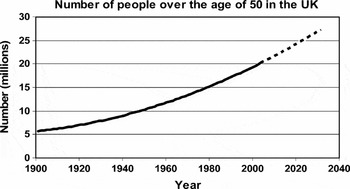
Life expectancy is rapidly increasing and as a consequence the global elderly population is growing (Figure 1). In the United Kingdom between 2002 and 2003 the older population increased at twice the rate of the total population: 0.8% for those over the age of 50 compared with a national growth rate of only 0.4% (www.statistics.gov.uk). Most worryingly, the fastest growing age group are also the most frail, those aged 80 and over, who now constitute 4.5% of the total population (2,749,507 individuals). In other countries this effect is more pronounced, for example over the same period Japan saw an increase of 2.9% in those aged over 65 compared with only a 0.1% rise in the population as a whole. These trends lead to serious socio-economic consequences. An expanded elderly population translates into increased age-associated diseases, which in turn represent a significant and increasing economic burden. People aged over 65 (just 16% of the total population) now account for 47% of total hospital and community health service spending in England (www.statistics.gov.uk).
Life expectancy is continuously rising and as a consequence the elderly population is growing. In 1901, 5.7 million people in the UK were over the age of 50. By 2003 this number had risen to 20 million and by 2031 it is predicted that 27.1 million over-50s will be living in the UK.

Impaired wound healing is one of the most common and costly age-associated pathological changes in the elderly population and represents a major area of unmet clinical need. Treatment of chronic non-healing wounds including leg ulcers, pressure ulcers and diabetic foot ulcers accounts for at least £4 billion of the UK's annual NHS expenditure (www.hospitalmanagement.net). This article focuses on issues related to wound healing in older people. Full details of the search methods are available from the corresponding author.
Structure of the skin
Our skin, the largest organ in the body, acts as a barrier to the external environment, maintains homeostasis by preventing fluid loss and regulating body temperature and mediates immune surveillance and sensory perception.Reference Wysocki1 Skin comprises two layers, a keratinized, stratified epidermis overlaying a supportive dermal layer composed of a collagen-rich extracellular matrix. The epidermis is multilayered; hemidesmosomes anchor the basal keratinocytes to the underlying basement membrane, a laminin and collagen IV rich extracellular matrix. Desmosomes, adherens and tight junctions interconnect neighbouring keratinocytes. Desmosomes have profound structural significance, attaching intracellularly to the keratin intermediate filament network to provide structural integrity. The basal epidermal layer is rich in stem cells, surrounded by proliferative transient amplifying cells. These transient amplifying cells move outwards through the suprabasal epidermal layers before undergoing terminal differentiation, a form of programmed cell death.Beneath the epidermis lies the supporting dermis, which supplies nutrients to the avascular epidermis while cushioning the underlying tissues from trauma. The dermis is divided into two layers, papillary and reticular. The papillary dermis, composed of loosely packed collagen fibres, lies immediately beneath the epidermis, whereas the reticular dermis, which resides beneath the papillary dermis and above the subcutaneous tissue, is composed of tightly packed collagen fibres and elastic fibres. The dermal extracellular matrix (ECM) in adult skin is complex, composed of collagen fibres (for strength), elastic fibres (for resilience) and glycosaminoglycans (for hydration). Buried within the dermis, but derived principally from the epidermis, are a number of appendages including hair follicles, sweat glands, sebaceous glands and lymph nodes.Reference Wysocki1 When the skin is injured it is essential to restore the protective barrier of the epidermis and remodel the damaged underlying dermal tissue.
Overview of acute wound healing
Upon wounding a number of overlapping highly co-ordinated processes occur to ensure effective tissue repair (Figure 2). Initially, blood vessels constrict preventing excessive blood loss and a fibrin clot forms a temporary barrier.Reference Clark2 Degranulation of platelets embedded in the fibrin clot releases pro-inflammatory cytokines and growth factors, which act as a chemotactic cue to initially recruit circulating inflammatory cells and later stimulate the migration of fibroblasts and keratinocytes.Reference Anitua, Andia, Ardanza, Nurden and Nurden3 Changes in endothelial cells lining blood vessels surrounding the wound allow macrophages and neutrophils to adhere to, roll and undergo diapedesis to exit blood vessels and enter the wounded tissue.Reference Schober and Weber4 In turn, recruited inflammatory cells release further cytokines and growth factors to heighten the immune response.Reference Gillitzer and Goebeler5 Neutrophils arrive at the wound site within minutes and begin to clear foreign debris and bacteria. Activated neutrophils release a cocktail of lysosomal enzymes and proteases that facilitate bacterial destruction and removal of damaged matrix. Following neutrophil infiltration, at around 4–7 days post-wounding in adults, monocytes enter the wound and differentiate into macrophages. Macrophages adhere to the ECM, become activated and produce a range of matrix metalloproteinases (MMPs) that degrade and remodel damaged ECM proteins. Macrophages release their own profile of growth factors and cytokines to modulate the earlier signals provided by neutrophils and degranulating platelets. It has recently been shown that macrophages are able to differentiate to a different lineage, named alternatively activated macrophages. These alternatively activated macrophages are instrumental to the resolution of inflammation, expressing molecules such as Ym1 and Arg1.Reference Ho and Sly6 In wounds where infection is minimal or absent, neutrophil infiltration subsides within a few days with macrophages persisting within the wound for several further days to phagocytose any remaining pathogenic material, cell debris and damaged extracellular matrix.Reference Martin, Reece, Maher and Reese7, Reference Fujiwara and Kobayashi8
The skin, providing a barrier to the external environment, comprises a keratinized, stratified epidermis overlying a supportive dermal layer composed of a collagen-rich extracellular matrix (A). When the skin is injured it mounts an immune response to combat infection, restores the protective barrier of the epidermis and re-models the damaged underlying dermal tissue (B).

The release of cytokines and growth factors from platelets, neutrophils and macrophages at the wound edge initiates a burst of cellular proliferation followed by a migration phase. Soon after injury, dermal fibroblasts at the wound site begin to proliferate and migrate to the newly formed granulation tissue where they begin to lay down a provisional collagen-rich matrix. Unwounded adult dermis is predominantly composed of type 1 collagen. However, upon wounding fibroblasts up-regulate integrins to adhere to the fibrin, fibronectin and vitronectin-rich wound matrix.Reference Eckes, Zigrino and Kessler9–Reference Midwood, Mao, Hsia, Valenick and Schwarzbauer11 Fibroblasts first lay down a predominantly type 3 collagen matrix and concomitantly secrete a mixture of matrix MMPs, including collagenases, stromelysins and gelatinases to degrade and remodel the existing extracellular matrix.Reference Salo, Mäkelä, Kylmäniemi, Autio-Harmainen and Larjava12 Following migration into the granulation tissue and stimulated by TGF-β1, a subset of fibroblasts differentiate into myofibroblasts.Reference Desmoulière, Geinoz, Gabbiani and Gabbiani13, Reference Vaughan, Howard and Tomasek14 These differentiated cells express α-smooth muscle actin and are capable of generating a contractile force to close the wound by the process of contracture.Reference Hinz15
Epidermal growth factors (EGFs), transforming growth factor-α and -β, heparin-binding epidermal growth factor (HB-EGF), fibroblast growth factor 7 (FGF7) and insulin-like growth factor (IGF-1) released from wound platelets, macrophages, fibroblasts and keratinocytes all act as keratinocyte motogens and/or mitogens promoting re-epithelialization.Reference Barrandon and Green16–Reference Haase, Evans, Pofahl and Watt21 Re-epithelialization requires the migration of keratinocytes directly through the granulation tissue, thus keratinocytes release tissue-type-specific plasminogen activator (tPA) and urokinase plasminogen activator (uPA) to convert plasminogen in the fibrin-rich clot to the enzyme plasmin, which digests a pathway for the migrating cells.Reference Grφndahl-Hansen, Lund, Ralfkiaer, Ottevanger and Danφ22–Reference Ossowski and Aguirre-Ghiso24 In addition, MMPs secreted by wound edge keratinocytes digest the extracellular matrix.Reference Pilcher, Dumin and Schwartz25–Reference Seiki27 To begin migration, hemidesmosomes which link the wound edge basal keratinocytes to the basement membrane are degraded.Reference Krawczyk and Wilgram28, Reference Litjens, de Pereda and Sonnenberg29 Approximately 24 h after injury, a surge of proliferation occurs behind the migrating front to compensate for the loss of migrating keratinocytes.Reference Garlick and Taichman30 Stem cells within the basal layer of the epidermis and hair follicles contribute to the re-epithelialization process.Reference Ito, Liu and Yang31 Migrating keratinocytes also deposit laminin, reforming the basement membrane. Once the opposing epithelial fronts meet, migration ceases and keratinocytes differentiate into a new stratified epidermis. A key aspect of tissue repair, which distinguishes it from ‘true’ regeneration, is that the newly formed tissue differs from native skin; notably, appendages such as hair follicles and sweat glands fail to re-form and the neo-epidermis is flat, lacking the undulations and rete ridges observed in normal skin.
Whilst the migration of keratinocytes restores the epithelial barrier, the formation of new blood vessels beneath the surface establishes a new vascular flow. The release of growth factors including FGF2, VEGF, TNFα and TGFα from damaged endothelial cells and recruited macrophages promotes angiogenesis by stimulating endothelial cell proliferation and migration.Reference Li, Zhang and Kirsner32 Up-regulation of αvβ3 integrins at the tips of sprouting capillaries is vital for angiogenesis to occur.Reference Brooks, Clark and Cheresh33 In addition, the secretion of MMPs from fibroblasts and keratinocytes permits invasion of endothelial cells into the wound.Reference Reed, Corsa, Kudravi, McCormick and Arthur34
The final and by far the longest stage of wound repair is the remodelling phase; this involves the degradation, synthesis, reorganization and stabilization of collagen fibres, which ultimately result in the formation of a connective tissue scar. In normal skin, collagen fibres are bundled together within the dermis in a highly organized pattern; however, after wounding damaged collagen fibres are deposited in characteristic parallel arrays. Throughout the remodelling phase the collagen content of the wound declines and the tensile strength increases via crosslinking of collagen fibres. As the wound matures, crosslinks become more complex, providing greater strength; however, wound tissue never achieves the strength of native skin. Central to the remodelling phase is the proteolytic activity of MMPs.Reference Moses, Marikovsky and Harper35, Reference Lobmann, Ambrosch, Schultz, Waldmann, Schiweck and Lehnert36
Skin ageing
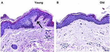
Aged skin is fundamentally different from the skin of younger individuals (Figure 3). A combination of intrinsic and extrinsic factors instigate the progressive loss of structural integrity and physiological function. Intrinsic ageing, a natural, genetically determined process, causes slow tissue degeneration, via a gradual shift from protein synthesis to protein degradation. External (extrinsic) ageing occurs through the accumulation of environmental damage, with exposure to ultraviolet (UV) radiation being the predominant external factor. In photo-protected skin endogenous reactive oxygen species (ROS), natural metabolic by-products, induce structural changes in DNA, proteins and lipids. In photo-aged skin, amplified ROS combined with direct UV effects on skin structural proteins leads to substantial changes in dermal extracellular matrix.
Age-related decline in the structural integrity of human skin. In young skin (A) the epidermis is connected to the underlying supportive dermis by rete ridges, finger-like projections that protrude into the dermis (shown by the arrow in A). As we age these rete ridges flatten, and contact between the epidermis and dermis is reduced (B). In addition, an age-associated decrease in the thickness of the epidermis and dermis is observed and the cellular content of the dermis declines.

In combination these factors lead to age-associated atrophy of both the epidermis and dermis; the dermo-epidermal junction, which is highly convoluted in young skin, becomes flattened and contact between epidermis and dermis deteriorates, rendering the skin more susceptible to traumatic injury.Reference Kurban and Bhawan37, Reference Branchet, Boisnic, Frances and Robert38 Decreased production and increased degradation of dermal collagen fibres, together with a progressive decline in number and organization of elastic fibres and reduced hydration, leads to a loss of skin elasticity.Reference Branchet, Boisnic, Frances and Robert38–Reference El-Domyati, Attia and Saleh41 These ECM changes, specifically degradation of dermal elastic fibres, cause the pronounced wrinkling observed in aged skin. Vascular endothelial cell proliferation declines with increasing age, and the vasculature within the dermis progressively atrophies. Similarly, an age-associated decline in the proliferative response of keratinocytes and fibroblasts occurs, with the cells eventually becoming senescent.Reference Swift, Kleinman and DePietro42, Reference Reed, Ferara and Vernon43 Thus the total number of fibroblasts, mast cells and macrophages present within the dermis declines over time.Reference Andrew, Behnke and Sato44 Epidermal melanocytes and Langerhans cell numbers are decreased in aged photo-protected skin, as are epidermal stem cells. In contrast, stem cell progeny keratinocytes, termed transit amplifying cells, are actually increased in number but substantially decreased in function with extended cell cycle duration.Reference Charruyer, Barland and Yue45 Age-related changes in skin appendages are also observed, including slower hair growth and decreased secretions from sweat and sebaceous glands.Reference Montagna and Carlisle46, Reference Pochi, Strauss and Downing47 These accumulative changes leave the elderly person increasingly susceptible to skin injuries and increasingly prone to poor healing.
Age-associated changes in wound healing
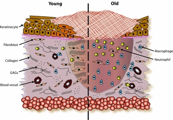
Our ability to heal wounds deteriorates as we age. Multiple factors contribute to this delayed healing, including alterations in immune response, angiogenesis, fibroblast function and keratinocyte migration (Figure 4).
Wound healing ability deteriorates with age. Wounds from elderly subjects display a heightened but ineffective immune response and reduced angiogenesis. In addition, reduced growth factor and cytokine levels lead to retarded re-epithelialization and depressed fibroblast function. A combination of decreased collagen synthesis and increased MMP activity leads to reduced collagen deposition.

Inflammatory phase
In aged mice and humans wound monocyte, macrophage and B-lymphocyte infiltration is delayed.Reference Ashcroft, Horan and Ferguson48, Reference Ashcroft, Horan and Ferguson49 Despite this delay, the total number of macrophages recruited to the wound actually increases with age.Reference Ashcroft, Horan and Ferguson49, Reference Swift, Burns, Gray and DiPietro50 Crucially, in aged individuals macrophage function is impaired, with growth factor production diminishing and phagocytic capacity reduced.Reference Swift, Kleinman and DePietro42, Reference Swift, Burns, Gray and DiPietro50 The functional significance of diminished macrophage activity in an elderly population has been demonstrated experimentally, where injection of aged mice with macrophages harvested from young mice is able to accelerate wound repair.Reference Danon, Kowatch and Roth51
Proliferative phase
Evidence for age-related changes in fibroblast function is less clear. Studies report reduced fibroblast proliferative capacity with increasing age, while other studies indicate that the mitogenic response of fibroblasts to cytokine and growth factor stimulation is unaltered.Reference Plisko and Gilchrest52–Reference Freedland, Karmiol, Rodriguez, Normolle, Smith and Garner54 Variable migration in aged fibroblasts has been associated with a reduction in α2β1 integrin function and a disorganized actin cytoskeleton.Reference Reed, Ferara and Vernon43 A key role of wound fibroblasts is to deposit and remodel ECM. While Freedland and co-workers report no change in synthesis of ECM proteins by fibroblasts extracted from young and old human skin following TGFβ treatment, a number of studies have demonstrated MMP over-expression in aged fibroblasts.Reference Reed, Ferara and Vernon43, Reference Ballas and Davidson55
An age-associated decrease in re-epithelialization rate has been demonstrated in multiple studies.Reference Swift, Kleinman and DePietro42, Reference Ashcroft, Horan and Ferguson48, Reference Holt, Kirk, Regan, Hurson, Lindblad and Barbul56 Keratinocyte migration and proliferation is stimulated by growth factors and cytokines. Numerous cytokines, growth factors and their receptors decrease with age, such as PDGF, TGFβ, KGF, EGF and EGFR, and may therefore mediate reduced keratinocyte proliferation and migration in aged wounds.Reference Ashcroft, Horan and Ferguson57 The response of keratinocytes to hypoxia has also been studied and whereas keratinocytes from young donors show an increase in cell migration when exposed to hypoxia, keratinocytes from elderly donors have depressed migratory ability.Reference Xia, Zhao, Tyrone, Chen and Mustoe58 In addition, aged keratinocytes fail to up-regulate MMP1, MMP9, TIMP1 and TGFβR1, each of which have important roles in the re-epithelialization process.Reference Xia, Zhao, Tyrone, Chen and Mustoe58
Angiogenesis
There is conflicting evidence regarding age-related changes in angiogenesis following wounding. Whereas Swift and co-workers showed a reduction in angiogenesis in excisional wounds from aged mice, Ashcroft and co-workers identified an initial delay in angiogenesis but an ultimate increase in angiogenesis in incisional wounds from aged mice.Reference Swift, Kleinman and DePietro42, Reference Ashcroft, Horan and Ferguson48 An in vitro study by Arthur and co-workers suggested that a reduction in angiogenic growth factors is responsible, in part, to an age-related decline in sprouting of microvessels.Reference Arthur, Vernon, Sage and Reed59 Subsequently, Swift and co-workers showed a decline in the production of growth factors that promote angiogenesis, including FGF2 and VEGF in wounds from aged mice.Reference Swift, Kleinman and DePietro42 In addition, their study showed a reduction in capillary growth in response to FGF2 in aged mice. Thus Swift and co-workers suggest that age-related decline in angiogenesis occurs through the combination of a reduction in angiogenic growth factor production and a decline in vascular endothelial cell responsiveness to growth factor stimulation.Reference Swift, Kleinman and DePietro42
Remodelling phase
Acute wounds from elderly humans have been reported to heal with an improved quality scar, as assessed by microscopic scar appearance (dermal architecture) more closely resembling that of normal skin than wounds from younger subjects.Reference Ashcroft, Horan and Ferguson48, Reference Ashcroft, Kielty, Horan and Ferguson60 This suggests that the aged wound phenotype may resemble a more regenerative form of healing. Unfortunately, despite improved appearance, the tensile strength of human scars declines with age.Reference Halasz61, Reference Sandblom62 Studies in animal models are unclear, with increased age linked to no change, decrease or even increase in mechanical strength.Reference Holm-Pedersen and Zederfeldt63, Reference Beck, DeGuzman, Lee, Xu, Siegel and Amento64
Hormonal regulation of wound healing
Over the last decade sex hormones have been demonstrated to play a fundamental role in the regulation of cutaneous wound healing. In humans the production of dehydroepiandrosterone sulfate (DHEAS), the common sex hormone precursor, and its metabolite dehydroepiandrosterone (DHEA) declines significantly with age.Reference Labrie, Bélanger, Cusan, Gomez and Candas65 DHEA is produced from cholesterol in the adrenal glands followed by local conversion into oestrogens and androgens. In humans, reduced systemic DHEA levels positively correlate with an increased risk of developing a chronic (non-healing) venous leg ulcer.Reference Mills, Ashworth, Gilliver, Hardman and Ashcroft66 In a murine incisional wound model, systemic DHEA treatment completely reverses the delayed wound healing phenotype exhibited by mice lacking systemic oestrogen.Reference Mills, Ashworth, Gilliver, Hardman and Ashcroft66 Moreover, local DHEA treatment reverses impaired wound healing in aged mice.Reference Mills, Ashworth, Gilliver, Hardman and Ashcroft66 DHEA administration may therefore act as an effective therapy for treatment of delayed healing wounds in older people.
Oestrogen
In both males and females oestrogen levels decline with age; whereas this decline occurs suddenly in females with the onset of menopause, in males the decline is more gradual. With life expectancy continuing to rise, the physiological implications of the postmenopausal period are becoming more pronounced. Oestrogen has a profound effect on skin ageing, exemplified by the accelerated decline in skin appearance that occurs in the perimenopausal years.Reference Verdier-Sévrain, Bonté and Gilchrest67 Oestrogen also has a remarkable beneficial influence on the rate of cutaneous wound healing. Topical oestrogen treatment in healthy men and women significantly increases the rate of wound healing.Reference Ashcroft, Greenwell-Wild, Horan, Wahl and Ferguson68 Moreover, evidence suggests that hormone replacement therapy (HRT) protects against the development of chronic wounds in postmenopausal women.Reference Margolis, Knauss and Bilker69, Reference Berard, Kahn and Abenhaim70 Our group have investigated the role of oestrogen during wound healing extensively using the ovariectomized mouse model. Ovariectomized mice, with surgically removed ovaries and thus depleted endogenous oestrogen levels, display retarded re-epithelialization, increased inflammation and reduced collagen deposition, a phenotype which can be reversed by systemic oestrogen administrationReference Ashcroft, Dodsworth and van Boxtel71–Reference Emmerson, Campbell, Ashcroft and Hardman74 (Figure 5). In addition, topical oestrogen treatment in aged humans has also been shown to increase collagen content of wounds.Reference Ashcroft, Greenwell-Wild, Horan, Wahl and Ferguson68
The beneficial influence of oestrogen on cutaneous wound healing. Depletion of systemic oestrogen levels by surgically removing the ovaries (OVX) significantly delays wound healing in a murine model. Ovariectomized mice exhibit larger wounds (B) with increased numbers of neutrophils (E) and macrophages (H) when compared with mice with intact ovaries (A, D and G). This delay can be entirely reversed by systemic oestrogen treatment of ovariectomized mice (C, F and I). Arrows indicate wound margins.

Oestrogen's beneficial effects on wound healing are mediated through a range of wound processes; it has a mitogenic effect on keratinocytes, increasing the rate of re-epithelialization;Reference Ashcroft, Greenwell-Wild, Horan, Wahl and Ferguson68 it regulates matrix turnover, promoting matrix deposition and inhibiting wound protease activity; it is anti-inflammatory, reducing wound levels of innate immune cells and promoting alternative macrophage activation. The effects on re-epithelialization are exemplified in a study that compared the rate of wound re-epithelialization in post-menopausal women who had never taken HRT to post-menopausal women who had taken HRT for at least 3 months; whereas those who refrained from taking HRT showed retarded re-epithelialization, re-epithelialization in post-menopausal women taking HRT occurred at a similar rate to that of pre-menopausal women.Reference Ashcroft, Dodsworth and van Boxtel71 Increased rate of re-epithelialization also occurs in aged humans topically treated with oestrogen.Reference Ashcroft, Greenwell-Wild, Horan, Wahl and Ferguson68 Furthermore, oestrogen treatment increases proliferation and migration of dermal fibroblasts, reverses diminished wound collagen deposition that occurs in post-menopausal women, and enhances wound strength.Reference Ashcroft, Greenwell-Wild, Horan, Wahl and Ferguson68, Reference Ashcroft, Dodsworth and van Boxtel71 The effects oestrogen has on the inflammatory response are striking; oestrogen treatment reduces neutrophil infiltration both in vitro and in vivo, an effect which occurs through reduced neutrophil chemotaxis and altered neutrophil adhesion molecules.Reference Ashcroft, Greenwell-Wild, Horan, Wahl and Ferguson68, Reference Lundgren75 Furthermore, in the absence of oestrogen classical macrophages prevail within the wound promoting inflammation; however, administration of oestrogen leads to the differentiation of monocytes into alternatively activated macrophages, thus promoting angiogenesis, wound repair and tissue remodelling.Reference Routley and Ashcroft76
We have recently demonstrated in silico the importance of oestrogen in age-associated delayed wound healing. At the level of gene expression, the majority of genes changed in delayed healing wounds from elderly subjects are oestrogen regulated (83% of down-regulated genes and 80% of up-regulated genes).Reference Hardman and Ashcroft73 A key downstream mediator of oestrogen signalling is macrophage migration inhibitory factor (MIF). Excessive inflammation and delayed wound healing in the oestrogen-deprived ovariectomized mouse model is entirely absent in ovariectomized MIF –/ – mice, thus implying that MIF is essential to mediate the detrimental effects of oestrogen deprivation, or conversely the beneficial effects of exogenous oestrogen, on wound repair.Reference Hardman, Waite, Zeef, Burow, Nakayama and Ashcroft72, Reference Emmerson, Campbell, Ashcroft and Hardman74, Reference Ashcroft, Mills and Lei77 MIF expression is directly inhibited by oestrogen and plays a detrimental role in healing, validated by the experimental demonstration of accelerated healing in ovariectomized wild-type mice after treatment with an anti-MIF antibody.Reference Ashcroft, Mills and Lei77 We have recently shown that the interaction between oestrogen and MIF varies in different wound cell types, adding an additional level of complexity.Reference Emmerson, Campbell, Ashcroft and Hardman74 These studies clearly implicate oestrogen as a major regulator of age-associated delayed wound healing.
Whilst the effects of oestrogen on cutaneous wound healing have been studied in detail, there are limited data regarding the influence of other female hormones, such as progesterone, on cutaneous tissue repair. Therapeutic HRT generally comprises both oestrogen and progesterone, with the progesterone present primarily to protect against unwanted uterine side-effects. However, the positive effects of HRT on wound healing in postmenopausal women may in part result from the effects of progesterone, while the delayed wound healing observed in mice subjected to ovariectomy and in postmenopausal women (or elderly men) may be partially due to reduced progesterone levels. Recently, progesterone treatment has been shown to reduce wound inflammation and promote alternatively activated macrophage recruitment in ovariectomized mice, consequentially resulting in improved wound healing.Reference Routley and Ashcroft76 Thus the beneficial potential of progesterone treatment for delayed healing wounds merits further investigation.
Androgens
Whilst oestrogen levels fall rapidly with increasing age, levels of testosterone slowly decrease or are maintained. In contrast to oestrogens, androgens have a deleterious effect on the rate of wound repair. Both humans and mice display a gender difference in repair, with acute wounds healing more slowly in males than females.Reference Ashcroft, Greenwell-Wild, Horan, Wahl and Ferguson68, Reference Taylor, Taylor and Smyth78 Moreover, in elderly males acute wound repair negatively correlates with increasing systemic testosterone levels, while being of the male gender is a strong positive risk factor for impaired wound healing in an elderly population.Reference Taylor, Taylor and Smyth78, Reference Ashcroft and Mills79 The adverse effects of androgens on wound healing are exemplified in the castrated mouse. In castrated mice, wound healing is accelerated, inflammation is dampened, pro-inflammatory cytokine production reduced, collagen deposition increased and the rate of re-epithelialization accelerated.Reference Ashcroft and Mills79 This improvement is reversed by the administration of androstenedione, the active androgen precursor.Reference Ashcroft, Mills and Flanders80 Furthermore, treatment of wild-type mice with the androgen receptor (AR) antagonist, flutamide, similarly increases the rate of wound healing.Reference Ashcroft and Mills79 In recent years, progress has been made in delineating the precise mechanisms by which androgens delay wound healing. In particular, Gilliver and co-workers demonstrated that inhibiting 5α-reductase, the enzyme which converts testosterone to 5α-dihydrotestosterone (DHT), a more active metabolite of testosterone, had the same effect as castration.Reference Gilliver, Ruckshanthi, Hardman, Nakayama and Ashcroft81 These data implicate DHT in the endogenous impairment of healing, primarily through a delayed re-epithelialization mechanism, as DHT reduces keratinocyte migration in vitro, an effect not seen in cultured fibroblasts treated with DHT.Reference Gilliver, Ruckshanthi, Hardman, Zeef and Ashcroft82
Therapeutic interventions
Current therapies available for treatment of chronic wounds in the elderly population are surprisingly limited, and in general offer partial benefits rather than complete cures. Moreover, treatments are generally based on the secondary effects, such as infection, rather than addressing the principal cause of delayed healing. Thus there is an urgent need to develop new therapies which are based on known molecular and cellular processes.
Oestrogenic compounds
Although systemic HRT has been used for many years to treat the adverse affects of the menopause, uncertainty over safety in recent years has led to a dramatic decrease in use.Reference Rossouw, Anderson and Prentice83–Reference Bluming and Tavris88 Two separate randomized controlled clinical trials that examined the effects of oestrogen in postmenopausal women had to be terminated early due to the health risks outweighing the benefits. These studies indicated an increased risk of heart disease, breast cancer, stroke and pulmonary embolism.Reference Rossouw, Anderson and Prentice83, Reference Anderson, Limacher and Assaf84 This has since led to a plethora of studies assessing the effects of selective oestrogen receptor modulators (SERMs) in the treatment of age-associated pathology.Reference Hardman, Emmerson, Campbell and Ashcroft89–Reference Nath and Sitruk-Ware94 SERMs are able to differentially act through each of the two oestrogen receptors to promote oestrogenic and/or anti-oestrogenic properties in a cell type-specific manner and hence theoretically bypass the detrimental effects of 17β-oestradiol.Reference McDonnell, Wijayaratne, Chang and Norris95 We recently demonstrated that in vivo treatment with tamoxifen or raloxifene (two SERMs in current clinical use) accelerated wound healing at a similar rate to 17β-oestradiol in ovariectomized mice. Both SERMs dampened neutrophil and macrophage wound infiltration and inhibited pro-inflammatory cytokine production whilst increasing the rate of re-epithelialization.Reference Hardman, Emmerson, Campbell and Ashcroft89 Further research in this exciting area should lead to new treatments for delayed wound healing in older people.
Growth factors and gene therapy
Over the past decade many studies have focused on the local or systemic administration of growth factors, including TGFβ1, basic FGF (bFGF) and platelet-derived growth factor (PDGF) to treat delayed healing wounds;Reference Shah, Foreman and Ferguson96–Reference Margolis, Morris and Papadopoulos100 however, whereas positive results have been observed in animal models, as yet, the benefits in the treatment of chronic wounds in humans have largely been disappointing. Major limitations to growth factor treatment of chronic wounds are the high protease levels in wound milieu which disrupt exogenously applied factors, and the fact that single growth factors are unable to stimulate all stages of wound repair; however, future treatment with a cocktail of growth factors may be beneficial. Moreover, topical growth factor application does not mimic the complicated temporo-spatial release of growth factors from platelets, inflammatory cells, fibroblasts and keratinocytes that occurs during healing. An alternative approach using gene therapy to express growth factors in the wound is being explored. This technique has been used successfully to deliver recombinant PDGF-BB and is the only US Food and Drug Administration (FDA) approved drug for chronic wound treatment.Reference Margolis, Cromblehome and Herlyn99–Reference Margolis, Morris and Papadopoulos100
Skin substitutes
Skin substitutes are being developed aggressively as an alternative approach to treatment of chronic wounds. These include the development of cultured epidermal autografts, dermal substitutes and tissue engineered skin. Cultured epidermal autografts are primarily used to treat burn patients; this technique involves culturing a patient's keratinocytes into large sheets, which are then grafted onto the wound. Whilst having the benefit of having an identical genetic background to the patient, the success rate is poor as epidermal sheets are extremely delicate and the graft rate extremely low (10–50%).Reference Horch, Kopp, Kneser, Beier and Bach101–Reference Hata102 The development of dermal substitutes has been more successful, with products now commercially available, e.g. Apligraf.Reference Dinh and Veves103–Reference Edmonds104 Such substitutes provide an artificial dermal layer, composed of provisional extracellular matrix, which facilitates epidermal migration and wound closure. In contrast, tissue engineering, using living cells, is being employed to engineer skin consisting of both a dermis and epidermis separated by a basement membrane. This technique has been used successfully to treat venous ulcers.
Conclusion
Profound changes occur to our skin as we age, rendering it both more susceptible to injury and less effective at injury repair. An expanding elderly population means the prevalence of non-healing chronic wounds is on the rise. At a basic level delayed healing involves an impaired immune response, diminished growth factor production, and perturbed fibroblast, keratinocyte and endothelial cell function. Whilst success has been made in identifying the processes that lead to delayed healing wounds, much work is still needed to determine the underlying molecular mechanisms that cause these defective processes. For example, using microarray technology we have identified novel gene candidates associated with processes such as matrix deposition, resolution of inflammation and epidermal differentiation all altered in wounds from elderly subjects.
One area we are currently very interested in is epidermal adhesion. Surprisingly, it is not yet known how the regulation of epidermal cell junctions is altered in chronic wounds from elderly subjects and the impact this has on healing. Identifying these important molecular mechanisms will provide a basis to develop more effective therapeutic strategies to treat chronic wounds in the elderly. In recent years we have shown that age-associated changes in the balance of sex hormones play a fundamental role in the regulation of cutaneous wound healing, with oestrogens acting beneficially and androgens detrimentally to the rate of repair. However, uncertainty over the safety of systemic HRT has left doubts over its use in the treatment of age-associated pathologies. While this has hindered the development of oestrogen-based therapeutics, emerging new compounds should provide a worthy alternative to HRT. In the case of wound healing our ongoing studies have identified a range of SERMs, including new experimental compounds, which will lead to new treatments to address this major area of unmet clinical need.