Introduction
The small spermatozoid nucleus is unique in that it contains a haploid genome and the DNA is associated to protamines, small highly basic proteins rich in Arg and Cys (Ward & Coffey, Reference Ward and Coffey1991). The protamine–DNA complex is highly condensed due to protamine–protamine disulfide bonds (Eddy, Reference Eddy, Knobil and Neill1988; Yanagimachi, Reference Yanagimachi1988). Once inside the egg, the sperm chromatin decondenses in order to share its genetic information (Bezanehtak & Swan, Reference Bezanehtak and Swan1999). A clue on the possible mechanism of chromatin decondensation was provided by a report indicating that in guinea pig spermatozoa the nuclear matrix contains actin, myosin, spectrin and cytokeratin (Ocampo et al., Reference Ocampo, Mondragón, Roa-Espitia, Chiquete-Félix, Salgado and Mújica2005). In addition, the heparin-mediated nuclear decondensation is retarded by either phalloidin (which stabilizes F-actin) or by 2,3-butanedione monoxime (a myosin ATPase inhibitor); these data again suggest that there is an active actin/myosin system in sperm nuclei (Ocampo et al., Reference Ocampo, Mondragón, Roa-Espitia, Chiquete-Félix, Salgado and Mújica2005). Other proteins that have been reported to participate in a motile actin/myosin system are the myosin light chain kinase (MLCK), calcineurin and actin (Pujol et al., Reference Pujol, Bosser, Vendrell, Serratosa and Bachs1993).
Calmodulin (CaM) (17 kDa) is widely distributed in nature (Stevens, Reference Stevens1982); its 148 aa sequence and its four Ca2+-binding sites are highly conserved (Tomlinson et al., Reference Tomlinson, Macneil, Walker, Ollis, Merritt and Brown1984). CaM controls a large number of processes, such as fertilization, contraction, motility, secretion, neurotransmission and metabolism (Stevens, Reference Stevens1982). The dynamics of actin/myosin complexes are controlled by CaM as follows: the Ca2+/CaM complex activates MLCK by binding near the carboxyl terminal (Vetter & Leclerc, Reference Vetter and Leclerc2003). The activated MLCK phosphorylates myosin light chain (MLC) at serine 19 (Adelstein, Reference Adelstein1980). Phosphorylated MLC undergoes a conformational change resulting in actin/myosin complex activation (Levinson et al., Reference Levinson, Moyer, Saggers and Ehrlich2004).
In mammalian spermatozoa, CaM has been detected in the acrosomal and post/acrosomal regions and in the flagellum (Jones et al., Reference Jones, Lenz, Palevitz and Cormier1980), together with several CaM-binding proteins (Noland et al., Reference Noland, Eldik, Garbers and Burgers1985). In guinea pig spermatozoa CaM was observed in the acrosomal and equatorial regions and along the flagellum; also, CaM migrates to the post acrosomal region in acrosome-reacted (AR) spermatozoa (Trejo & Mújica, Reference Trejo and Mújica1990; Moreno-Fierros et al., Reference Moreno-Fierros, Hernández, Salgado and Mújica1992). CaM was also found in the sperm plasma membrane, in perinuclear material and in the free vesicles formed during AR (Hernández et al., Reference Hernández, Trejo, Espinosa, González and Mújica1994). To date, CaM has not been observed in the nuclei of spermatozoa.
In order to determine whether the previously detected dynamic actin/myosin system has a physiological role in the sperm nucleus, it is necessary to search for the presence of proteins known to be involved in the control of this system. One likely candidate would be CaM. With this in mind, it was decided to search for CaM and MLC in the nuclei of guinea pig spermatozoa. In addition, it was decided to explore the effect of different CaM antagonists on the decondensation of spermatozoid nuclei.
Materials and Methods
Antibodies and reagents
All reagents were of analytical quality. Trizma base, dl-dithiothreitol (DTT), hexadecyltrimethylammonium bromide (CTAB), sucrose, HEPES, heparin, hematoxylin Harris, Hoechst stain solution 33258, Tween 20, Triton X-100, ATP, nocodazole, cytochalasin B, glycerol, glycine, 2-β-mercaptoethanol, creatine phosphokinase, cycloheximide, N-(6-aminohexyl)-1-naphthalenesulfonamide hydrochloride (W5), calmidazolium chloride, N-(6-aminohexyl)-5-chloro-naphthalenesulfonamide hydrochloride (W7), Coomassie brilliant blue, Ponceau S solution, human chorionic gonadotropin (HCG), protein A agarose, ethylenediaminetetraacetic acid (EDTA), sodium orthovanadate (Na3VO4), sodium molybdate (Na2MoO4), sodium fluoride (NaF), calcium ionophore A23187, FITC–phalloidin were from Sigma Chemical Co.; DNase I and Complete™ tablets, a mixture of protease inhibitors, were bought from Roche; sodium dodecyl sulfate (SDS) was obtained from BDH; acrylamide/Bis acrylamide, TEMED, ammonium persulfate, a protein assay kit, nitrocellulose membranes and MW markers were from Bio-Rad; Brij 36-T was from Canamex, S.A.; fat-free milk was purchased from Baden, S.A.; ethanol, acetone, methanol, formaldehyde, NaCl, KH2PO4, MgSO4, KCl, Na2HPO4 and NH4Cl were from J.T. Baker; dimethyl sulfoxide (DMSO), isopropyl alcohol were from Merck; the enhanced chemiluminescence reagent (ECL) and immunogold silver staining (IGSS) quality gelatin were from Amersham; Medium grade LR White resin kit was bought from London Resin. Cooper grids, Formvar®, phosphotungstic acid were purchased from Electron Microscopy Sciences.
Antibodies
Anti-myosin light chain monoclonal antibodies (Clone MY-21; cat M4401) were from Sigma Chemical Co. The anti-actin monoclonal antibody was kindly provided by Dr Manuel Hernández (Department of Cell Biology, CINVESTAV-IPN, México). This reagent is a monoclonal antibody against actin (Diaz-Barriga et al., Reference Diaz-Barriga, Carrizales, Yañez, Hernández, Robles, Palmer and Saborio1989) and there after tested in diverse occasions (Pérez et al., Reference Pérez, Sánchez, Vidali, Hernández, Lara and Sánchez1994; Pastén-Hidalgo et al., Reference Pastén-Hidalgo, Hernández-Rivas, Roa-Espitia, Sánchez-Gutiérrez, Martínez-Pérez, Monrroy, Hernández-González and Mújica2008). For the polyclonal anti-CaM antibody, calmodulin was obtained from bovine testis and purified by affinity chromatography as described in Dedman & Kaetzel (Reference Dedman, Kaetzel, Means and O'Malley1983). CaM was subjected to SDS-PAGE in the presence and in the absence of Ca2+ and the ultraviolet absorbance spectrum was used to assess CaM purity. Using the pure CaM preparation, anti-CaM antibodies were prepared in sheep. Antibodies were purified by affinity chromatography in a CaM–sepharose column (Dedman et al., Reference Dedman, Welsh and Means1978). The antibody titer was determined by enzyme-linked immunoabsorbent assay (ELISA). These antibodies have been tested (Trejo & Mújica, Reference Trejo and Mújica1990; Hernández et al., Reference Hernández, Trejo, Espinosa, González and Mújica1994). Antibodies against myosin were produced in rabbit using pure guinea pig skeletal muscle myosin (Margossian & Lowey, Reference Margossian and Lowey1982) and purified by affinity chromatography on a myosin–sepharose column and titrated by ELISA. These antibodies have been tested in muscle and in nuclear matrices, obtaining similar results to those reported for a commercial antibody (Ocampo et al., Reference Ocampo, Mondragón, Roa-Espitia, Chiquete-Félix, Salgado and Mújica2005). TRITC-labelled secondary goat anti-rabbit, rabbit anti-sheep, goat anti-mouse antibodies and horseradish peroxidase (HRP)-labelled secondary goat anti-rabbit and goat anti-mouse antibodies were from Jackson Immune Research Laboratories, Inc.; G protein labelled with HRP and gold labelled were obtained from Sigma Chemical Co.
Guinea pig sperm and sperm nuclei isolation
Cauda epididymis and vas deferens spermatozoa were obtained as in Trejo & Mújica (Reference Trejo and Mújica1990) and centrifuged, washed twice in 154 mM NaCl at 600 g for 3 min and counted in a Neubauer chamber (Mújica & Valdes-Ruiz, Reference Mújica and Valdes-Ruiz1983). Spermatozoa were resuspended in 50 mM Tris–HCl pH 7.5 (1 × 10⁸ cells/ml), then 100 μl of a commercial mixture of protease inhibitors (Complete™: one tablet dissolved in 5 ml of distilled water) was added. The sperm suspension was treated with Brij 36-T (Brij spermatozoa) at 1.2% final concentration (Juárez-Mosqueda & Mújica, Reference Juárez-Mosqueda and Mújica1999) and incubated for 5 min on ice, for plasma membrane, nuclear membrane and acrosome solubilization. Brij spermatozoa were collected and washed three times at 600 g for 3 min in 1 ml Tris–Complete™ each time. For nuclei isolation, Brij spermatozoa in Tris–Complete™ were treated with fresh 25.4 mM DTT and incubated for 15 min on ice. In order to solubilize the perinuclear theca-flagellum, 2.22% CTAB (final concentration) was added (Hernández-Montes et al., Reference Hernández-Montes, Iglesias and Mújica1973). The insoluble fraction containing DTT/CTAB nuclei was washed three times in 1 ml Tris–Complete™ as above. Nuclear purity was assessed with a Zeiss optical microscope, Axioscop 2. The DTT/CTAB nuclei were divided: an aliquot was fixed (v/v) in 3% formaldehyde for 1 h and glass slides were prepared for indirect immunofluorescence; a second aliquot was used for western blotting and a third sample was used in decondensation assays.
Nuclear matrix isolation
Nuclear matrices were prepared as described previously (Ocampo et al., Reference Ocampo, Mondragón, Roa-Espitia, Chiquete-Félix, Salgado and Mújica2005). Briefly, 1 × 10⁸ DTT/CTAB-nuclei/ml of Tris–Complete™ pH 7.5, were mixed with 1 ml of 1 M NaCl and incubated for 30 min on ice twice. Each time, nuclei were collected at 600 g for 3 min and suspended in 1 ml Tris–Complete™. NaCl supernatants were saved and filtered in 0.45 μm filters and concentrated (3500 g for 50 min at 4 °C) in Amicon ultratubes. Nuclei were resuspended in 1 ml Tris–Complete™ (35 × 106 nuclei/ml), 5 IU heparin and incubated at 37 °C for 1 min. Then the sample was diluted with 2 ml Tris–Complete™ and centrifuged (600 g for 3 min). Supernatants were passed through 0.45 μm filters and concentrated as before. Heparin-treated nuclei were incubated for 30 min in 1 ml Tris–Complete™ (1 × 10⁸ nuclei/ml), 50 IU DNase I, 10 mM MnCl2 at 37 °C. Nuclei were collected at 600 g for 3 min for a second DNase I treatment; supernatants were processed as above. The pellet (sperm nuclear matrices) from 1 × 10⁸ nuclei was resuspended in 1 ml Tris–Complete™, solubilized with 0.5% SDS (final concentration) and protein was concentrated as above. After filtration (0.45 μm membrane), all samples were concentrated in Amicon ultratubes at 3500 g for 50 min at 4°C. Protein concentration was determined as in Lowry et al., (Reference Lowry, Rosebrough, Farr and Randall1951). Samples were used for electrophoresis and western blotting.
SDS-PAGE and western blotting
DTT/CTAB nuclei, nuclear matrices suspended in Tris–Complete™ and supernatants from the different solutions used to obtain the nuclear matrices were collected. All samples were diluted with 0.5 volume sample buffer (500 mM Tris pH 6.8, 10% glycerol, 10% SDS, 0.05% 2-β-mercaptoethanol and 0.01% bromphenol blue) and boiled for 5 min (Laemmli, Reference Laemmli1970). Samples were subjected to electrophoresis in 15% polyacrylamide–SDS gels and transferred to nitrocellulose membranes (Towbin et al., Reference Towbin, Staenhelin and Gordon1979). CaM transfer was done at 4 °C for 20 h at 20 volts. The buffer used was: 25 mM potassium phosphate (pH 7), 25 mM sodium phosphate (pH 7), 12 mM Tris, 192 mM glycine, 20% methanol (Hincke, Reference Hincke1988). Nitrocellulose membranes were immunostained as previously described (Moreno-Fierros et al., Reference Moreno-Fierros, Hernández, Salgado and Mújica1992). Antibodies were appropriately diluted with blocking solution, containing 5% fat-free milk in TBS-T (150 mM NaCl, 100 mM Tris–HCl pH 7.6 plus 0.1% Tween 20). Primary antibodies used were: anti-calmodulin (1:100) and anti-myosin light chain (1:200). HRP-labelled secondary antibodies were diluted 1:4000 in blocking solution. Three controls were: (a) sample stained without the primary antibody, only with the secondary antibody; (b) sample incubated with preimmune sera instead of the primary antibody; and (c) for CaM an additional control was to incubate the antibody with a 10-fold molar excess purified CaM. HRP was developed by chemiluminescence ECL kit.
Immunoprecipitation
Protein A–agarose (5 μl) was incubated with 0.4 μg anti-myosin antibody for 5 min at 4 °C. Then, 1 mg nuclear matrix sperm protein (see above) was added. The mixture was kept overnight at 4 °C under constant agitation. Antibody–protein complexes were recovered by centrifugation (5000 g for 5 min). Then, the samples were washed two times with RIPA buffer (20 mM Tris–HCl, 316 mM NaCl, 2 mM EDTA, 20 mM sodium orthovanadate, 20 mM sodium molibdate, 50 mM sodium fluoride and 1% Triton X-100, pH 7.5). The pellet was resuspended in 50 mM Tris–HCl pH 7.5 (50 μl) plus 25 μl of Laemmli sample buffer and boiled for 5 min (Laemmli, Reference Laemmli1970). Protein was subjected to electrophoresis in 10% polyacrylamide–SDS gels and transferred to nitrocellulose membranes for western blotting (see above). The primary antibody was anti-actin, appropriately diluted (1:100) with blocking solution: 5% fat-free milk in TBS-T. HRP-labelled appropriate secondary antibody (1:4000) was used. HRP was developed by chemiluminescence ECL kit. As a positive control of the myosin–actin interaction, we used muscle extract. The muscle extract was prepared from guinea pig skeletal muscle as described above for myosin antibodies.
Myosin light chain, calmodulin and F-actin detection in DTT/CTAB sperm nuclei using indirect immunofluorescence and FITC–phalloidin for revealing F-actin
In DTT/CTAB nuclei, MLC and CaM were detected by indirect immunofluorescence (Moreno-Fierros et al., Reference Moreno-Fierros, Hernández, Salgado and Mújica1992). Primary antibodies were diluted with blocking solution (3% BSA in PBS): for MLC detection, an anti-MLC monoclonal antibody diluted 1:200 was used and for CaM detection an anti-CaM polyclonal antibody 1:50. TRITC-labelled secondary antibodies were diluted 1:500 in blocking solution. Controls were: (a) sperm nuclei incubated only with the secondary antibody; (b) sperm nuclei incubated with preimmune sera instead on the primary antibody; and (c) for CaM an additional control was to incubate the antibody with a 10-fold molar excess purified CaM. For F-actin staining, DTT/CTAB nuclei were incubated with FITC–phalloidin (10 μ g/ml) for 30 min (Moreno-Fierros et al., Reference Moreno-Fierros, Hernández, Salgado and Mújica1992). Samples were observed in a Confocal microscope (Leica, TCS SP2 Confocal Laser Scanning Microscope).
Immunogold localization of calmodulin in nuclear matrix and whole spermatozoa
Nuclear matrices were fixed in Karnovsky (Reference Karnovsky1965) and adsorbed onto Formvar carbon-coated grids. A drop was placed on 200-mesh coated grids and left for 15 min before drawing the excess sample off. Aldehyde groups were blocked by incubating the grids in a drop of 50 mM NH4Cl for 10 min and rinsing with PBS. Samples were then treated with blocking solution: 3% immunogold silver staining (IGSS) quality gelatin in PBS (Ursitti & Wade, Reference Ursitti and Wade1993) for 30 min. The primary antibody was anti-CaM diluted 1:10 with blocking solution. G-protein, coupled to 5 nm colloidal gold particles was used. Then, samples were stained with 0.02% phosphotungstic acid and micrographed and examined in a JEOL JEM 2000 EX-100S electron microscope. As a negative control we examined nuclear matrices incubated only with G-protein where no immunogold staining was detected.
Whole sperm were fixed in 4% paraformaldehyde for 1 h at room temperature. Samples were washed with PBS and dehydrated in gradually increased concentrations of ethanol for 30 min each, infiltrated into one volume of LR White and one volume 100% ethanol for 1 h, then into pure resin overnight at 4 °C, embedded in pure LR White resin and polymerized under UV light at 4 °C during 24 h. For immunogold staining, thin sections obtained in a Reichert Jung ultramicrotome were mounted on formvar-carbon-coated nickel grids and sequentially floated on PBSMT (PBS added with 0.05% Tween 20 plus 1% free-fat milk). Grids were incubated with anti-CaM antibodies (diluted 1:10 in PBSMT) during 2 h at room temperature and then for 12 h, at 4 °C. Grids were thoroughly washed with PBSMT and incubated with G-protein (diluted in PBSMT) coupled to 5 nm gold particles. Negative control samples incubated only with the G-protein were performed. All sections were stained with 2% uranyl acetate and examined and micrographed in a JEOL JEM 2000 EX-100S electron microscope as mentioned above.
Effect of the calmodulin antagonists W5, W7 and calmidazolium on heparin-mediated nuclear decondensation
DTT/CTAB nuclei, 35 × 106/ml in 50 mM Tris pH 7.5 were treated or not (control) with 100 μM W5, 100 μM W7 or 10 μM calmidazolium for 30 min at room temperature. These inhibitor concentrations have been reported by others (Berruti et al., Reference Berruti, Anelli and Camatini1985). Afterwards, 5 IU heparin was added and aliquots were withdrawn at 20, 40, 60, 120 and 240 s and fixed (v/v) with 3% formaldehyde for 1 h. After fixation, nuclei were collected centrifuging at 600 g for 3 min. Pellets were resuspended in 50 mM NH4Cl and incubated 15 min at room temperature. Subsequently, nuclei were washed twice with PBS and once with distilled water as above. Smears from each sample were laid on glass slides, stained with Harris hematoxylin (Luna, Reference Luna1963) and observed using an Olympus BX40 microscope, ×1000 magnification, micro-photographed with a digital camera (Hitachi model KP-D50) and captured in software Imaging System AnalySIS 3.0 GmbH, for morphometric analysis.
Effect of the calmodulin antagonists W5, W7 and calmidazolium on X. laevis egg extract-mediated nuclear decondensation
Preparation of egg extracts from X. laevis
Extracts from X. laevis eggs were obtained as described by Hutchinson et al., (Reference Hutchison, Cox and Ford1988) with slight modifications. At three month intervals mature frogs were stimulated to lay eggs by a first injection of 100 IU human chorionic gonadotropin into their dorsal lymph sacs, then after 5 h a second injection of 500 IU human chorionic gonadotropin was performed; 17 h later the eggs were harvested. Eggs were collected in saline water (110 mM NaCl) at 21 °C and then incubated in de-jellying solution (5 mM DTT, 110 mM NaCl, 20 mM Tris–HCl, pH 8.5) for 5 min. Following removal of the jelly coats, the eggs were rinsed three times in saline and examined. Eggs were activated with the Ca2+ ionophore A23187 (5 μg/ml) for 5 min (Blow & Laskey, Reference Blow and Laskey1986). Next, the eggs were rinsed twice in ice-cold extraction buffer (110 mM KCl, 5 mM MgCl2, 20 mM HEPES (pH 7.5), 2 mM 2-β-mercaptoethanol) containing protease inhibitors (Complete™ 100 μl/ml). Excess buffer was removed from the egg suspension and the sample was centrifuged at 10000 g for 20 min at 4 °C. After centrifugation a stratified extract was obtained consisting of a yolk pellet, a soluble phase and a lipid cap. The soluble phase was removed and mixed with cytochalasin B (50 μg/ml final concentration). This material was centrifuged a second time as above. Samples were stored at –70 °C in the presence of Complete™ (protease inhibitor mixture), plus 5% glycerol and used for activation by ATP treatment.
ATP treatment of egg extracts
The egg extracts (1 ml) were mixed with 0.15 volume of ELB (1 mM DTT, cycloheximide (1 μg/ml), 250 mM sucrose) and nocodazol (1:500). The mixture was centrifuged at 10 000 g for 20 min at 4 °C. Next, 1 mM ATP, 20 μl/ml PC (10 mM sodium phosphate, pH 7) and 1 μl/ml creatin kinase (50 μg/ml) were added and incubated for 5 min (Leno & Laskey, Reference Leno and Laskey1991). Egg extracts were used for nuclear decondensation.
Nuclear decondensation assay
DTT/CTAB nuclei, 18 × 106/ml in 50 mM Tris pH 7.5 were treated or not with 100 μM W5, 100 μM W7 or 10 μM calmidazolium for 30 min at room temperature. Then samples were subjected to centrifugation at 600 g for 3 min and the pellet was resuspended in 1 ml of treated egg extract (see above). These samples were incubated at 37 °C and aliquots were withdrawn at 1, 3, 6, 10, 30, 120 and 240 min, fixed (v/v) and stained with: 8 μg/ml Hoechst 33258, 7.4% formaldehyde, 200 mM sucrose, 10 mM HEPES, pH 7.6. From each sample, smears were prepared on glass slides and observed in an Olympus IX70 microscope, ×1000 magnification and micro-photographed with a digital camera (Color View 12) and captured using the software Imaging System AnalySIS 3.0 GmbH, for morphometric analysis.
Morphometric analysis
Nuclei morphometric analysis was performed with the software Imaging System AnalySIS 3.0 GmbH. The evaluated parameters were area and diameter. Comparisons between treatments were performed by unpaired t-test. All results are representative of at least three different experiments. Results comparing three replicates are expressed as the mean ± standard deviation. In each determination 40 heparin-treated nuclei or 40 egg extract treated nuclei were evaluated. Significance levels for both were set at p < 0.001.
Results
In guinea pig sperm calmodulin was detected in whole nuclei and in the nuclear matrices
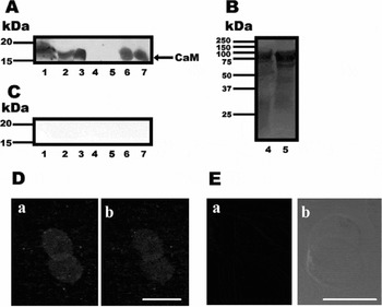
Highly purified, membrane-free nuclei were obtained by spermatozoid treatment with DTT/CTAB, followed by extensive washing. Then, the nuclear matrices were isolated using sequential protein extraction treatments: (1) high NaCl concentration; (2) heparin; and (3) DNase I. The proteins extracted after each different treatment, as well as those retained in either whole nuclei or nuclear matrices, were analysed by SDS-PAGE, transferred to nitrocellulose membranes and subjected to western blotting using CaM antibodies; these antibodies detected a 17 kDa protein (Fig. 1A). CaM was detected in whole nuclei (Fig.1A, lane 1) and in the isolated nuclear matrices (Fig. 1A, lane 7). Two positive CaM controls were used: testis homogenate (Fig. 1A, lane 2) and pure CaM (Fig. 1A, lane 3). In the extracted proteins, CaM was detected only after the DNase I treatment (Fig. 1A, lane 6). In contrast, CaM was not detected in the nuclear NaCl extracts (Fig. 1A, lane 4) or in the heparin extracts (Fig. 1A, lane 5). Thus, CaM remained associated to the nuclear matrix even after diverse extraction procedures, suggesting that it was not a contaminant from the cytosol. In a silver-stained gel, it was observed that both the NaCl and the heparin treatment extracted some proteins from the nucleus (Fig. 1B, lanes 4 and 5, respectively). The antibody specificity was confirmed as follows: (1) a competitive inhibition assay, where the anti-CaM antibody was treated with 10-fold molar excess of purified CaM did not show any bands (Fig. 1C); (2) omission of the primary antibody resulted in absence of any band (data not shown); or (3) using preimmune serum instead of the primary antibody which was negative too (data not shown).
Calmodulin (CaM) identification in guinea pig sperm nuclei by western blotting and indirect immunofluorescence. (A) Positive expression of CaM was observed in whole nuclei sperm proteins (lane 1). Also, in the positive controls: mouse testis (lane 2), pure CaM (lane 3). Nuclear proteins extracted with: DNase I (lane 6) and nuclear matrices (lane 7). No expression of CaM was observed in NaCl (lane 4) and heparin (lane 5) extracted proteins; although positive proteins bands were detected in silver stained gel (B). (C) Negative control; no immunoreactive bands were detected in samples assayed (lanes 1–7) when the first antibody was competed with a 10 fold molar excess of pure CaM. (D) Immunolocalization of CaM in whole nuclei (DTT/CTAB) of guinea pig sperm, confocal microscopy image (projection) (a) and optical sections (b). (E) Negative control; nuclei were treated without the first antibody, no fluorescence was observed (a). Phase contrast image (b). Bar: 8 μm.

CaM was also detected in DTT/CTAB nuclei by indirect immunofluorescence and confocal microscopy projection (Fig. 1D, a). CaM was observed in whole nuclei as a fine granulated fluorescence. In optical sections, in the middle of the nucleus, CaM gave the same image (Fig. 1D, b). The negative controls were: (1) samples in which the primary antibody was omitted (Fig. 1E, a), no fluorescence was observed; phase contrast image (Fig. 1E, b); (2) the primary antibody was competed with an excess (10 fold) of pure CaM protein; or (3) preimmune serum was used instead of the primary antibody, none of the negative controls exhibited fluorescence (data not shown).
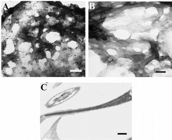
Nuclear matrices were subjected to immunogold staining using a polyclonal anti-CaM antibody. Heavy labelling of the nuclear matrix was observed (Fig. 2A). The negative control was a sample in which the primary antibody was omitted and showed severe reduction of the gold label (Fig. 2B). In addition, when thin sections of whole sperm cells were immunogold stained, CaM was revealed within the nucleus and to a lesser extent elsewhere (Fig. 2C).
Immunogold detection of CaM in nuclear matrices and whole sperm cells. Nuclear matrices were treated with anti-CaM antibodies. Appropriate gold-labelled (5 nm particles) G-protein was used. (A) CaM detection was abundant in the nuclear matrices. (B) Negative control, nuclear matrices incubated only with G-protein, where no immunogold staining was detected. Bars: 200 nm. (C) Thin sections from guinea pig spermatozoa embedded in LR White resin incubated with anti-CaM antibodies, the image shows positive CaM detection inside nuclei. Bar: 500 nm.

Guinea pig sperm nuclei contain F-actin and the myosin light chain
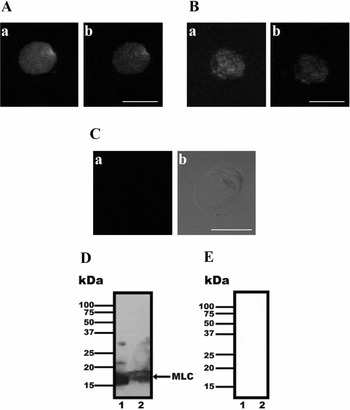
In DTT/CTAB nuclei, F-actin was observed by confocal microscopy after staining with FITC–phalloidin (Fig. 3A, a). In optical sections, the same fluorescence image was observed (Fig. 3A, b). MLC was also detected by indirect immunofluorescence and confocal microscopy (Fig. 3B, a). The image exhibits granulated fluorescence throughout the nucleus. In optical sections, the same granulated fluorescence was observed (Fig. 3B, b). In negative controls, in which the primary antibody was omitted or incubated with preimmune serum instead of the primary antibody, no fluorescence was observed (Fig. 3C, a); we also include the phase contrast image of the same sample (Fig. 3C, b).
In guinea pig sperm whole nuclei: F-actin localization by FITC–phalloidin stain and myosin light chain (MLC) localization and identification by indirect immunofluorescence and western blotting. (A) Confocal microscopy image (projection) showing F-actin in whole DTT/CTAB nuclei of guinea pig sperm (a) and optical sections (b). (B) Confocal microscopy image (projection) showing the immunolocalization of MLC in whole nuclei of guinea pig sperm (a) and optical sections (b). (C) Negative control; nuclei were treated with preimmune sera instead the first antibody, no fluorescence was observed (a). Phase contrast image (b). Bar: 8 μm. D: DTT/CTAB nuclei positive expression of MLC is shown. Lane 1: positive control (mouse testis); lane 2: nuclear sperm proteins of guinea pig. (E) Negative control; not immunoreactive band was detected when the first antibody was omitted.

The presence of MLC in DTT/CTAB nuclei was confirmed by western blotting (Fig. 3D, lane 2). As with the positive control, testis homogenate (Fig. 3D, lane 1) was used. In both the control and the sample, the anti-MLC antibody detected a wide band spanning from 17 to 20 kDa, which is in the range specified by the manufacturer (Sigma Chemical Co.) and expected for MLC (Wagner, Reference Wagner1982). In a negative control in which the primary antibody was omitted, no bands were detected (Fig. 3E) in nuclear proteins (lane 2) neither testis homogenate (lane 1). A second negative control was done with preimmune sera instead of the primary antibody, it give also a negative result (data not shown).
Actin-myosin interaction was observed in guinea pig sperm nuclei matrices
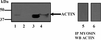
Guinea pig spermatozoid nuclei contain actin and myosin (Ocampo et al., Reference Ocampo, Mondragón, Roa-Espitia, Chiquete-Félix, Salgado and Mújica2005). The interaction between these proteins would be a strong indication that they play a physiologic role in nuclei. To test this hypothesis, an SDS-extract from nuclear matrices was treated with an anti-myosin antibody plus protein A agarose. The immunoprecipitate was subjected to SDS-PAGE, transferred to a nitrocellulose membrane and analysed with an anti-actin antibody. A 37–50 kDa protein band was detected (Fig. 4, lane 1), indicating that actin co-precipitated with myosin. In supernatants of the immunoprecipitate no bands were detected (Fig. 4, lane 2). In the muscle extract used as a control, a band of 45 kDa was revealed by the anti-actin antibody (Fig. 4, lane 3) also in whole nuclei spermatozoa a band of 45 kDa was detected (Fig. 4, lane 4). Negative controls performed without the primary antibody or without the primary antibody but in the presence of preimmune sera, did not show positive bands (Fig. 4, lane 6); an additional control was performed incubating nuclear matrix extracts with Protein A agarose alone (without anti-myosin antibody) and revealed for actin. Under these conditions no bands were detected (Fig. 4, lane 5).
Myosin association to actin in the nuclear matrix of guinea pig spermatozoa. Nuclear matrices were dissolved in 0.5% SDS and immunoprecipitated (IP) using anti-myosin antibody. The precipitate and the supernatant were electrophoresed and analysed by western blotting with anti-actin antibodies. Lane 1: nuclear matrices (IP); lane 2: supernatant (IP); lane 3: muscle homogenate (positive control); lane 4: whole nuclei; lane 5: negative control incubating nuclear matrices extract with protein A agarose, but without anti-myosin antibody and lane 6: negative control without the primary antibody, actin was not detected.

The heparin-mediated sperm nuclei decondensation is inhibited by CaM antagonists
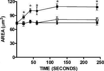
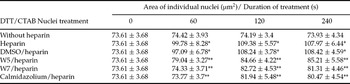
In order to define whether CaM participates in nuclear decondensation, we measured the effect of different CaM antagonists (W5, W7 and calmidazolium in DMSO) on the heparin-mediated nuclear decondensation. The diluent (DMSO) plus heparin was assayed as a control. Non treated nuclei remained stable up to 240 s (Fig. 5, ·) heparin promoted significant nuclei decondensation at 20 s (p < 0.001) and a further increase was observed to become stable at 240 s (Fig. 5, ◾). Calmidazolium inhibited decondensation completely (Fig. 5, ◽). Decondensation was evaluated at 60 up to 240 s after heparin addition measuring the area (Table 1) and diameter (data not shown) of individual nuclei. Before treatment, the mean area of the nuclei was 73.61 μm2 and the highest stable decondensation was 109.38 μm2. All the CaM antagonists were effective inhibitors of nuclear decondensation.
Rate of heparin-mediated nuclei decondensation in the presence and in the absence of the calmodulin antagonist calmidazolium (10 μM). Reaction mixture as in Table 1. Aliquots were taken at the times indicated in the figure. Nuclear decondensation was evaluated up to 240 s of treatment. From 20 s onwards heparin promoted significant (*p < 0.001) nuclei decondensation. · Without heparin, ◾ heparin and ◽ heparin/calmidazolium.

Heparin-mediated decondensation of sperm nuclei. Effect of calmodulin antagonists W5 (100 μM), W7 (100 μM) and calmidazolium (10 μM).

DTT/CTAB nuclei (35 × 106/ml) in 50 mM Tris pH 7.5, were incubated for 30 min with calmodulin antagonists (or without) or DMSO (antagonist's diluent); then added with 5 IU heparin. At zero time and after 60, 120 and 240 s heparin treatment, samples were fixed with 1.5% formaldehyde in PBS (final concentration). Smears from each sample were stained on glass slides and subjected to morphometric analysis (see Materials and methods). All results are representative of at least three different experiments. Data are the means ± standard deviation of three replicates.
*p < 0.001 vs non-heparin-treated nuclei.
**p < 0.001 vs heparin or DMSO/heparin-treated nuclei.
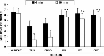
After longer incubation times, heparin treated sperm nuclei disappeared from view, which probably indicates that nuclear structures became unstable (Fig. 6), such that after 10 min of heparin treatment, the original 6.3 × 10⁶ sperm nuclei decreased to 1.3 × 10⁶ nuclei. An 80% decrease. This was not mediated by proteases, as addition of Complete™ (a mixture of protease inhibitors) did not prevent nuclei disappearance (data not shown). In contrast, in the presence of the CaM antagonists, the heparin-mediated loss of sperm nuclei remained low, at about 20%; sperm nuclei numbers were as follows: in the presence of: W5, 5.7 × 10⁶; W7, 5. 3 × 10⁶ and calmidazolium 5.7 × 10⁶ (Fig. 6). Thus, it seems that in addition to inhibiting decondensation, or perhaps as a consequence of this inhibition, CaM antagonists also inhibit the heparin-promoted loss of sperm nuclei.
Number of DTT/CTAB sperm nuclei remaining after heparin treatment in the presence and in the absence of calmodulin antagonists. Three independent experiments were performed. Data are the means from samples fixed at 4 and 10 min of heparin treatment. *p < 0.001 vs sample without heparin. **p < 0.001 vs Tris/heparin or DMSO/heparin. CDZ, calmidazolium.

The Xenopus laevis egg extract-mediated decondensation of sperm nuclei is inhibited by different CaM antagonists
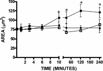
The heparin decondensation method suggested that CaM participates in nuclear decondensation. To further analyse this possibility, we decided to test a second method to decondense nuclei which is perhaps more physiological: the X. laevis egg extract-mediated sperm nucleus decondensation. In this model, we also tested the effect of each of three different CaM antagonists: W5, W7 and calmidazolium in DMSO. The results were different to those obtained with heparin because the X. laevis extract promoted a much lower rate of decondensation and in addition treated nuclei did not disappear, even at very long incubation times. In the non-treated controls, nuclei remained stable up to 240 min of experimentation (Fig. 7, ·). In nuclei treated with the X. laevis extract, nuclei remained stable for up to 6 min of incubation; then, at 10 min significant decondensation (p < 0.001) was observed which increased up to 2 h and then remained constant (Fig. 7, ◾). Calmidazolium inhibited decondensation (Fig. 7, ◽).
Rate of Xenopus laevis egg-extract-mediated nuclei decondensation in the presence and in the absence of the calmodulin antagonist calmidazolium (10 μM). Reaction mixture as in Table 2. Aliquots were taken at the times indicated in the figure. Nuclear decondensation was evaluated up to 240 min of treatment. From 10 min onwards X. laevis egg-extract promoted significant (*p < 0.001) nuclei decondensation. · Without egg extract, ◾ egg extracts and ◽ egg extract/calmidazolium.

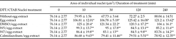
Other CaM antagonists were tested; it was observed that each CaM antagonist inhibited the X. laevis extract-mediated nuclear decondensation, as determined by measuring the area (Table 2) and diameter (data not shown) of individual nuclei at 10 up to 240 min. The highest decondensation value was observed at 120 min of treatment, where an area of 125.42 μm2 was measured. The CaM antagonist sensitivity of the egg extract-mediated nuclear decondensation indicated that this process is mediated by CaM. In addition, it was observed that the egg extract treatment did not cause disappearance of decondensed sperm nuclei.
Xenopus laevis egg extract-mediated decondensation of sperm nuclei. Effect of calmodulin antagonists W5 (100 μM), W7 (100 μM) and calmidazolium (10 μM).

DTT/CTAB nuclei (18 × 106/ml) in 50 mM Tris pH 7.5 were incubated for 30 min with (or without) calmodulin antagonists or DMSO (antagonists diluent). Nuclei were washed and further incubated in 1 ml Xenopus laevis activated egg extract for zero, 10, 30, 120 and 240 min and fixed. Samples were stained and morphometric nuclei analysis was done as indicated in Materials and Methods. All results are representative of at least three different experiments. Data are the means ± standard deviation of three replicates.
*p < 0.001 vs non-extract-treated nuclei.
**p < 0.001 vs egg extract-treated nuclei or DMSO/egg extract-treated nuclei.
Discussion
When the Ca2+ concentration increases in a given cell compartment, four Ca2+ ions bind to calmodulin (CaM) activating it. Then, the 4Ca2+/CaM complex binds and activates the myosin light chain kinase (MLCK). Activated MLCK phosphorylates the myosin light chain (MLC) at serine 19. Once activated, myosin binds F-actin, forming a dynamic, motile system (Adelstein, Reference Adelstein1980; Sellers, Reference Sellers2000). The MLCK-mediated MLC phosphorylation produces a conformational change in the actin/myosin complex, which in turn causes contraction (Stull et al., Reference Stull, Tansey, Tang, Word and Kamm1993).
In non-muscle cells, actin/myosin complexes are involved in processes such as cytokinesis and migration (Adelstein et al., Reference Adelstein, Conti and Pato1980). In the nuclei of different cell types, CaM is involved in a number of functions such as DNA replication and repair (Vendrell et al., Reference Vendrell, Aligué, Bachs and Serratosa1991). The presence of CaM-binding proteins in the nucleus of neural cells has been observed (Pujol et al., Reference Pujol, Bosser, Vendrell, Serratosa and Bachs1993). Also, in rat-hepatocyte nuclei, proteins such as MLCK, caldesmon, spectrin and actin have been detected and their presence indirectly suggests that CaM might participate in nuclear decondensation (Bachs et al., Reference Bachs, Lanini, Serratosa, Coll, Bastos, Aligué, Rius and Carafoli1990).
Our group reported that: (a) in guinea pig sperm nuclei, there are cytoskeleton proteins (spectrin and cytokeratin) and CaM binding proteins (actin and myosin); and (b) actin and myosin participate in the heparin-mediated decondensation of nuclei (Ocampo et al., Reference Ocampo, Mondragón, Roa-Espitia, Chiquete-Félix, Salgado and Mújica2005). Here, CaM was detected in the whole nucleus and in the nuclear matrix of guinea pig sperm (Figs. 1 and 2). The 17 kDa band revealed has a MW similar to that previously reported (Crivici & Ikura, Reference Crivici and Ikura1995; Putkey et al., Reference Putkey, Kleerekoper, Gaertner and Waxham2003). CaM is deeply embedded in the nucleus, strongly suggesting that this is not a cytoplasmic contaminant. That is, after thorough washing, pure DTT/CTAB nuclei retained CaM, which was not released by either the NaCl treatment or the heparin treatment. Only the DNase I treatment succeeded in partially extracting CaM from the nuclear matrices.
To assess the physiological role of CaM in nuclei, we followed the effect of different CaM antagonists (W5, W7 and calmidazolium) on two nuclear decondensation models: (a) heparin (Table 1 and Fig. 5); and (b) X. laevis egg extracts (Table 2 and Fig. 7). Heparin has been suggested to promote decondensation by competing with DNA for protamines (Bertanzon et al., Reference Bertanzon, Stevens, Toniolo and Bonora1981). Egg extracts have been reported to cause nuclei decondensation, probably mimicking the physiological process with more accuracy (Lohka & Masaui, Reference Lohka and Masaui1983). CaM antagonists inhibited decondensation by either the heparin or the egg extract with a value of p < 0.001.
The egg extract-mediated decondensation is perhaps more physiological. This idea would explain the high stability observed in the decondensed nuclei, which in the heparin model were highly unstable (Fig. 6). The nuclei loss observed in the heparin-treated samples was not mediated by proteases, as addition of a protease inhibitor mixture (Complete™, Roche) did not protect the nuclei.
The identification of actin in several nuclear complexes has led to suggestions that it participates in diverse nuclear activities including chromatin remodelling (Olave et al., Reference Olave, Reck-Peterson and Crabtree2002), transcription (Philimonenko et al., Reference Philimonenko, Zhao, Iben, Dingova, Kysela, Kahle, Zentgraf, Hofmann, Lanerolle, Hozak and Grummt2004) and nucleocytoplasmic traffic (Bettinger et al., Reference Bettinger, Gilbert and Amberg2004). However, in the cell nucleus no F-actin had been detected leading to suggestions that actin forms only short filaments (Pederson & Aebi, Reference Pederson and Aebi2003). However, we did detect F-actin in isolated whole nuclei (DTT/CTAB nuclei) from guinea pig spermatozoa (Fig. 3A). We also detected MLC in the whole nucleus (Fig. 3B, D) and identified an interaction of actin with myosin in the nuclear matrix (Fig. 4). Thus, it may be proposed that in the guinea pig sperm nucleus there is a complete contractile actin/myosin system, where CaM would activate nuclei decondensation through phosphorylation of MLC.
Acknowledgements
We are grateful to Dr Saul Villa Treviño and Dra Leticia Moreno Fierros for providing e-microscope facilities (Olympus). The staff of the Unidad de Microscopia Electrónica (UME), CINVESTAV-IPN, QFB Sirenia González and QFB Lourdes Rojas, for providing electron microscopy facilities. The technical assistance of TSU Esau Emilio Promontor Gómez, M.C. Ana Lilia Roa Espitia, Víctor Cortés and QBP Blanca Estela Reyes is acknowledged. This work was supported by a doctoral fellowship from Consejo Nacional de Ciencia y Tecnología (CONACyT)-México (no. 173791) and Instituto de Ciencia y Tecnología del Distrito Federal to Armando Zepeda-Bastida and funded by the grants no. 41725-Q and 59176 (CONACyT) to AM. The authors declare that there is no conflict of interest that would prejudice the impartiality of this scientific work.