Introduction
African animal trypanosomiasis (AAT) is a parasitic disease caused by protozoans of the genus Trypanosoma (OIE Terrestrial Manual, 2013) and causes economic losses of 4.5 billion US dollars per year (Oluwafemi et al., Reference Oluwafemi, Ilemobade and Laseinde2007), as a result of direct (mortality, production losses, costs of prophylactic and curative trypanocidal drugs) and indirect losses due to crop production decay and agricultural workers' involvement (deficiency of animal protein diets) (Harberd, Reference Harberd1988; Angara et al., Reference Angara, Ismail and Ibrahim2014). In Somalia, the economic impact of AAT has been estimated at 88 million US dollars (Mohamed and Dairri, Reference Mohamed Ahmed and Dairri1987). Although the National Tsetse and Trypanosomiasis Control Project (NTTCP) has been established in the 1980s (Mohamed and Dairri, Reference Mohamed Ahmed and Dairri1987), and a tsetse and trypanosomiasis (T & T) control project has been funded by the International Committee of the Red Cross (ICRC) in some villages of Shabelle and Jubba regions (ICRC, 2017), no other control measures or wide coverage area to reduce the losses from trypanosomiasis have been implemented following the collapse of the central government of Somalia in 1991 (Salah, Reference Salah2016). Effective and sustainable T & T control projects will contribute to improving livestock health that enhances the production and livelihood of dependent families (Swallow, Reference Swallow2000).
Examination of thick and thin peripheral blood or buffy coat films stained with Giemsa stains or fresh wet blood or buffy coat smears has been used for the detection and identification of Trypanosome species (Rosenblatt, Reference Rosenblatt2009; OIE Terrestrial Manual, 2013). However, these direct parasitological techniques lack sensitivity and specificity (Mattioli and Faye, Reference Mattioli and Faye1996; Picozzi et al., Reference Picozzi, Tilley, Fèvre, Coleman, Magona, Odiit, Eisler and Welburn2002). Therefore, molecular methods provide multi-species-specific detection of trypanosomes in a single polymerase chain reaction (PCR) (Salim et al., Reference Salim, Bakheit, Kamau, Nakamura and Sugimoto2011) and have been used in epidemiological studies (Desquesnes and Dávila, Reference Desquesnes and Dávila2002; Picozzi et al., Reference Picozzi, Tilley, Fèvre, Coleman, Magona, Odiit, Eisler and Welburn2002; Kouadio et al., Reference Kouadio, Sokouri, Koffi, Konaté, Ahouty, Koffi and N'Guetta2014; Hassan-Kadle et al., Reference Hassan-Kadle, Ibrahim, Nyingilili, Yusuf, Vieira and Vieira2019). Moreover, the pan-PCR techniques for Trypanosomes would reduce the cost of PCR three to five times (Njiru et al., Reference Njiru, Constantine, Guya, Crowther, Kiragu, Thompson and Dávila2005).
In Somalia, AAT has been reported in different domestic ruminants by standard trypanosome detection methods (STDM) (Macchioni and Abdullatif, Reference Macchioni and Abdullatif1985; Mohamed and Dairri, Reference Mohamed Ahmed and Dairri1987; Dirie et al., Reference Dirie, Wallbanks, Molyneux and Bornstein1988a, Reference Dirie, Wardhere and Farah1988b; Ainanshe et al., Reference Ainanshe, Jennings and Holmes1992). A previous study has found T. congolense (Schoepf et al., Reference Schoepf, Mustafa Mohamed and Katende1984; Dirie et al., Reference Dirie, Wardhere and Farah1988b) and T. vivax infecting sheep (Dirie et al., Reference Dirie, Wardhere and Farah1988b). Both Trypanosome species have also been reported in cattle from southern Somalia (Moggridge, Reference Moggridge1936; Dirie et al., Reference Dirie, Wallbanks, Molyneux and Bornstein1988a). Additionally, a recent study has confirmed infections with Trypanosoma evansi and Trypanosoma simiae in camels through DNA sequencing (Hassan-Kadle et al., Reference Hassan-Kadle, Ibrahim, Nyingilili, Yusuf, Vieira and Vieira2019). Moreover, the human African trypanosomiasis (sleeping sickness) is unknown in Somalia, despite the presence of Glossina pallidipes, a known vector of the disease in other areas of East Africa with the practice of transhumance (Harberd, Reference Harberd1988). The widespread infestation of Trypanosome vectors in Somalia is one of the main obstacles to the development of the livestock industry which supports the livelihood of 12.3 million Somali communities living in an area of 640 000 km2 (Bernacca, Reference Bernacca1967; UNFPA, 2014). However, there is a lack of data on AAT after the civil war of the 1990s. Hence, the present study aimed to assess the prevalence of Trypanosoma spp. in cattle, goats and sheep from Afgoye and Jowhar districts of Somalia.
Materials and methods
Study area
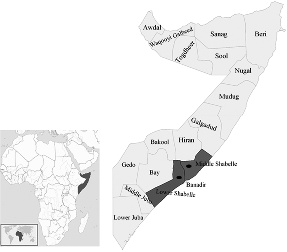
The present study was carried out in Afgoye (2°08′47.67″N 45°07′08.11″E) and Jowhar (2°46′38.72″N 45°30′05.85″E) districts, Somalia (Fig. 1), areas are known to be infested by tsetse and other biting flies (Hursey, Reference Hursey1985; Mohamed and Dairri, Reference Mohamed Ahmed and Dairri1987; Dirie et al., Reference Dirie, Walbanks, Aden, Bornstein and Ibrahim1989).
Map of Somalia showing the location of blood samples. Sampled regions are highlighted in green (Middle Shabelle) and blue (Lower Shabelle), and the dots indicate the approximate location of the sampled districts (Jowhar and Afgoye). The figure was generated and modified using QGIS software version 2.18.19.

Sampling
From November 2017 to February 2018, which represents the dry season in Somalia, a total of 614 ruminants from Afgoye (n = 304) and Jowhar (n = 310) were evaluated. Blood samples were collected by jugular venipuncture of cattle (n = 202; 183 females and 19 males), sheep (n = 206; 190 females and 16 males) and goats (n = 206; 191 females and 15 males). Five millilitres were placed into EDTA tubes for microscopical detection of Trypanosomes and preparation of blood spots on filter paper (Whatman no.4, Whatman, Springfield Mill, UK) for PCR analysis (Ahmed et al., Reference Ahmed, MacLeod, Hide, Welburn and and Picozzi2011).
Parasitological diagnosis of Trypanosoma spp.
All blood samples were evaluated for the presence of Trypanosoma spp. by the buffy coat technique (BCT), as previously described (Murray et al., Reference Murray, Murray and Mc Intyre1977). The BCT was considered positive when Trypanosomes could be visually detected regardless of the species.
DNA extraction and PCR for Trypanosoma spp.
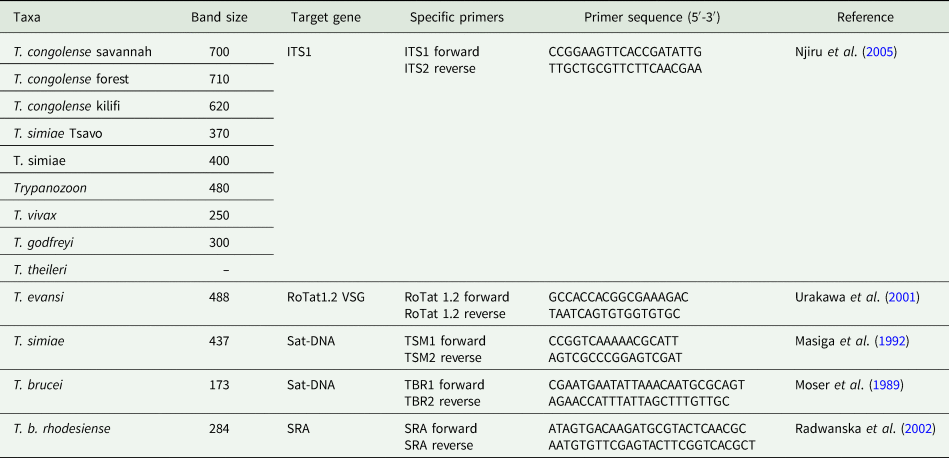
DNA was extracted from all 614 blood spots by Chelex® 100 (Sigma–Aldrich, St. Louis, USA), as previously described (Ahmed et al., Reference Ahmed, MacLeod, Hide, Welburn and and Picozzi2011). Five different PCR assays were used to detect Trypanosoma spp.: (i) internal transcribed spacer 1 (ITS1)-PCR that amplifies the ITS1 region (Njiru et al., Reference Njiru, Constantine, Guya, Crowther, Kiragu, Thompson and Dávila2005), which is known to vary in size within trypanosome species, except members of Trypanozoon species (T. brucei brucei, T. brucei rhodesiense, T. brucei gambiense, T. evansi, and T. equiperdum), and therefore differentiates Trypanosomes by their ITS sizes, (ii) TBR-PCR specific for T. brucei (Moser et al., Reference Moser, Cook, Ochs, Bailey, McKane and Donelson1989), (iii) human-serum-resistance-associated (SRA)-PCR targeting the SRA gene uniquely specific for T. brucei rhodesiense (Radwanska et al., Reference Radwanska, Chamekh, Vanhamme, Claes, Magez, Magnus, De Baetselier, Büscher and Pay2002), (iv) RoTat 1.2 PCR which amplifies the Rode Trypanozoon antigenic type (RoTat) 1.2 variable surface glycoprotein (VSG) gene fragment specific for the detection of T. evansi (Urakawa et al., Reference Urakawa, Verloo, Moens, Büscher and Majiwa2001), and (v) TSM-PCR for detection of T. simiae (Masiga et al., Reference Masiga, Smyth, Hayes, Bromidge and Gibson1992).
All samples were initially screened for Trypanosoma spp. by the ITS1-PCR assay (Njiru et al., Reference Njiru, Constantine, Guya, Crowther, Kiragu, Thompson and Dávila2005). Samples positive for Trypanozoon subspecies were further tested for T. brucei using TBR-PCR (Moser et al., Reference Moser, Cook, Ochs, Bailey, McKane and Donelson1989) and RoTat 1.2 PCR for the presence of T. evansi (Urakawa et al., Reference Urakawa, Verloo, Moens, Büscher and Majiwa2001). Positive samples on the TBR-PCR were further screened for T. brucei rhodensiense by SRA-PCR (Radwanska et al., Reference Radwanska, Chamekh, Vanhamme, Claes, Magez, Magnus, De Baetselier, Büscher and Pay2002). Finally, all T. simiae positive samples on the ITS1-PCR assay were further subjected to the TSM-PCR (Masiga et al., Reference Masiga, Smyth, Hayes, Bromidge and Gibson1992). The PCR primers used in this study are shown in Table 1. A camel sample is known to be positive for T. evansi (Hassan-Kadle et al., Reference Hassan-Kadle, Ibrahim, Nyingilili, Yusuf, Vieira and Vieira2019) was used as a positive control, in all ITS1, TBR and TSM PCR runs, while brucei rhodesiense DNA (kindly donated by Dr Enock Matovu, Makerere University, Uganda) was used in SRA-PCR assay. Nuclease-free water was used as a negative control in all reactions. The amplified PCR products were analysed by electrophoresis in a 1.5% agarose gel, stained with ethidium bromide and visualized by UV illuminator (UVITEC™ Cambridge, UK).
PCR primers used in the present study

Data management and analysis
Either Chi-square or Fisher's exact test was used to assess the association of the individual variables (district and species) with Trypanosoma spp. infection. Odds ratio (OR), 95% confidence intervals (95% CI) and P values were calculated, and results were considered significant when P ⩽ 0.05. Data were compiled and analysed by Statistical Package for Social Sciences (SPSS) version 25 (IBM Corp., Armonk, NY, USA).
Results
Parasitological diagnosis of Trypanosoma spp.
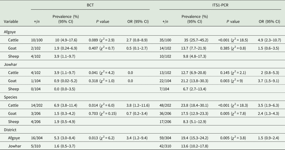
A total of 21/614 (3.4%; 95% CI: 2.1–5.2%) ruminants were positive for Trypanosome species by BCT. Cattle were more likely to be infected by Trypanosoma spp. than sheep (OR: 3.8; χ 2 = 6.0, P = 0.01). Ruminants reared in Afgoye district were more likely to be infected to Trypanosoma spp. than those reared in Jowhar district (OR: 3.4, 95% CI: 1.2–9.4, P = 0.01) (Table 2).
Prevalence of Trypanosoma spp. in cattle, goats and sheep from Afgoye and Jowhar districts, Somalia

+, number of positive animals; n, number of samples; 95% CI, 95% confidence interval; OR, odds ratio.
Molecular diagnosis of Trypanosoma spp.
Overall, 101/614 (16.4%, 95% CI: 13.6–19.6%) ruminants tested positive for Trypanosoma spp. by the ITS1-PCR assay.
The highest prevalence of Trypanosoma spp. was observed in cattle (48/202; 23.8%) followed by goats (36/206; 17.5%) and sheep (17/206; 8.3%) (Table 2). The ITS1-PCR assay has detected 27/101 (26.7%; 95% CI: 18.4–36.5%) ruminants single infected by Trypanosomes. Additionally, coinfections with different trypanosomes were found in 74/101 (73.3%; 95% CI: 63.5–81.6%) ruminants, with coinfections mainly observed in cattle (35/48; 72.9%) and goats (26/36; 72.2%). The prevalence of Trypanosoma spp. by ITS1-PCR within each ruminant species evaluated are summarized in Table 3.
Single and coinfections with Trypanosoma spp. in cattle, goats and sheep in Somalia

+, number of positive animals; n, number of samples; T.v, T. vivax; T.c, T. congolense; T.g, T. godfreyi; T.s, T. simiae.
Cattle (OR: 3.5, 95% CI: 1.9–6.3, P < 0.001) and goats (OR: 2.4, 95% CI: 1.3–4.3, P = 0.005) were more likely to be infected with Trypanosoma spp. than sheep. Ruminants reared in Afgoye district were likely to be infected by Trypanosoma spp. than those in Jowhar district (OR: 1.5, 95% CI: 0.9–2.4, P = 0.005). The highest prevalence of Trypanosome infection was recorded in cattle and goats from Afgoye and Jowhar districts, respectively (Table 2).
Ninety-one ITS1-PCR Trypanozoon-positive samples were further tested by species-specific PCR to characterize the T. evansi and T. brucei. As a result, four out of 91 (4.4%) ruminants tested positive for T. brucei by the TBR-PCR assay, while the remaining 87/91 (95.6%) ruminants tested positive for T. evansi by the RoTat 1.2 PCR assay. All the four T. brucei-positive samples have tested negative for T. b. rhodesiense by the SRA-PCR assay. Additionally, two PCR assays were used to detect T. simiae in the present study: the ITS1-PCR was able to detect 19/101 (18.8%) ruminants, while 12/19 (63.2%) ruminants tested positive by the TSM-PCR assay.
Discussion
To the author's knowledge, this is the first study to combine BCT and molecular detection of Trypanosome species in ruminants from Somalia. The overall prevalence recorded by ITS1-PCR was significantly higher than the one observed with the BCT technique (P < 0.001, χ 2 = 58.3). This was expected due to the known higher sensitivity of molecular methods, as previously reported (Angwech et al., Reference Angwech, Nyeko, Opiyo, Okello-Onen, Opiro, Echodu, Malinga, Njahira and Skilton2015; N'Djetchi et al., Reference N'Djetchi, Ilboudo, Koffi, Kaboré, Kaboré, Kaba, Courtin, Coulibaly, Fauret, Kouakou, Ravel, Deborggraeve, Solano, De Meeûs, Bucheton and Jamonneau2017).
Herein, overall, 3.4% ruminants were positive to Trypanosoma spp. by using the BCT technique. Previous studies performed in ruminants from Somalia have shown prevalence rates ranging from 4% to 28.6% by STDM with a record of T. congolense, T. vivax and T. brucei (Moggridge, Reference Moggridge1936; Schoepf et al., Reference Schoepf, Mustafa Mohamed and Katende1984; Dirie et al., Reference Dirie, Wallbanks, Molyneux and Bornstein1988a, Reference Dirie, Wardhere and Farah1988b; Ainanshe et al., Reference Ainanshe, Jennings and Holmes1992). Considering that T. brucei and T. evansi are morphologically indistinguishable (Gibson, Reference Gibson2003), it is important to consider that previous studies performed in Somalia may have misclassified the Trypanosoma species detected in ruminants.
In the present study, 16.4% ruminants tested positive for Trypanosoma spp. by the ITS1-PCR assay, which is in agreement with a previous study that has reported a prevalence of 17.2% in Tanzania (Simwango et al., Reference Simwango, Ngonyoka, Nnko, Salekwa, Ole-Neselle, Kimera and Gwakisa2017). However, our finding is lower than that reported in Sudan (57.7%) (Osman et al., Reference Osman, Njahira, Skilton, Ibtisam and A/Rahman2016) and Uganda (41%) (Angwech et al., Reference Angwech, Nyeko, Opiyo, Okello-Onen, Opiro, Echodu, Malinga, Njahira and Skilton2015), but it is higher than that reported in Zambia (7.5%) (Laohasinnarong et al., Reference Laohasinnarong, Goto, Asada, Nakao, Hayashida, Kajino, Kawazu, Sugimoto, Inoue and Namangala2015). Differences among the prevalence of Trypanosome species may be explained by the population studied and management, and tsetse seasonal dynamics (rainy vs dry season).
Previous studies have shown that sheep is more frequently infected by Trypanosoma species than goats under natural conditions, suggesting that goats are more refractory to Trypanosome infections than sheep (Masiga et al., Reference Masiga, Okech, Irungu, Ouma, Wekesa, Ouma, Guya and Ndung'u2002; Ng'ayo et al., Reference Ng'ayo, Njiru, Kenya, Muluvi, Osir and Masiga2005). In the present study, Trypanosoma spp. infection rates were significantly higher in cattle (23.8%; P < 0.001, χ 2 = 18.3) and goats (17.5%; P = 0.005, χ 2 = 7.8) than in sheep (8.3%), with goats being 2.4 times more likely to be infected by these protozoa than sheep. This may be attributed by the host susceptibility to Trypanosome infections and the skin coat suitability for fly feeding (Murray et al., Reference Murray, Morrison and Whitelaw1982; Wilkowsky, Reference Wilkowsky, Florin-Christensen and Schnittger2018).
Trypanosoma evansi (86.1%) followed by T. vivax (45.5%) were the predominant Trypanosoma species detected in ruminants from the studied areas. This may be due to the camel grazing in the area and the presence of other biting flies which may accelerate the mechanical transmission of the parasite (Mossaad et al., Reference Mossaad, Ismail, Ibrahim, Musinguzi, Angara, Xuan, Inoue and Suganuma2020). The T. simiae and T. godfreyi were detected in cattle and goats, which may be linked to the presence of warthog (Phacochoerus africanus) and bushpig (Potamochoerus larvatus somaliensis) in the studied districts (data not shown).
Although a previous study has detected T. simiae in sheep from Kenya (Ng'ayo et al., Reference Ng'ayo, Njiru, Kenya, Muluvi, Osir and Masiga2005), herein all sheep tested negative for this protozoon. Wild pigs constitute a reservoir for T. simiae and T. godfreyi infections to ruminants (Hamill et al., Reference Hamill, Kaare, Welburn and Picozzi2013). To the author's knowledge, this is the first study to detect T. simiae and T. godfreyi in cattle and goats from Somalia.
In the present study, ruminants reared in Afgoye district were 1.5 times more likely to be infected by Trypanosoma spp. than those in Jowhar district (P = 0.005, χ 2 = 3.8). Both districts are located along with the Shabelle river, where tsetse and other biting flies are often abundant (Hursey, Reference Hursey1985; Mohamed and Dairri, Reference Mohamed Ahmed and Dairri1987; Dirie et al., Reference Dirie, Walbanks, Aden, Bornstein and Ibrahim1989). However, in Jowhar, livestock owners repel flies with smoke from smouldering cattle dung around the animal shelters with cattle being grazed during the night, when flies are inactive, while goats and sheep are grazed during the day. This probably explains why cattle (12.7%) Trypanosome infection rate was lower than goats (21.2%), with goats being 3.7 times more likely to be infected by Trypanosoma spp. than sheep (P = 0.003, χ 2 = 9). Conversely, in Afgoye, cattle, goats and sheep are grazed during the day, when flies are active, and no other local strategies for controlling T & T have been implemented. This possibly explains why cattle Trypanosome infection rate (35%) was higher than goats (13.7%) and sheep (9.8%), with cattle being 4.9 times more likely to be infected by Trypanosoma spp. than sheep (P < 0.001, χ 2 = 18.5). Thus, this may explain why the overall Trypanosome prevalence rate in Afgoye was higher than in Jowhar district.
Small ruminants constitute a key component of livestock in many regions of East Africa. A previous study has shown that sheep and goats may harbour T. b. rhodesiense, the causative agent of sleeping sickness or human African trypanosomiasis (Ng'ayo et al., Reference Ng'ayo, Njiru, Kenya, Muluvi, Osir and Masiga2005). Additionally, previous studies have also implicated that cattle may act as a reservoir of Trypanosomes to humans (Onyango et al., Reference Onyango, Van Hoeve and De Raadt1966; N'Djetchi et al., Reference N'Djetchi, Ilboudo, Koffi, Kaboré, Kaboré, Kaba, Courtin, Coulibaly, Fauret, Kouakou, Ravel, Deborggraeve, Solano, De Meeûs, Bucheton and Jamonneau2017). In the present study, all animals tested with the SRA primer set were tested negative for T. b. rhodesiense. Considering that the T. b. rhodesiense vector, Glossina pallidipes, has been reported in Somalia (Mohamed and Dairri, Reference Mohamed Ahmed and Dairri1987), associated with the fact that this Trypanosome species is present in ecologically similar areas of neighbouring Kenya (Rutto and Karuga, Reference Rutto and Karuga2009) and Ethiopia (Baker et al., Reference Baker, McConnell and Hady1970), further studies should focus on the detection of T. b. rhodesiense in the country.
Concluding remarks
The present study has shown that AAT is prevalent in cattle, goats and sheep from Afgoye and Jowhar districts of Somalia, with the identification of Trypanosoma evansi, T. brucei, T. vivax, T. congolense, T. godfreyi and T. simiae. Coinfections with at least two Trypanosome species were also reported in all ruminants in this study. This is the first study on the molecular detection of Trypanosoma spp. in Somali ruminants as well as detection of T. simiae and T. godfreyi in cattle and goats in Somalia. Further large-scale studies and sustainable control programmes against trypanosomes and its vectors are needed in the country. In addition, studies should also focus on the possibility of detecting of T. brucei rhodesiense, HAT causative agent, in the country.
Acknowledgements
The authors would like to thank Hassan Hussein and Abdiwali Mohamud at ARTC lab, Abrar University for the assistance of blood samples collection and kind cooperation of animal owners who participated in this study. We also acknowledge Dr Enock Matovu (Makerere University, Uganda) for the donation of T. b. rhodesiense DNA to be used as a positive control at the Vector and Vector Borne Diseases Institute, Tanzania. The Brazilian National Council of Scientific and Technological Development (CNPq) provided a fellowship of research productivity (PQ) to Dr Rafael Vieira.
Financial support
This study was supported by the Abrar University, Somalia (grant number AURG04012017). The sponsor had no role in the design of the study, collection, analysis and interpretation of data, writing the manuscript and the decision to submit the article for publication.
Conflicts of interest
The authors declare there are no conflicts of interest.
Ethical standards
This study was approved by the ethical committee of Abrar University, Somalia (reference number AU/ARTC/EC/04/2017). All cattle, goats and sheep owners gave consent to sample their animals.