Introduction
Pulmonary regurgitation and residual right ventricular outflow tract obstruction are common residual lesions following tetralogy of Fallot repair. While pulmonary regurgitation is usually well tolerated, the resulting chronic right ventricular volume overload contributes to progressive right ventricular remodelling and the eventual onset of clinical symptoms. Reference Geva1–Reference Sasson, Houri and Raucher Sternfeld6 Moreover, left ventricular remodelling has also been observed in adolescents and young adults who have undergone repair for tetralogy of Fallot. Reference Andrade, Jerosch-Herold and Wegner7,Reference Li, Yu, Lai, Wong and Cheung8 Cardiac MRI is the gold standard method to assess right and left ventricular size and function in children with CHD. Reference Geva9
Pulmonary valve replacement is the standard treatment for symptomatic patients with severe pulmonary regurgitation after tetralogy of Fallot repair. A partial reversal of right ventricular dilatation after pulmonary valve replacement occurs and is associated with reduced right ventricular volume and improved ejection fraction. However, the optimal timing of pulmonary valve replacement for asymptomatic patients is still unclear, with definitive evidence of an associated reduction in subsequent adverse cardiac events such as ventricular arrhythmia or mortality still lacking in adult survivors of tetralogy of Fallot repair who have undergone subsequent pulmonary valve replacement. Reference Jp, T. and La2,Reference Pe, Mp and Ca10,Reference Dm, Ci and C.11
As a result, the current guideline-directed indications for pulmonary valve replacement in adults with tetralogy of Fallot repair and pulmonary regurgitation are based on the presence of active symptoms and the attainment of a consensus right ventricular volume threshold measured by cardiac MRI. Reference Stout, Daniels and Aboulhosn4,Reference Marelli, Beauchesne and Colman12 While these time-dependent changes of right and left ventricular volumes in the paediatric population with tetralogy of Fallot repair are interpreted according to body surface area, a definitive trajectory of ventricular volume dilation over time leading to subsequent pulmonary valve replacement has not yet been determined for children. Therefore, our objective was to assess whether the assessment of left and right ventricular adaptation over time, measured by cardiac MRI of children with prior tetralogy of Fallot repair, would demonstrate a differential trajectory in patients later destined to require pulmonary valve replacement during childhood. We hypothesised that the temporal trajectory of early ventricular dilation would differ based on the type of residual lesion and would be distinctive in patients later destined to attain a volume-threshold indication for pulmonary valve replacement.
Material and methods
Study design
This retrospective single-centre study included patients with tetralogy of Fallot who underwent complete repair before 24 months of age, with at least one subsequent cardiac MRI study after the completed surgical correction. The institutional review board approved this study and waived individual informed consent (CHLA-21-00110, approval date: 4/23/21).
Surgical records were identified in the Heart Institute’s Society of Thoracic Surgeons database at Children’s Hospital Los Angeles. The database was queried for surgeries occurring between 1 January 2000 and 31 December 2020, in patients with an umbrella diagnosis of “TOF” or “Tetralogy of Fallot” who underwent primary surgical repair without prior catheter intervention. Primary repair was defined as including completed ventricular septal defect closure with or without surgical intervention on the right ventricular outflow tract or pulmonary valve. We excluded patients with pulmonary atresia and those with major aortopulmonary collateral arteries. Vital status and the location of initial surgery were not determinant factors for inclusion in the final cohort.
Data collection
This included baseline cohort demographics; staged or primary surgical repair; type of complete repair including valve sparing, transannular patch, or right ventricular to pulmonary artery conduit; presence of associated CHD; concomitant genetic/syndromic diagnoses, and additional surgical/interventional procedures. Echocardiographic, electrocardiographic, and cardiac MRI data were derived from reports of clinically indicated studies.
Defining residual lesion after tetralogy of Fallot repair
Residual lesions of the pulmonary valve were categorised by assessing the clinical description, echocardiographic reports, cardiac MRI reports, and any available catheterisation data: we referenced the last available imaging studies in patients who did not require pulmonary valve replacement, or the last available imaging studies prior to pulmonary valve replacement in those who did, to categorise the nature and severity of post-repair residual pulmonary valve physiology.
All post-tetralogy of Fallot repair patients were anticipated to have residual pulmonic regurgitation, stenosis, or a combination of these. Residual physiologic status was assessed using echocardiographic, cardiac catheter, and MRI evaluations, assigning patients to one of four mutually exclusive groups: moderate or worse pulmonary valve regurgitation; significant right ventricular to pulmonary artery pathway stenosis with only mild regurgitation; both stenosis and moderate or worse regurgitation (mixed physiology); or neither of the foregoing (normal physiology). For analysis, these groups were subsequently consolidated into two: those with moderate or worse pulmonary valve regurgitation and those without. The following imaging methodology was applied.
Pulmonary regurgitation severity was categorised based on colour Doppler flow mapping as mild, moderate, moderate to severe, or severe/free regurgitation based on American Society of Echocardiography guidelines. Reference Zoghbi, Adams and Bonow13 Peak right ventricular systolic pressure was estimated by tricuspid regurgitant jet velocity using continuous wave Doppler. The presence of a residual stenotic lesion was indicated by the spectral Doppler mapping of right ventricular outflow tract peak velocity using continuous wave Doppler mapped from the subpulmonic region or the main pulmonary artery. The left ventricular systolic function was assessed by fractional shortening. In the absence of available echocardiographic data prior to pulmonary valve replacement, cardiac MRI conducted prior to pulmonary valve replacement was used to categorise the residual lesion, as well as to assess progressive ventricular remodelling.
All cardiac MRI studies performed on these patients were reviewed. These were performed on a Philips 1.5T Achieva, 1.5T General Electric CVi, or Philips 3T Ingenia scanners using torso or cardiac phased array coils appropriate to body habitus. White blood cine steady-state free precession imaging was performed in axial, long axis, four chamber, short axis, and right ventricular outflow tract views. Flow assessments using phase contrast cardiac MRI were performed across the aortic root and the main and branch pulmonary arteries. Assessment of left and right ventricular volumes was performed by manual segmentation of short-axis cine steady-state free precession images at end-diastole and end-systole. End-diastolic and end-systolic volumes were calculated by use of Simpson’s rule for each ventricle, and from these volumes, stroke volume and ejection fraction were calculated, with inclusion of the papillary muscles in the blood pool. Arterial blood flow was calculated from phase contrast images by use of a semiautomatic vessel edge-detection algorithm with operator correction, if necessary. Pulmonary valve regurgitant fraction was calculated as percentage of backward flow over forward flow. Software packages for volumetric and flow analyses included Medis, Philips Intellispace Portal, and Circle 42. All volume and flow measurements were indexed for body surface area to account for body size and growth. Additionally, guidelines for pulmonary valve replacement use indexed right ventricular volumes as thresholds for surgical intervention in asymptomatic patients. Reference Stout, Daniels and Aboulhosn4
Where available, catheter-derived right ventricular pressure of greater than half of the systemic systolic pressure indicated the presence of a residual stenotic lesion. Reference Feltes, Bacha and Beekman14,Reference King, Braun, Goldblatt, Liberthson and Weyman15 The clinically indicated 12-lead surface electrocardiogram at the same or most proximate time point prior to pulmonary valve replacement was interrogated, and QRS duration was determined manually.
Clinical endpoint
The clinical end points of pulmonary valve replacement or death/cardiac transplantation were assessed, based on the age of the patient at these time points. The technique for pulmonary valve replacement was categorised as requiring a surgical valved conduit, a surgical prosthetic or bioprosthetic valve, or a transcatheter valve implantation. Patients were referred for pulmonary valve replacement procedure by their treating physician, with the intervention indicated based on currently recognised clinical symptoms and/or right ventricular volume parameters at that time. The indication for surgical valve replacement was typically based on the right ventricular end-diastolic indexed volume exceeding 160 ml/m2, as an institutional policy, or evidence of symptoms with subjective right ventricle dilation of greater than moderate degree by echocardiography, if MRI assessment was not feasible. Reference Stout, Daniels and Aboulhosn4
Statistical analysis
Continuous variables are reported as medians and interquartile ranges, and categorical variables are presented as frequencies and percentages. The Mann−Whitney U test was used for continuous data and Fisher’s exact test for categorical variables. Paired data were assessed with the Wilcoxon signed-rank test. All comparison tests were 2-tailed, and an alpha critical value < 0.05 was considered statistically significant. For outcome analysis, patients were assessed based on whether or not they required pulmonary valve replacement during follow-up.
The relationship between time, measured as years of age at cardiac MRI, and ventricular volumes, right ventricular end-diastolic volume to left ventricular end-diastolic volume ratio, ventricular ejection fraction, pulmonary regurgitation fraction, and left ventricular mass derived from cardiac MRI was analysed using linear mixed-effects models. A random-effects structure with time was used to account for correlations between repeated measurements in the same patient and irregularly timed measurements. The model was adjusted for sex and the presence or absence of a pure regurgitant residual lesion after complete repair. Reference Hagdorn, Beurskens and Gorter16 To assess longitudinal changes, time represented as age in years of the patient was log-transformed in years log2(log base 2) to account for “non-linear” rapidly progressive ventricular dilation, which was frequently observed following 3 years after tetralogy of Fallot repair, and for the possibility of an observed plateau in right ventricular volume enlargement occurring 5–7 years after tetralogy of Fallot repair. Reference Mertens17,Reference Bhat, Mercer-Rosa and Fogel18 An interaction effect between pulmonary valve replacement during the follow-up and time in log2 years of age was investigated to assess the differences over time in the cardiac MRI metrics in the groups of patients who did and did not require pulmonary valve replacement during follow-up. Temporal changes of cardiac MRI data were described as means and 95% confidence intervals. Two subgroup analyses were performed as sensitivity analyses to examine the consistency of time (years log2 of age) as a predictor of ventricular changes compared to the entire cohort. The first subgroup analysis focused exclusively on children who underwent serial cardiac MRI (two or more tests). The second subgroup analysis was performed by stratifying the population based on the type of residual physiology after repair (regurgitant, stenotic, mixed, and normal physiology). The models also included time in age log2 years as a predictor, adjusted for sex, and interaction effect analysis.
We calculated weighted Pearson correlation coefficients (r) between the right and left ventricular volumes and right and left ejection fraction measured by cardiac MRI to assess the strength of correlation between variables and the interaction between left and right ventricular remodelling. Pearson r values were weighted based on the number of available cardiac MRI at each imaging visit. Imaging visit = #cardiac MRI (e.g. imaging visit 1 = the first cardiac MRI after the complete repair; imaging visit 2 = the second cardiac MRI after the complete repair, and so on).
All analyses were performed using a complete-case scenario, and no imputation methods were used to handle missing data. Analyses were performed using Stata (StataCorp, 2017; Stata Statistical Software: Release 15; College Station, TX: StataCorp LLC)
Results
Patient characteristics
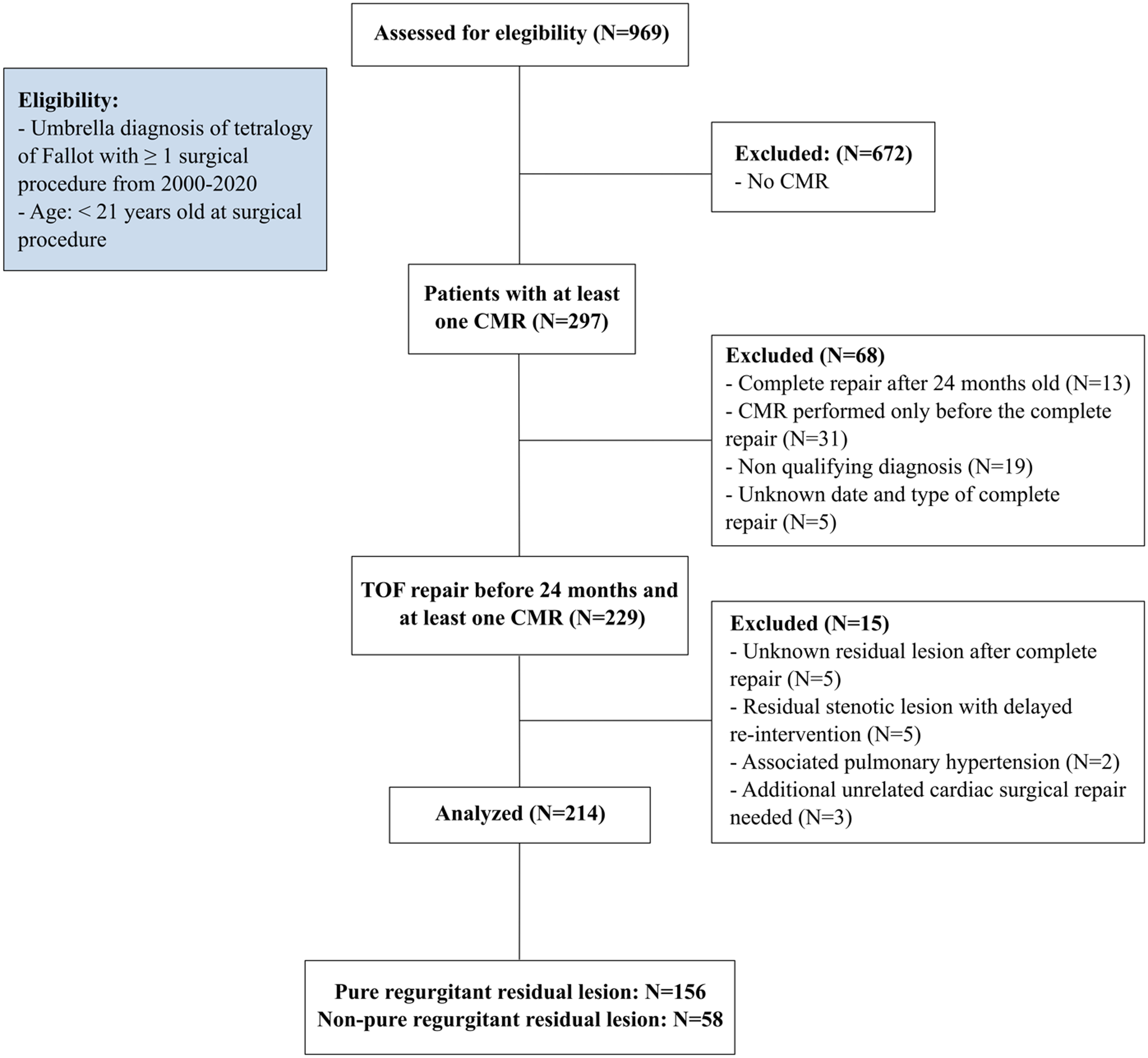
Of 969 subjects screened by diagnostic code in the hospital health information system database, 214 were included in the final analysis (Figure 1). Most exclusions were on the basis of not having undergone any cardiac MRI assessment. One hundred and thirty-nine patients (64.8%) were male, with a median age at complete repair of 6 months (4–9). Forty-nine patients (22.9%) had a staged surgical repair, with the Blalock–Thomas–Taussig shunt being the most common initial operation (N = 45/49, 91.8%). The most frequent complete repair technique was a transannular patch in 162 patients (75.7%), followed by a valve-sparing repair in 37 patients (17.3%) and a right ventricular to pulmonary artery conduit placement in 15 patients (7%). One hundred and forty-two patients (66.4%) of our cohort progressed to require eventual pulmonary valve replacement during the study time frame at a median age of 12 years (9–15.6), with only three of these receiving a transcatheter pulmonary valve, and the rest undergoing surgical pulmonary valve replacement. The median follow-up time following the index complete repair was 10.7 years (7.9–14.4).
Patient selection flow sheet. Abbreviations: CMR = cardiac magnetic resonance; PH = pulmonary hypertension; RVOT = right ventricle outflow tract.

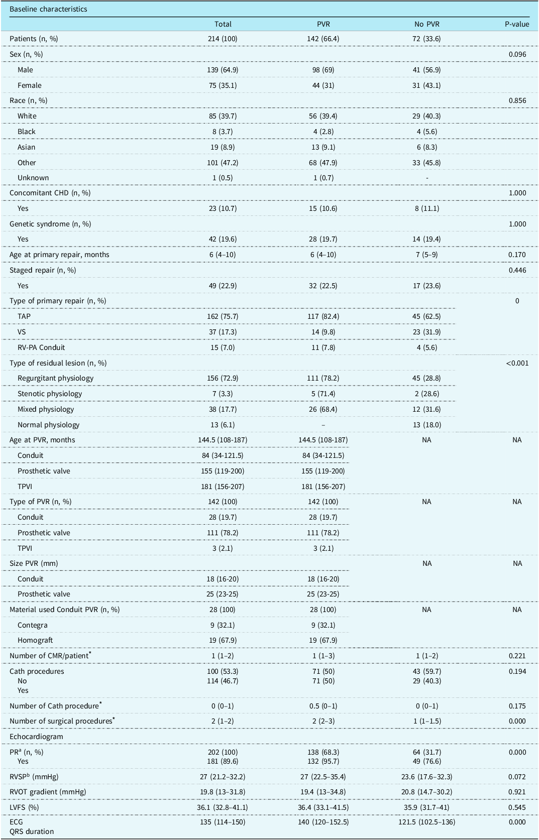
Table 1 describes the baseline patient characteristics: we identified that a higher proportion of patients later requiring pulmonary valve replacement had undergone complete repair via a transannular patch technique (82.4% vs. 62.5%, P < 0.001). Residual physiologic status was assessed as moderate or worse regurgitation in 156 (72.9%). Stenosis with only mild regurgitation was assessed in 7 (3.3%). Combined moderate or worse regurgitant and stenotic physiology was present in 38 (17.7%), and normal physiology was present in 13 (6.1%). Individuals presenting with regurgitant, stenotic, or mixed physiology were more likely to have a pulmonary valve replacement in comparison with those with normal physiology (78.2% vs. 71.4% vs. 68.4% vs. 0%, P < 0.001, for regurgitant, stenotic, mixed, and normal physiology respectively). A longer QRS duration (140 ms [120–152.5] vs. 121.5 [102.5–136], P < 0.001) was also evident in this group. One patient (0.5%) died following pulmonary valve replacement due to postoperative neurologic complications. No patients required cardiac transplantation.
Baseline characteristics by pulmonary valve replacement during follow-up

ECG = electrocardiogram; LVSF = left ventricular shortening fraction; NA = non-applicable; PR = pulmonary regurgitation; PVR = pulmonary valve replacement; RVOT = right ventricle outflow tract; RV-PA = right ventricle-pulmonary artery; TAP = transannular patch; TPVI = transpulmonary valve implanted; VS = valve sparing. Continuous data presented in median and interquartile range (IQR), and categorical data presented in frequencies and proportions. a Moderate or more. b Estimated by the tricuspid regurgitation jet. *Mean and (SD) for (1) number of cardiac MRI: 1.9 (1.3); (2) Cath procedures: 0.7 (1.0); (3) Surgical procedures: 2.0 (0.7).
Cardiac MRI assessment
We reviewed 382 cardiac MRI studies from 213 patients, with one patient excluded as an outlier, due to extreme early dilation of the right ventricle, soon after repair of tetralogy of Fallot with absent pulmonary valve syndrome using a right ventricular to pulmonary artery conduit. In 141 patients, MRI studies both before and after pulmonary valve replacement were available for review. Seventy-two patients who had not yet required pulmonary valve replacement also underwent cardiac MRI evaluation. The median age at the first cardiac MRI was 9.4 years (6–12.2), and for those with serial cardiac MRI studies, the median age at last cardiac MRI was 13.9 (9.7–17). Data from up to four serial MRI studies on the same patient were included in our analysis. Overall, the median number of cardiac MRI studies per patient was 1 (1–2).
The weighted Pearson correlation analysis of right and left ventricular end-diastolic and end-systolic volumes showed a moderate positive correlation with each other (r = 0.56, P < 0.0001; r = 0.50, P < 0.0001). Likewise, the right and left ventricular ejection fractions also demonstrated a moderate positive correlation (r = 0.50, P = 0.008).
Stages of ventricular remodelling
After complete repair and prior to pulmonary valve replacement
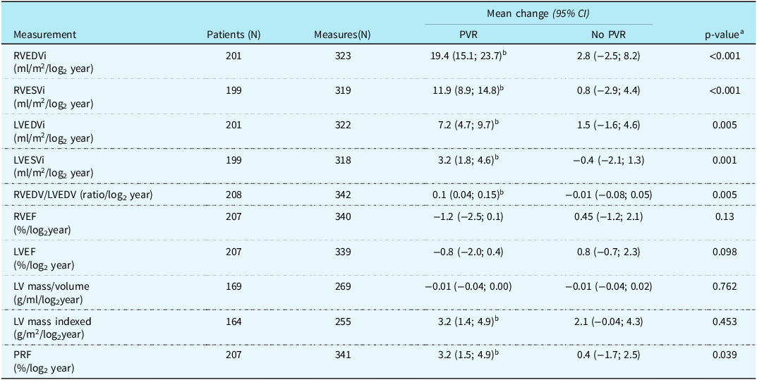
We analysed 323 cardiac MRI studies from 201 patients, performed after complete repair and prior to pulmonary valve replacement. Seventy of these had no subsequent intervention on the pulmonary valve (follow-up time: 10.5 years [7.1–13.1]), and 131 later underwent pulmonary valve replacement (follow-up time: 11.5 years [8.1–15.7]) during the study period. Table 2 describes the observed changes in ventricular volume, ejection fraction, pulmonary regurgitation fraction, and left ventricular mass modelled over time by log2 years of age. The interpretation of log2 years of age is that a 1-unit increase corresponds to a doubling of the original value of age in years. For instance, an increase from 1 log2 year to 2 log2 years represents a doubling of age from 2 years to 4 years.
Ventricular adaptation over time using age in log2years as time variable at CMR exams performed before PVR procedure in the group that later had a surgical intervention at the pulmonary valve and compared with the group that had no PVR. Data points are mean changes per log2 year and 95% confidence interval

CI = confidence interval; LVEDVi = left ventricle end-diastolic volume indexed; LVEF = left ventricle ejection fraction; LVESVi = left ventricle end-systolic volume indexed; RVEDV/LVEDV = right ventricle end-diastolic volume/left ventricle end-diastolic volume; RVEDVi = right ventricle end-diastolic volume indexed; RVESVi = right ventricle end-systolic volume indexed; RVEF = right ventricle ejection fraction; PVR = pulmonary valve replacement; PRF = pulmonary regurgitation fraction. Results are derived from a linear mixed-effect model with RV and LV volumes as dependent variables and time (log base 2 age at CMR), sex, regurgitant residual lesion, PVR, and PVR × time as independent variables. a P-value for the difference between the patients who had PVR during the follow-up and those who did not. b The slope is significantly (p-value < 0.05) different from 0.
In this analysis, we compared the temporal change of cardiac MRI parameters in those patients who later required a pulmonary valve replacement against those who did not. We detected a more rapid dilation by log2 years for the right and left ventricular volumes, an increase right ventricular end-diastolic volume to left ventricular end-diastolic volume ratio, an increase in indexed left ventricular mass, and an increase in pulmonary regurgitation fraction in the group that later had pulmonary valve replacement. In contrast, there was no change over time in ventricular volumes, mass, or pulmonary regurgitation fraction in the group that did not later require pulmonary valve replacement.
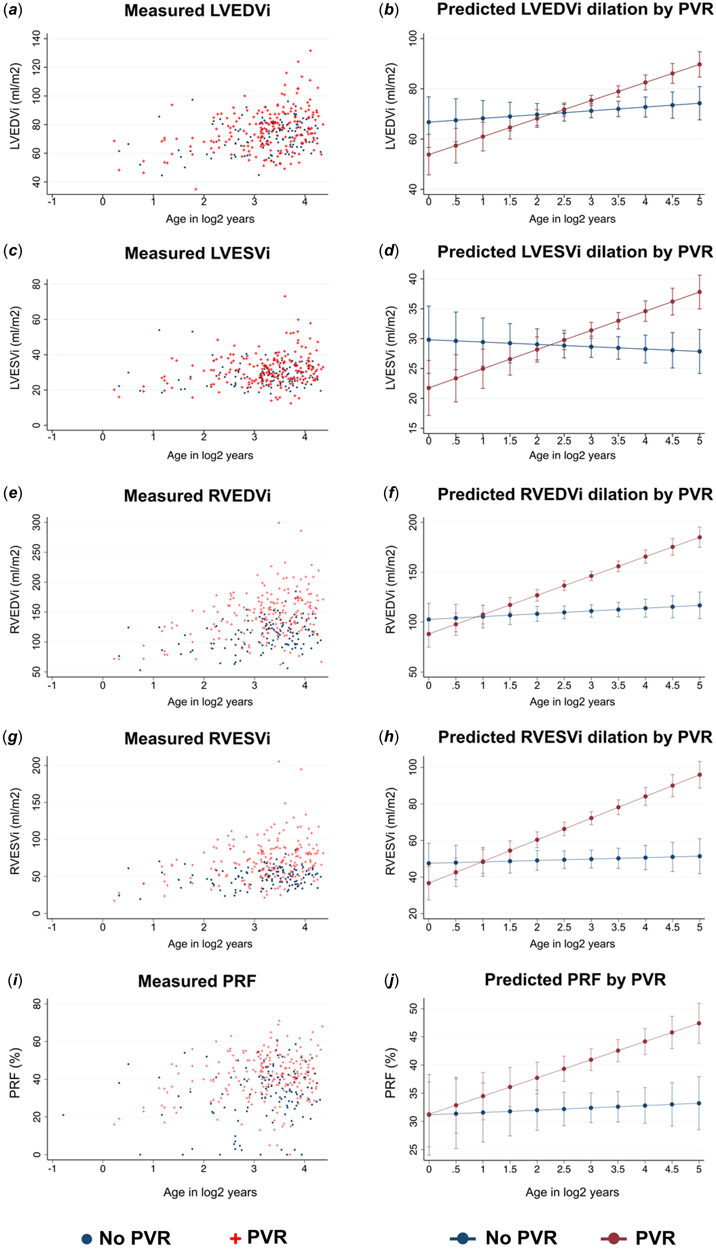
Figure 2 demonstrates the individual data points of right and left ventricular volumes and pulmonary regurgitation fraction, along with the linear representation of the mean change over log-transformed age, in these parameters for the two groups of patients. Our modelling presented in Figure 2 suggested that in children as young as 4 years of age (i.e. log2(4 years) = 2), a right ventricular end-diastolic or end-systolic volume index greater than 120 ml/m2 and 60 ml/m2, respectively, was associated with later progression to require pulmonary valve replacement during the follow-up (Figure 2f and 2h). In addition, we observed that for patients 11 years of age or older (e.g. log2(11 years) = 3.5) at the time of assessment, a left ventricular end-diastolic or end-systolic volume index greater than 80 ml/m2 and 33 ml/m2, respectively, was also prominently associated with subsequent pulmonary valve replacement occurring (Figure 2b and 2d).
Measured and predicted temporal change of RV and LV volumes and PRF in patients with rTOF. The graphics ( a ), ( c ), ( e ), ( g ), and I represent the measured LV EDVi, LV ESVi, RV EDVi, RV ESVi, and PRF over time measured in log2years (e.g. 2 log2years = 4 years old). In the graphics ( b ), ( d ), ( f ), ( h ), and ( j ), the blue line represents the estimated slope of change over time (log2 years) in patients that had no PVR during follow-up, and the red line represents the slope for patients that had PVR determined using linear mixed-effects modelling adjusted by sex and pure regurgitant residual lesion, and 95% confidence interval (CI) for each group (PVR and no PVR).

In the subgroup analysis including only patients with serial (2 or more) cardiac MRI studies (n = 76), we confirmed the above findings: a more rapid increase in the right and left ventricular volumes in the group who later required pulmonary valve replacement was observed (Supplemental material Table S1). Similarly, a reduction in the right ventricular ejection fraction and an increase in the pulmonary regurgitation fraction occurred in those who later required pulmonary valve replacement. In the group of patients who did not require pulmonary valve replacement, there was no observed change in ventricular volumes, ejection fraction, left ventricular mass index, and pulmonary regurgitation fraction.
In the subgroup analysis assessing the change over time of cardiac MRI metrics stratified by type of residual lesion, we detected a more rapid increase in right ventricular volumes and right ventricular end-diastolic volume to left ventricular end-diastolic volume ratio for both regurgitant and mixed residual physiology in the group who later required pulmonary valve replacement, with the dilation of right ventricular volumes being strongest in the regurgitant group compared to the mixed residual lesion group. There was also a similar trend in the increase of left ventricular volumes, although this did not reach statistical significance. The stratified analysis was not performed for patients presenting with stenotic physiology and normal physiology due to limited sample size (Supplemental material Tables S2 and S3).
After pulmonary valve replacement
We assessed 22 patients who had cardiac MRI studies performed before and after pulmonary valve replacement, at a median age of 11 years (5.7–13.7) and 16.7 years (12.5–19.2), respectively. Pulmonary valve replacement resulted in a significant reduction in pulmonary regurgitant fraction, right ventricular end-diastolic volume to left ventricular end-diastolic volume ratio, and in right ventricular end-systolic and end-diastolic volumes, resulting in an unchanged right ventricular ejection fraction. Left ventricular end-systolic and end-diastolic volumes were also unchanged (Supplemental material Table S4).
Single-point and serial MRI studies performed only after pulmonary valve replacement were available in 27 patients, with a median age of 11.2 years (5.7–14.1) at the time of pulmonary valve replacement (Supplemental material Table S5). The median age at first cardiac MRI was 14 years (10.5–16) and was 17 years (10.5–19.5) at the most recent cardiac MRI. Of 27 patients studied at that time, 20 (74%) had only one cardiac MRI, and 7 (26%) patients had serial evaluations (range 2–3). While the sample size with serial studies was small, no progressive change in indexed right ventricular end-systolic or end-diastolic volume and right ventricular end-diastolic volume to left ventricular end-diastolic volume ratio was evident. There was some evidence of a progressive reduction over time in indexed left ventricular end-diastolic volume, which decreased by –10.5 ml/m2/log2years (–18.14, –2.91; P = 0.007), with no change in left ventricular end-systolic volume or increase in left ventricular ejection fraction. There was also an increase in left ventricular mass by 8.05 g/m2/log2years (1.82, 14.28; P = 0.011) following pulmonary valve replacement in these patients. We found no further ongoing change in systolic or diastolic right ventricular volumes or in right ventricular ejection fraction. The left ventricular mass-volume ratio also remained unchanged over time.
Discussion
In this retrospective cohort study of children with repaired tetralogy of Fallot, changes over time assessed by cardiac MRI were evident for both right and left ventricular volumes, left ventricular mass index, and pulmonary regurgitation fraction. Prior to pulmonary valve replacement, the rate of change of right and left ventricular remodelling was far more pronounced in patients destined to later require pulmonary valve replacement during their follow-up period, which extended to a median of 10.7 years (7.9–14.4) after the primary repair. Based on our data, the separation of remodelling trajectory over time in the pulmonary valve replacement group, versus the no pulmonary valve replacement group, may occur from as young as 4 years of age, suggesting that early and serial evaluation by cardiac MRI could inform eventual prognosis in these children. If a different rate of progression of dilation is identifiable, such patients could be followed more closely, and pulmonary valve replacement could be considered at a younger age. Further work is needed to determine whether this is in fact a distinct phenotype amongst the post-repair tetralogy of Fallot population in childhood. In addition, our patients who underwent transannular patch repair and presented with a longer QRS duration were more likely to require pulmonary valve replacement later during follow-up. This observation aligns with previous studies comparing patients with transannular patch repair to those with valve-sparing techniques, which demonstrated a higher number of pulmonary valve replacements in the former group. Reference Blais, Marelli and Vanasse19 Furthermore, preoperative QRS duration has been reported to be associated with markedly elevated right ventricular volumes before pulmonary valve replacement in patients with repaired tetralogy of Fallot. Reference Cocomello, Sinha, Gonzalez Corcia, Baquedano, Benedetto and Caputo20
Right ventricular remodelling
Previous studies in adolescents with repaired tetralogy of Fallot using serial cardiac MRI have identified absolute duration of time from complete repair, greater degree of pulmonary regurgitation, and greater initial right ventricular volume as risk factors for progressive right ventricular dilation. Reference Bhat, Mercer-Rosa and Fogel18 However, controversy persists regarding the inevitability of progressive right ventricular dilation in adult survivors of repaired tetralogy of Fallot. While some longitudinal studies have shown an association between time and right ventricular volume increment, others have found no obvious temporal progression, even in the presence of severe pulmonary regurgitation. Reference Bhat, Mercer-Rosa and Fogel18,Reference Buddhe, Shah and Lai21–Reference T., F. and M.24 This discrepancy might be due to ascertainment bias in studies focusing on adult populations, as patients requiring pulmonary valve replacement prior to age 18 may have been excluded. Our data suggest that an early trajectory of right ventricular dilation over time is present in some paediatric patients but not all, influencing the selection of children and adolescents for pulmonary valve replacement.
Right ventricular remodelling stratified by residual lesion physiology
In the subgroup analysis stratifying the patients by type of residual lesion physiology (mixed and regurgitant lesions), we detected significant right ventricular volume dilation over time in both groups. However, an increase in right ventricular volumes over time was more pronounced in the group of patients presenting with regurgitant postoperative lesions. In a previous study assessing the impact of right ventricular pressure overload in patients with tetralogy of Fallot aged older than 8 years and young adults, researchers found an association between right ventricular pressure overload and smaller right ventricular volume. In that study, a mild residual right ventricular outflow tract gradient of 15–30 mmHg did not protect against the need for later pulmonary valve replacement in patients with significant pulmonary regurgitation, while a right ventricular outflow tract gradient > 25 mmHg was associated with arrhythmias and cardiac death. Reference Latus, Stammermann and Voges25
Left ventricular remodelling
For the left ventricle, estimates of progression of dilation following tetralogy of Fallot repair are less well documented. Our study found that left ventricular volumes increased over time, with a corresponding increase in left ventricular mass index, particularly in patients who later required pulmonary valve replacement. Previous studies have reported a lower left ventricular mass/volume ratio in children and adults with repaired tetralogy of Fallot, potentially due to the skewing effect of eccentric left ventricular remodelling in those with marked dilation. Reference Andrade, Jerosch-Herold and Wegner7 Additionally, a previous study evaluating healthy children aged 0–18 years demonstrated a progressive increase in left ventricular mass with age. Reference van der Ven, Sadighy and Valsangiacomo Buechel26
The pertinent physiology is hinted at by Gnanappa et al., who reported an increase in net pulmonary forward flow, with an increased left ventricular stroke volume and greater left ventricular dimension in patients with more severe right ventricular dilation. Reference Gnanappa, Celermajer, Zhu, Puranik and Ayer27 Taken together, these data support the notion that progressive right ventricular dilation is likely to result in progressive dilation of the left ventricle either by a series effect of increasing net forward flow or by a parallel effect of ventricular-ventricular interaction in repaired tetralogy of Fallot patients, as was suggested by others who have correlated the systolic function of the right and left ventricle in this population. Reference Davlouros, Kilner and Hornung28–Reference Kempny, Diller and Orwat30 It is possible that both mechanisms are relevant. We believe that our finding that left ventricular volume and mass did not progress in the subset of patients who did not require pulmonary valve replacement during the paediatric age range indicates that not all children with repaired tetralogy of Fallot are at equal risk for this outcome. This is supported by an earlier study of adults following repaired tetralogy of Fallot without pulmonary valve replacement during follow-up that found no progression of left ventricular volumes over time during the study period. Reference Wijesekera, Raju and Precious22
Pulmonary regurgitant fraction
Our study demonstrated a progressive increase in the pulmonary regurgitation fraction over time in patients who would later require pulmonary valve replacement, whereas previous data have been inconsistent: Luijnenburg et al. affirmed this observation in children and adults with repaired tetralogy of Fallot; however, Bhat et al. did not observe such an association. Reference Bhat, Mercer-Rosa and Fogel18,Reference Luijnenburg, Helbing and Moelker31 Conceptually, a close relationship between pulmonary regurgitation fraction and right ventricular remodelling after repaired tetralogy of Fallot is plausible and implies significant coupling of right ventricular and pulmonary arterial diastolic function. The reason for such heterogeneity in this patient population requires further study.
Right ventricular systolic function
Systolic dysfunction of the right ventricle post-tetralogy of Fallot repair as indicated by reduced right ventricular ejection fraction on cardiac MRI has been proposed as an indication for pulmonary valve replacement. Reference Stout, Daniels and Aboulhosn4,Reference Baumgartner, De Backer and Babu-Narayan32 It is of interest to know whether this is a progressive phenomenon or not: indeed, we did not detect a temporal change in right and left ventricular ejection fraction, either in patients who required pulmonary valve replacement or those who did not when it was assessed in the complete study cohort. However, for children with serial cardiac MRI assessment, right ventricular ejection fraction did decrease over time in the group that later required pulmonary valve replacement. Previously published studies have presented different results regarding the right ventricular ejection fraction change over time, with both no significant deterioration and a decline over time being described. Reference Goo33,Reference Buechel, Dave and Kellenberger34 This discrepancy may be related to the heterogeneity of the disease or differences in study population age.
Changes after pulmonary valve replacement
Following pulmonary valve replacement, we observed a marked reduction in pulmonary regurgitation fraction and a short-term improvement in indexed right ventricular volume. However, improved left ventricular remodelling was less pronounced, raising concerns about its ability to fully reverse-remodel post-intervention. Our data suggest a more gradual, progressive improvement in indexed left ventricular end-diastolic volume, with an increase in indexed left ventricular mass over time. After the initial assessment following pulmonary valve replacement, we did not detect any further changes in right ventricular volumes. A previous study assessing longitudinal changes in right and left ventricular volumes and ejection fraction over age in adults with tetralogy of Fallot found that prior pulmonary valve replacement was not associated with changes in right and left ejection fraction or right ventricular end-diastolic volumes. Reference Ruperti-Repilado, Haag and Fischer35
Limitations
Our study is limited in that it is retrospective and based on a single-centre experience. In addition, cardiac MRI evaluations were clinically driven and not universally obtained, with limited objective criteria for an MRI, relying instead on individual clinician judgement regarding symptom progression and echocardiographic right ventricular dilation. This raises the possibility of selection bias and increases the possibility of our data being skewed towards the more severe spectrum of right ventricular dilation. In that regard, we would expect this cohort to represent those with more obvious post-tetralogy of Fallot repair right ventricular changes, which is in keeping with the relatively high proportion of patients requiring pulmonary valve replacement during follow-up compared with a previous study that reported 35.7% of pulmonary valve replacement after primary repair. Reference Sabate Rotes, Eidem and Connolly36 Given the nature of the study, it was not possible to objectively assess clinical symptoms in our analysis.
Conclusions
We conclude that a distinctive adverse trajectory of right and left ventricular remodelling can be identified in patients quite early during the follow-up of tetralogy of Fallot repair in childhood, distinguishing patients who are likely to require pulmonary valve replacement before adulthood. A strong association was confirmed between the transannular patch repair technique and the need for paediatric pulmonary valve replacement, as well as with longer QRS duration in the post-repair course. Important left ventricular dilation was also noted in these patients and found to reverse more slowly after pulmonary valve replacement. Therefore, early and serial assessment of these changes by MRI should be conducted with a view to determine the optimal timing of pulmonary valve replacement and to preserve ventricular function in adolescence and young adulthood. The optimal timing of initial MRI surveillance might begin as young as age 5, given the divergence in indexed right ventricle volume we observed in the patient groups, but this remains to be determined by further prospective study. We believe this will also be influenced by the development of expandable catheter deployed bioprosthetic valves that are able to age with the patient.
Supplementary material
The supplementary material for this article can be found at https://doi.org/10.1017/S1047951125001891.
Acknowledgements
None.
Financial support
This research received no specific grant from any funding agency in the public, commercial, or not-for-profit sectors.
Competing interests
There are no financial conflicts of interest to disclose.
Ethical standard
CHLA-21-00110, approved on 4/23/2021.