Introduction
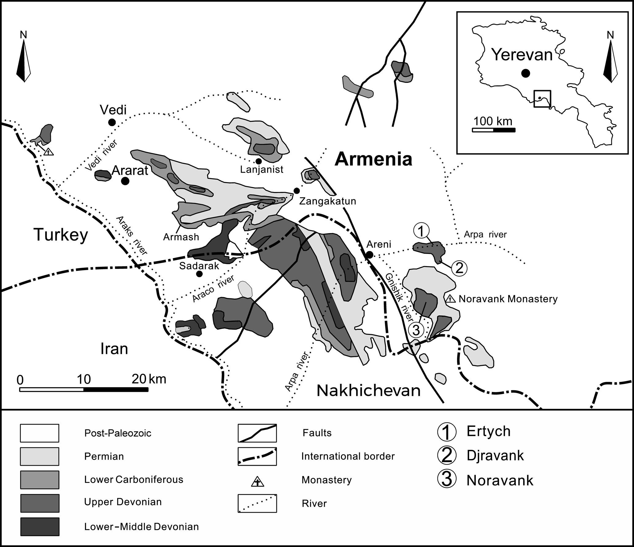
The Upper Devonian sequences of the Lesser Caucasus (or Transcaucasia, Fig. 1) are remarkable in terms of their completeness and richness in fossil remains (Abrahamyan, Reference Abrahamyan1957, Reference Abrahamyan, Mkrtchian, Vardaniants, Gabrielian, Maghakian and Paffenholz1964; Arakelyan, Reference Arakelyan, Mkrtchian, Vardaniants, Gabrielian, Maghakian and Paffenholz1964; Alekseeva et al., Reference Alekseeva, Afanasjeva, Grechishnikova, Oleneva and Pakhnevich2018a, Reference Alekseeva, Afanasjeva, Grechishnikova, Oleneva and Pakhnevichb). They have attracted the interest of pioneer geologists, such as the famous German geologist Hermann Abich, who first described Devonian outcrops and a new spiriferide species (‘Spirifer’ orbelianus) from Armenia in Reference Abich1858 (Serobyan et al., in press). Upper Devonian–lower Carboniferous brachiopods were systematically studied by Abrahamyan during the years 1949 to 1974. One of her most important publications dates back to 1957 in which she described and illustrated 36 brachiopod species (including eight new ones) from the Famennian–Tournaisian interval. These species belong to the orders Athyrida, Orthida, Orthotetida, Productida, Rhynchonellida, and Spiriferida. In the same publication, she also presented a biostratigraphic scheme based on brachiopods wherein part of the lower Famennian was characterized by the ‘Cyrtospirifer’ orbelianus taxon-range Zone (Fig. 2). The latter brachiopods were partly revised by Abrahamyan (Reference Abrahamyan and Akopian1974), in addition to newly studied Frasnian species. However, despite the extensive taxonomic studies led by Abrahamyan (Reference Abrahamyan1957, Reference Abrahamyan and Akopian1974) and more recently by Alekseeva et al. (Reference Alekseeva, Afanasjeva, Grechishnikova, Oleneva and Pakhnevich2018a, Reference Alekseeva, Afanasjeva, Grechishnikova, Oleneva and Pakhnevichb), who mainly focused on brachiopods from Nakhichevan (Fig. 1), the taxonomy and biostratigraphy of lower Famennian brachiopod assemblages from Armenia still remain poorly understood. This is especially true for the rhynchonellide and athyride brachiopods, which are very common in the Upper Devonian–lower Carboniferous sequences of this area. Their study can provide valuable insights into Late Devonian changes in brachiopod diversity and paleobiogeographic distribution. This paper revises the descriptions of the rhynchonellides and atrypides in the lower Famennian ‘Cyrtospirifer’ orbelianus Zone on the basis of newly collected material from three distinct localities in central Armenia (Fig. 1) and discusses their biostratigraphy and paleobiogeographic significance.
Schematic geological map and distribution of the Upper Paleozoic sequences in the Lesser Caucasus (central Armenia and Nakhichevan), including the location of the three sections mentioned in the present study (modified after Serobyan et al., Reference Serobyan, Grigoryan, Mottequin, Mayilyan, Crônier and Danelian2019).

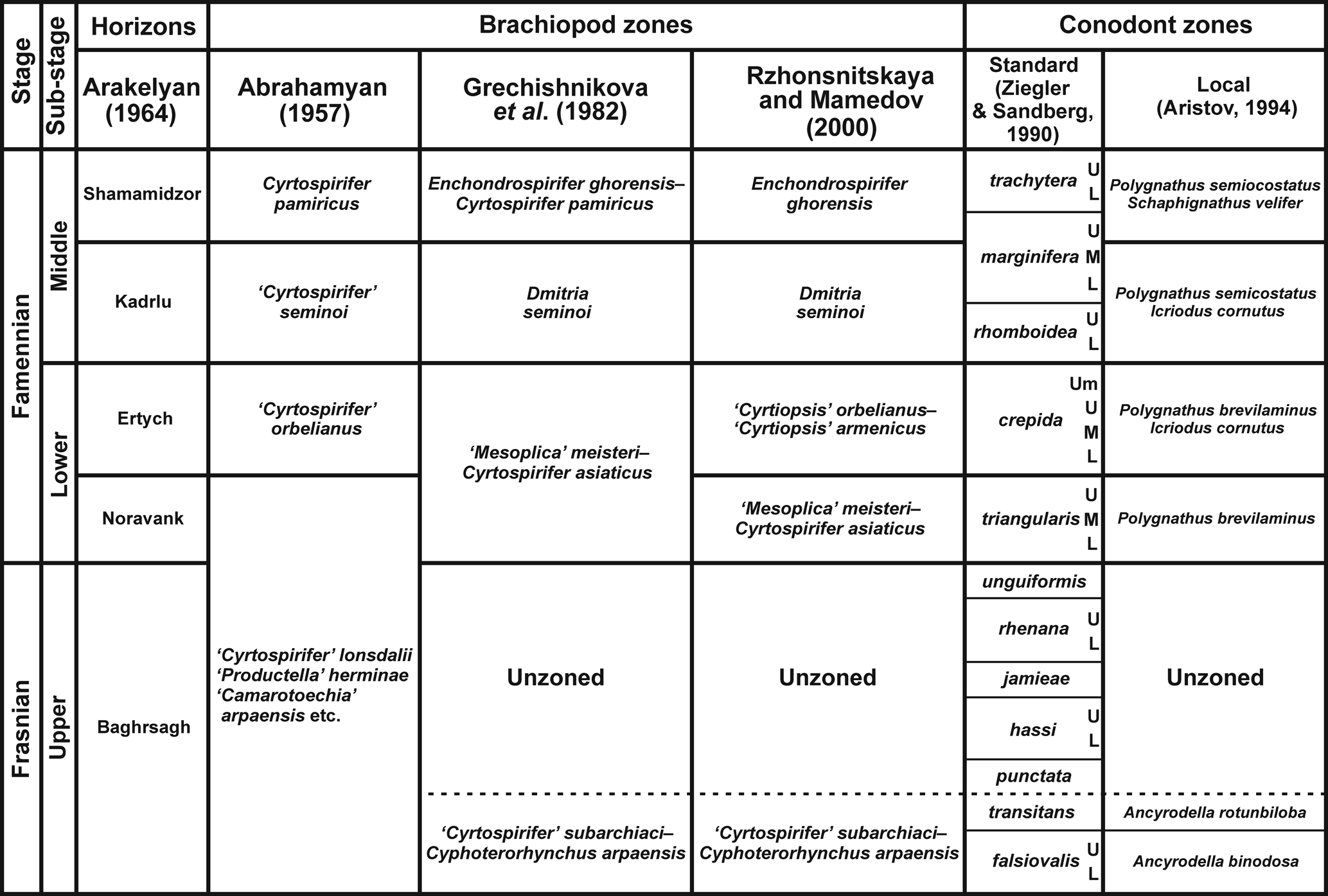
Biochronostratigraphic framework of the upper Frasnian–middle Famennian sedimentary sequences in the Lesser Caucasus, including the brachiopod biozones established by Abrahamyan (Reference Abrahamyan1957) in central Armenia and by Grechishnikova et al. (Reference Grechishnikova, Aristov, Reitlinger and Chizhova1982) and Rzhonsnitskaya and Mamedov (Reference Rzhonsnitskaya and Mamedov2000) in Nakhichevan. It also includes the regional conodont zonation established by Aristov (Reference Aristov1994) in Nakhichevan and the standard conodont zones of Ziegler and Sandberg (Reference Ziegler and Sandberg1990).

Geological setting
The Upper Devonian sequences of Armenia are composed of mixed carbonate-siliciclastic sediments that accumulated on a shallow water platform. They consist essentially of marly and sandy biogenic limestones, rich in brachiopods, and intercalations of quartzites and shales. Geodynamically speaking, they are part of the South Armenian Block (SAB), a microcontinent that detached from Gondwana during the Triassic and Jurassic (Sosson et al., Reference Sosson, Rolland, Müller, Danelian, Melkonyan, Kekelia, Adamia, Babazadeh, Kangarli, Avagyan, Galoyan, Mosar, Sosson, Kaymakci, Stephenson, Bergerat and Starostenko2010). The ~1500 m thick Upper Devonian–lower Carboniferous sedimentary sequences in Armenia were subdivided by Abrahamyan (Reference Abrahamyan, Mkrtchian, Vardaniants, Gabrielian, Maghakian and Paffenholz1964) and Arakelyan (Reference Arakelyan, Mkrtchian, Vardaniants, Gabrielian, Maghakian and Paffenholz1964) into a number of horizons (e.g., Baghrsagh, Noravank, Ertych, Fig. 2) that are difficult to recognize lithologically because they were defined based on brachiopod biozones. Consequently, they have a biostratigraphic rather than lithostratigraphic significance (see Serobyan et al., Reference Serobyan, Grigoryan, Mottequin, Mayilyan, Crônier and Danelian2019).
Because of their abundance in these sequences, brachiopods were recognized to be useful for stratigraphic correlations and used to support the extensive mapping effort planned by the Soviet authorities for the area after World War II. Rzhonsnitskaya (Reference Rzhonsnitskaya1948) first established a rudimentary biostratigraphical scheme based on brachiopods, which was much improved by Abrahamyan (Reference Abrahamyan1957), who introduced a continuous biozonation characterized by marker species or species assemblages (Fig. 2). The ‘Cyrtospirifer’ orbelianus Zone of Abrahamyan (Reference Abrahamyan1957) is a taxon-range biozone covering part of the lower Famennian. Grechishnikova et al. (Reference Grechishnikova, Aristov, Reitlinger and Chizhova1982) and Rzhonsnitskaya and Mamedov (Reference Rzhonsnitskaya and Mamedov2000) applied Abrahamyan's zonal scheme in Nakhichevan, while improving and complementing it for some intervals. Figure 2 correlates the above-mentioned brachiopod zones with the regional conodont zonation of Aristov (Reference Aristov1994), which was established in Nakhichevan, and the standard conodont zonation of Ziegler and Sandberg (Reference Ziegler and Sandberg1990).
Materials and methods
Most of specimens illustrated and investigated herein were collected during several field seasons organized in 2018 and 2019. The material was collected from the ‘Cyrtospirifer’ orbelianus Zone of Abrahamyan (Reference Abrahamyan1957) because it crops out well in three distinct sections of central Armenia (Ertych, Djravank, and Noravank; Fig. 1). The ‘Cyrtospirifer’ orbelianus Zone can be correlated with the ‘Cyrtiopsis’ orbelianus-’Cyrtiopsis’ armenicus Zone of Rzhonsnitskaya and Mamedov (Reference Rzhonsnitskaya and Mamedov2000), and is considered coeval to the crepida standard conodont Zone of Ziegler and Sandberg (Reference Ziegler and Sandberg1990). The material sampled in the field was complemented with specimens that were collected during the 1940s to 1980s by Abrahamyan and Arakelyan, both from Armenia and Nakhichevan.
All ground specimens were selected among recently collected material because historical specimens cannot be sectioned; thus, the latter have been used only for measurements. Statistical analysis (i.e., ANCOVA—test for comparison of several models of simple linear regression) was applied in order to determine if the regression line slopes of our four samples are significantly different. Statistical analysis was performed under the free PAST 3 software. The internal morphology of specimens was investigated by using the standard technique of serial sections and acetate peels. Almost complete articulated specimens were coated with magnesium oxide before being photographed with a Canon EOS 700D camera. Afterwards, in order to enhance contrast and brightness, all images were further processed using Adobe Photoshop CS6. Specimens selected for scanning electron microscopy were coated with gold and a ZEISS EVO Scanning Electron Microscope was used to image micro-ornamentation.
Repositories and institutional abbreviations
All illustrated material is housed at the Geological Museum of the Institute of Geological Sciences of the National Academy of Sciences of Armenia, Yerevan (IGSNASRAGM/PS), unless otherwise stated. The prefix PS indicates the Laboratory of Paleontology and Stratigraphy. Part of the studied material is housed at the public paleontological collection of the University of Lille (USTL), France. The prefix RBINS is used for some specimens deposited at the Royal Belgian Institute of Natural Sciences, Brussels. Specimens cited in Brice (Reference Brice1967) are stored at the Laboratoire de Paléontologie de la Faculté Libre des Sciences des Lille (collections De Lapparent and Brice). The material studied by Erlanger (Reference Erlanger1993) is housed at the Borissiak Paleontological Institute of the Russian Academy of Sciences, Moscow and is catalogued under the prefix PIN and collection no. 3744.
Systematic paleontology
The supraspecific classification follows Savage et al. (Reference Savage, Manceñido, Owen, Carlson, Grant, Dagys, Sun and Kaesler2002) and Savage (Reference Savage and Kaesler2007) for the Order Rhynchonellida instead of the one adopted by Sartenaer (Reference Sartenaer2001, Reference Sartenaer2003), wherein the Subfamily Ripidiorhynchinae Savage, Reference Savage, Copper and Jin1996 is elevated to the family level with Ripidiorhynchus Sartenaer, Reference Sartenaer1966 as its only representative, except otherwise stated. The supraspecific classification also follows the one suggested by Alvarez and Rong (Reference Alvarez, Rong and Kaesler2002) for the Order Athyrida. The term Athyrida is used here according to the recommendation made by Copper and Jin (Reference Copper and Jin2017).
Order Rhynchonellida Kuhn, Reference Kuhn1949
Superfamily Rhynchotrematoidea Schuchert, Reference Schuchert and Eastman1913
Family Trigonirhynchiidae Schmidt, Reference Schmidt1965
Subfamily Trigonirhynchiinae Schmidt, Reference Schmidt1965
Genus Sartenaerus Özdikmen, Reference Özdikmen2008
Type species
Camarotoechia baitalensis Reed, Reference Reed1922.
Remarks
Camarotoechia baitalensis was selected by Sartenaer (Reference Sartenaer1970) as the type species of his new genus Centrorhynchus. Although Savage et al. (Reference Savage, Manceñido, Owen, Carlson, Grant, Dagys, Sun and Kaesler2002) placed this genus in the subfamily Hemitoechiinae, Nicollin and Brice (Reference Nicollin and Brice2004, p. 440) noted that it should be reassigned to the subfamily Trigonirhynchiinae because its septalium is posteriorly covered. Finally, because the generic name introduced by Sartenaer (Reference Sartenaer1970) had already been used by Luehe (Reference Luehe1911) for a genus of parasitic worms (Acanthocephala), Özdikmen (Reference Özdikmen2008) introduced the new genus name Sartenaerus for Centrorhynchus sensu Sartenaer (Reference Sartenaer1970). Mottequin et al. (Reference Mottequin, Brice and Legrand-Blain2014) indicated that the genus Centrorhynchus (= Sartenaerus) first occurs in the middle Famennian, as observed in Western Europe (Mottequin and Brice, Reference Mottequin and Brice2016), but the brachiopod record from Armenia establishes that it was already present in the lower part of this stage.
Sartenaerus baitalensis (Reed, Reference Reed1922)
Figures 3, 4
- Reference Reed1922
Camarotoechia baitalensis Reed, p. 94, pl. 14, figs. 11–21.
- Reference Reed1922
Camarotoechia baitalensis comitata, var. nov.; Reed, p. 97, pl. 14, figs. 24–25; pl. 15, figs. 1–3a.
- Reference Reed1922
Camarotoechia baitalensis transversalis, var. nov.; Reed, p. 97, pl. 14, figs. 22, 23.
- Reference Serobyan, Grigoryan, Mottequin, Mayilyan, Crônier and Danelian2019
Sartenaerus baitalensis; Serobyan et al., p. 9 (see for a complete synonymy).
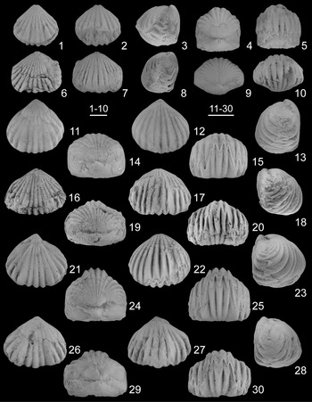
Sartenaerus baitalensis (Reed, Reference Reed1922) from the lower Famennian ‘Cyrtospirifer’ orbelianus Zone in Armenia. (1–5) IGSNASRAGM 3900/PS 3006 (Ertych section), almost complete juvenile specimen in ventral, dorsal, lateral, posterior, and anterior views; (6–10) IGSNASRAGM 3901/PS 3007 (Ertych section), partly exfoliated juvenile specimen in ventral, dorsal, lateral, posterior, and anterior views; (11–15) IGSNASRAGM 3902/PS 3008 (Djravank section), almost complete specimen in ventral, dorsal, lateral, posterior, and anterior views; (16–20) IGSNASRAGM 3903/PS 3009 (Djravank section), partly exfoliated specimen in ventral, dorsal, lateral, posterior, and anterior views; (21–25) IGSNASRAGM 3904/PS 3010 (Noravank section), articulated specimen in ventral, dorsal, lateral, posterior, and anterior views; (26–30) IGSNASRAGM 3905/PS 3011 (Noravank section), articulated specimen in ventral, dorsal, lateral, posterior, and anterior views. Scale bars = 5 mm (1–10), 10 mm (11–30).

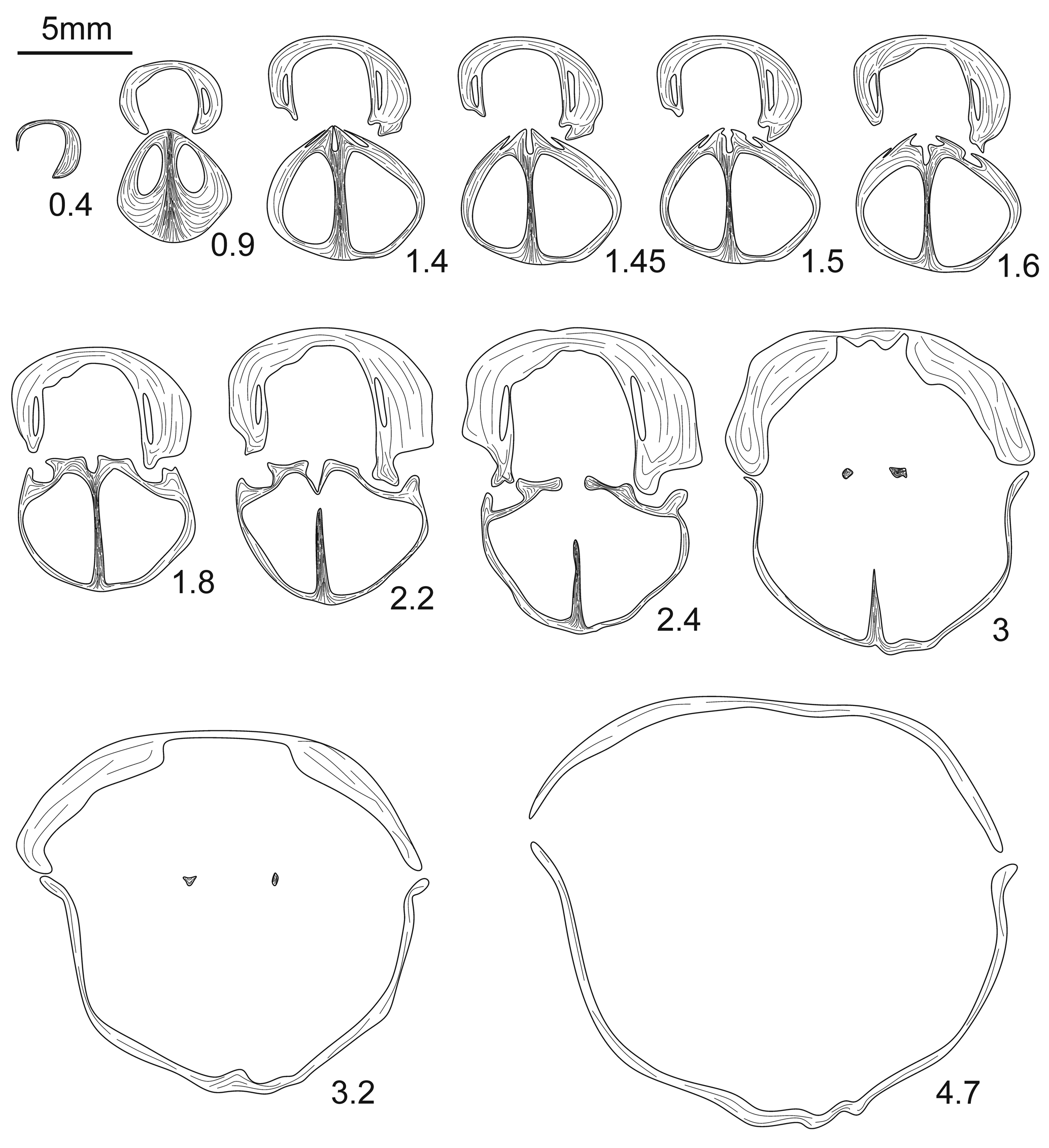
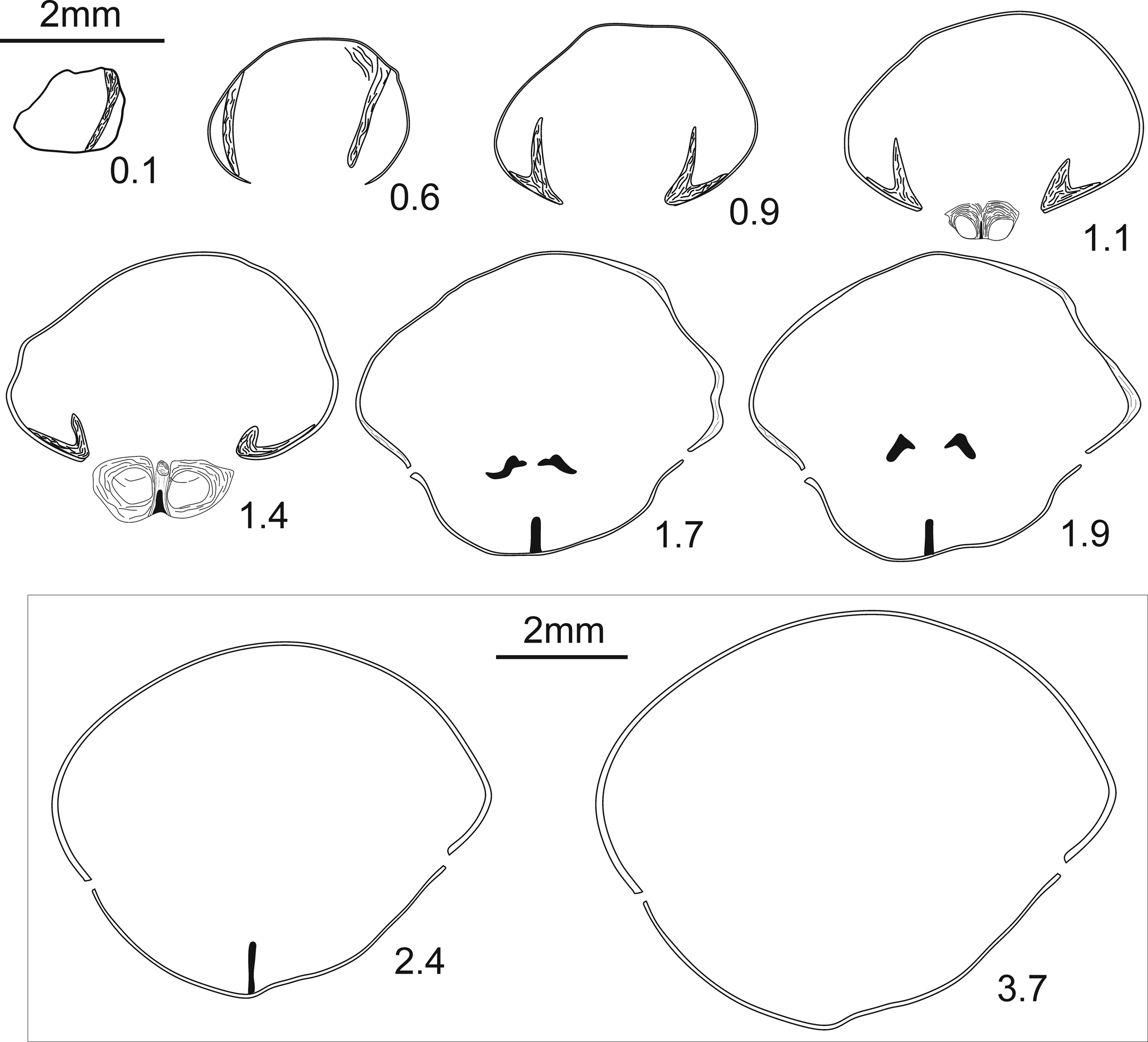
Transverse serial sections of Sartenaerus baitalensis (Reed, Reference Reed1922) from the ‘Cyrtospirifer’ orbelianus Zone in Armenia. Numbers refer to distances in mm from the tip of the ventral umbo. The ground specimen corresponds to the one illustrated on Figure 3.11–3.15 (IGSNASRAGM 3906/PS 3012).

Holotype
Reed (Reference Reed1922) did not designate a holotype among the specimens he illustrated. Savage et al. (Reference Savage, Manceñido, Owen, Carlson, Grant, Dagys, Sun and Kaesler2002) subsequently designated as lectotype the specimen illustrated in Reed (Reference Reed1922, pl. 14, fig. 11a, b).
Occurrence
This species was first described by Reed (Reference Reed1922) from the Famennian (the lowest and uppermost parts excluded; Sartenaer, Reference Sartenaer1970) of Pamir (Tajikistan). In Armenia, it was first reported by Abrahamyan (Reference Abrahamyan1957) in the lower Famennian ‘Cyrtospirifer’ orbelianus Zone. This is consistent with the record of this species in Nakhichevan (Mirieva, Reference Mirieva2010).
Description
Shell medium-sized (reaching 26 mm in width, 25.4 mm in length, and 23.8 mm in thickness) (Table 1), wider than long, strongly dorsibiconvex, rounded subpentagonal to transversally ovate in outline; widest more anteriorly to mid-length; highest slightly posteriorly to the front (in adults); anterior margin slightly emarginate to straight; anterior commissure highly zigzagged and strongly uniplicate.
Measurements in mm and ratios of Sartenaerus baitalensis (Reed, Reference Reed1922). Abbreviations: W = width of the shell, L = length of the shell, T = thickness of the shell, Ws = width of the sulcus.

Ventral valve inflated with convex flanks sloping steeply towards zigzagging lateral commissures; highest at anterior margin, then decreasing gently or maintained towards the posterior margin; umbo wide, inflated; beak large, prominent, suberect; foramen not observed; palintropes low, concave; sulcus shallow to moderately deep, wide, inconspicuously originating at umbo, well delimited by bounding ribs, flat to slightly round-bottomed at front; tongue high to very high, wide, with sharp borders, subtrapezoidal in outline, strongly bent dorsally.
Dorsal valve strongly inflated, strongly curved in posterior and lateral profile views with convex flanks sloping steeply towards lateral commissures; highest slightly posteriorly to the front, then moderately to strongly decreasing towards the posterior margin; beak obtuse, covering the delthyrium; fold starting at umbo, moderately high, well defined, becoming relatively higher anteriorly.
Ornamentation of coarse, strong, angular ribs with rounded apices arising from beaks; three ribs in sulcus with one median rib usually stronger and four ribs on fold with two opposed ribs usually stronger, 6–7 ribs per flank; ribs wider than interspaces.
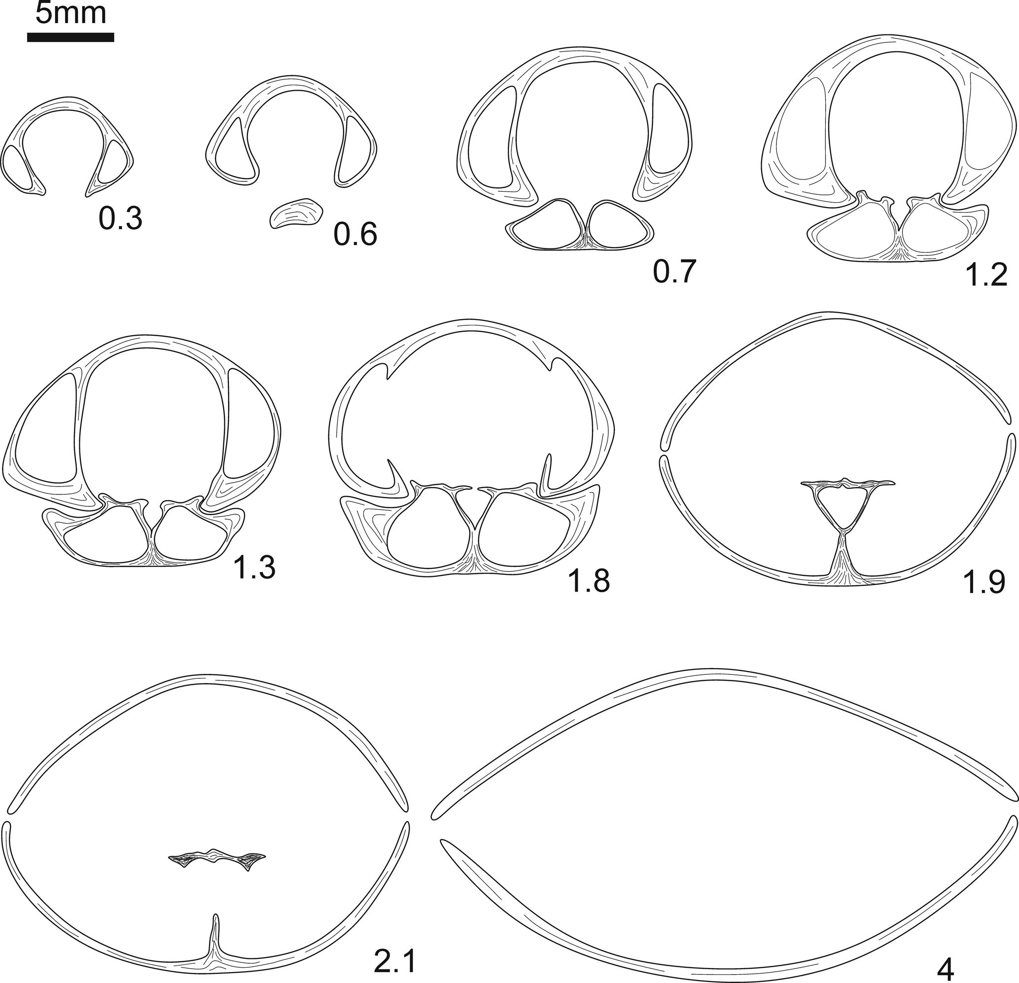
Ventral valve interior (Fig. 4) with strong and relatively short dental plates, subparallel posteriorly then converging ventrally toward anterior; teeth strong, crenulated; ventral muscle field flabellate anteriorly then becoming hastate or subtrapezoidal (Fig. 4, serial sections 2.2–3); lateral apical cavities strongly filled in by callosities.
Dorsal valve interior with long and quite high septum, extending up to 15% of the valve length; septalium narrow, relatively short, without cover; dental sockets large; hinge plates long, divided immediately anteriorly to septalium; crural bases horizontal.
Materials
Thirty-four articulated specimens, six ventral and four dorsal valves, 12 articulated specimens, two ventral and three dorsal valves come from the Ertych section; 11 articulated specimens, two ventral and two dorsal valves from the Djravank section; and 11 articulated specimens, two ventral valves from the Noravank section.
Ontogeny
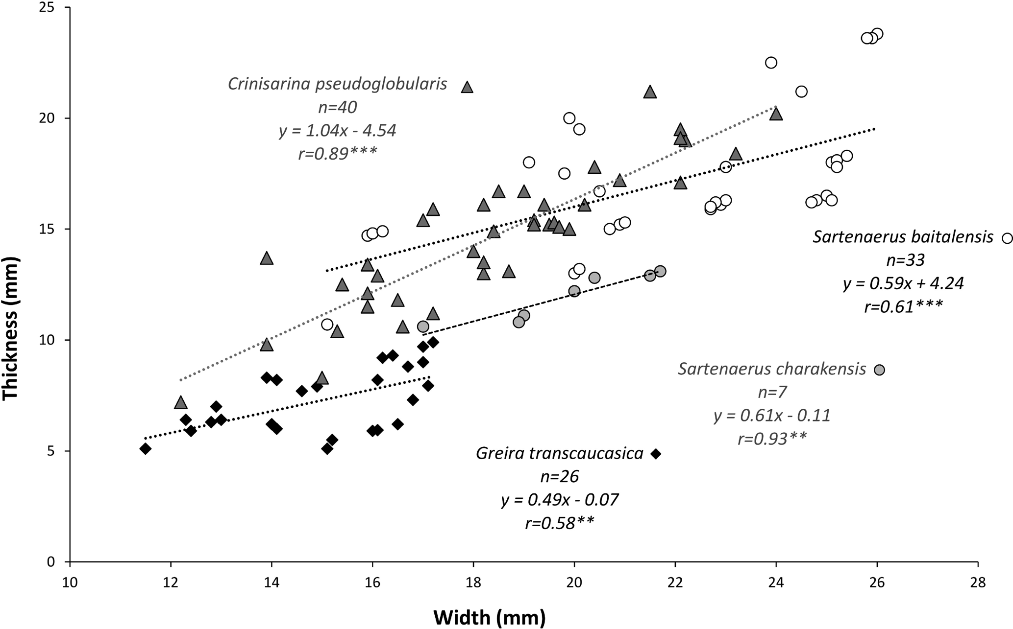
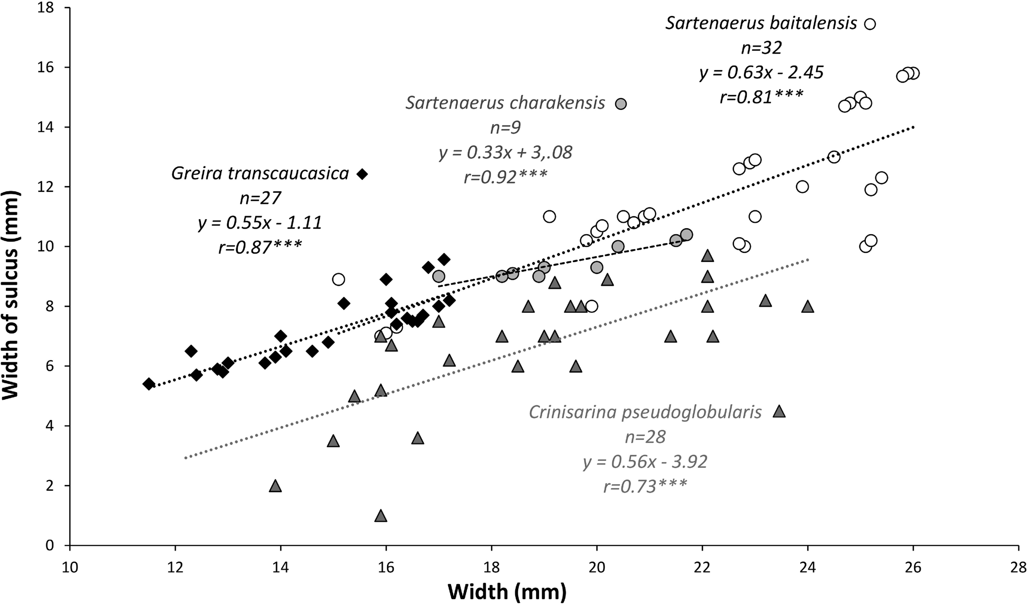
Juveniles display all external morphological characters that are typical for this species with the exception of their maximum height, which is at about mid-length of the shell, while the maximum height of adults is situated close to the front. Our collections contain juvenile specimens, which may be split into two groups based on their width/thickness ratio; some of them (Fig. 3.1–3.5) are more inflated than their relatively older representatives. Scatter plots of length versus width, thickness versus width, and width of sulcus versus width are presented in Figures 16–18. The relative proportions of Sartenaerus baitalensis represented by sufficient material remain constant throughout ontogeny (Figs. 16–18), as is reflected by the linear regression lines and the significant probability values (linear regression: y = ax + b; significant probability value: p < 0.01 whatever the degree of development of individuals). To complete the scatterplots, the measurements (in mm) of numerous individuals of S. baitalensis are also presented in Table 1.
Remarks
Sartenaer (Reference Sartenaer1970) erected the genus Centrorhynchus (= Sartenaerus Özdikmen, Reference Özdikmen2008) based on the illustrations of Camarotoechia baitalensis in Reed's (Reference Reed1922) publication, as well as his own appreciation of a few specimens of this species coming from upper (?) Famennian strata of Pamir, which were offered to him by Rzhonsnitskaya in January 1964 during his stay in St. Petersburg and now stored at the RBINS. It is pertinent to highlight here that the internal structure of the type material of Sartenaerus baitalensis was poorly known because neither Reed (Reference Reed1922) nor Sartenaer (Reference Sartenaer1970) had provided any drawings of serial sections. Our material is externally indistinguishable from Reed's species. This is also true for the internal features, with the single exception that none of our eight ground specimens displayed the connectivum that was reported by Sartenaer (Reference Sartenaer1970) for this species. However, as noted by Brice (Reference Brice1967, Reference Brice1971) for S. charakensis, this delicate structure is not always preserved. Only the serial sectioning of topotypic material of S. baitalensis will shed light on the real development of its connectivum.
Sartenaerus charakensis (Brice, Reference Brice1967)
Figures 5.1–5.10, 6
- Reference Lapparent and Le Maître1963
Camarotoechia cf. baitalensis Reed; de Lapparent and Le Maître, p. 188.
- Reference Brice1967
‘Ptychomaletoechia’ charakensis, Brice, p. 95, pl. 8, figs. 1–6; text-figs. 2, 3.
- Reference Brice and Lang1968
‘Ptychomaletoechia’ charakensis; Brice and Lang, p. 120.
- Reference Brice, Colleau and Lapparent1969
‘Ptychomaletoechia’ charakensis; Brice et al., p. 1596.
- Reference Sartenaer1970
Centrorhynchus charakensis; Sartenaer, p. 12.
- Reference Brice1971
Ptychomaletoechia (?) charakensis; Brice, p. 22, pl. 1, figs. 4a–e, 5a–d, 6a–e; text-fig. 9A.
- Reference Brice, Mistiaen and Rohart1999
Centrorhynchus charakensis; Brice et al., table 6.
- non Reference Grechishnikova, Aristov, Reitlinger and Chizhova2000
Centrorhynchus charakensis; Jafarian, p. 226, table 1; pl. 2, fig. 3a–c.
- Reference Brice and Deville2007
Centrorhynchus charakensis; Brice and Deville, p. 11.
- Reference Mirieva2010
Ptychomaletoechia charakensis; Mirieva, p. 74, table 1.
- Reference Grechishnikova and Levitskii2011
Centrorhynchus charakensis; Grechishnikova and Levitskii, p. 22, table 1.
- Reference Alekseeva, Afanasjeva, Grechishnikova, Oleneva and Pakhnevich2018a
Sartenaerus charakensis, Pakhnevich in Alekseeva et al., p. 899, pl. 6, fig. 1; text-fig. 46.
- Reference Mottequin and Brice2019
Centrorhynchus charakensis; Mottequin and Brice, table S1.
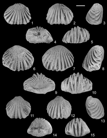
(1–10). Sartenaerus charakensis (Brice, Reference Brice1967) from the lower Famennian ‘Cyrtospirifer’ orbelianus Zone in Armenia. (1–5) IGSNASRAGM 3907/PS 3013 (Ertych section), partly exfoliated specimen in ventral, dorsal, lateral, posterior, and anterior views; (6–10) IGSNASRAGM 3908/PS 3014 (Djravank section), almost complete specimen in ventral, dorsal, lateral, posterior and anterior views. (11–15) Porthmorhynchus? sp. from the lower Famennian ‘Cyrtospirifer’ orbelianus Zone of the Noravank section, IGSNASRAGM 3909/PS 3015, almost complete specimen in ventral, dorsal, lateral, posterior, and anterior views. Scale bar = 5 mm for all.

Holotype
An articulated specimen (AF 3GK 40) from the middle-upper Famennian of Ghor Province, Afghanistan (Brice, Reference Brice1967, pl. 8, fig. 3a–e).
Occurrence
Although Brice (Reference Brice1967) considered initially that this species occurs in upper Frasnian–lower Famennian strata of Afghanistan, she later specified (Brice, Reference Brice1971) that Sartenaerus charakensis is restricted to the lower to upper Famennian interval and that it appears to be rare above the Dmitria seminoi brachiopod Zone (see Fig. 2). Mirieva (Reference Mirieva2010) recorded the presence of S. charakensis in Nakhichevan within the ‘Cyrtiopsis’ orbelianus-‘C.’ armenicus and Dmitria seminoi brachiopod zones of Rzhonsnitskaya and Mamedov (Reference Rzhonsnitskaya and Mamedov2000). Afterwards, Grechishnikova and Levitskii (Reference Grechishnikova and Levitskii2011) reported the latter species in younger deposits of Nakhichevan, more precisely in the Enchondrospirifer ghorensis-Cyrtospirifer pamiricus Zone. This study establishes for the first time the presence of this species within the lower Famennian ‘Cyrtospirifer’ orbelianus Zone of Abrahamyan (Reference Abrahamyan1957) in Armenia.
Description
Shell medium-sized (attaining 21.7 mm in width, 16.2 mm in length, and 13.1 mm in thickness) (Table 2), wider than long, sharply dorsibiconvex, transversally elliptic in outline; widest in the anterior third of the valve (slightly to the posterior of the anterior of the shell); anterior margin slightly emarginate to straight; anterior commissure strongly uniplicate.
Measurements in mm and ratios of Sartenaerus charakensis (Brice, Reference Brice1967). Abbreviations: W = width of the shell, L = length of the shell, T = thickness of the shell, Ws = width of the sulcus.

Ventral valve slightly convex in posterior and lateral profile views, with flanks sloping gently towards the lateral commissures; highest in the posterior third of the valve, then decreasing gently towards the anterior margin; umbo slightly inflated; beak and foramen unobserved; sulcus wide, moderately deep, starting at mid-length or slightly posteriorly, well delimited by bounding ribs, flat-bottomed at the front; tongue high, wide, subtrapezoidal in outline, perpendicular to the commissural plane or bent dorsally.
Dorsal valve markedly inflated with convex flanks sloping moderately to steeply towards the lateral commissures; highest more posteriorly to the front, then decreasing progressively towards the posterior margin; fold moderately high, inconspicuously originating at some distance from the umbo, round-topped at the front.
Ornamentation of coarse, angular ribs, originating at beaks and becoming fainter towards lateral cardinal extremities; 9–10 ribs on the flanks, three ribs in sulcus, 3–4 ribs on fold; ribs wider than interspaces.
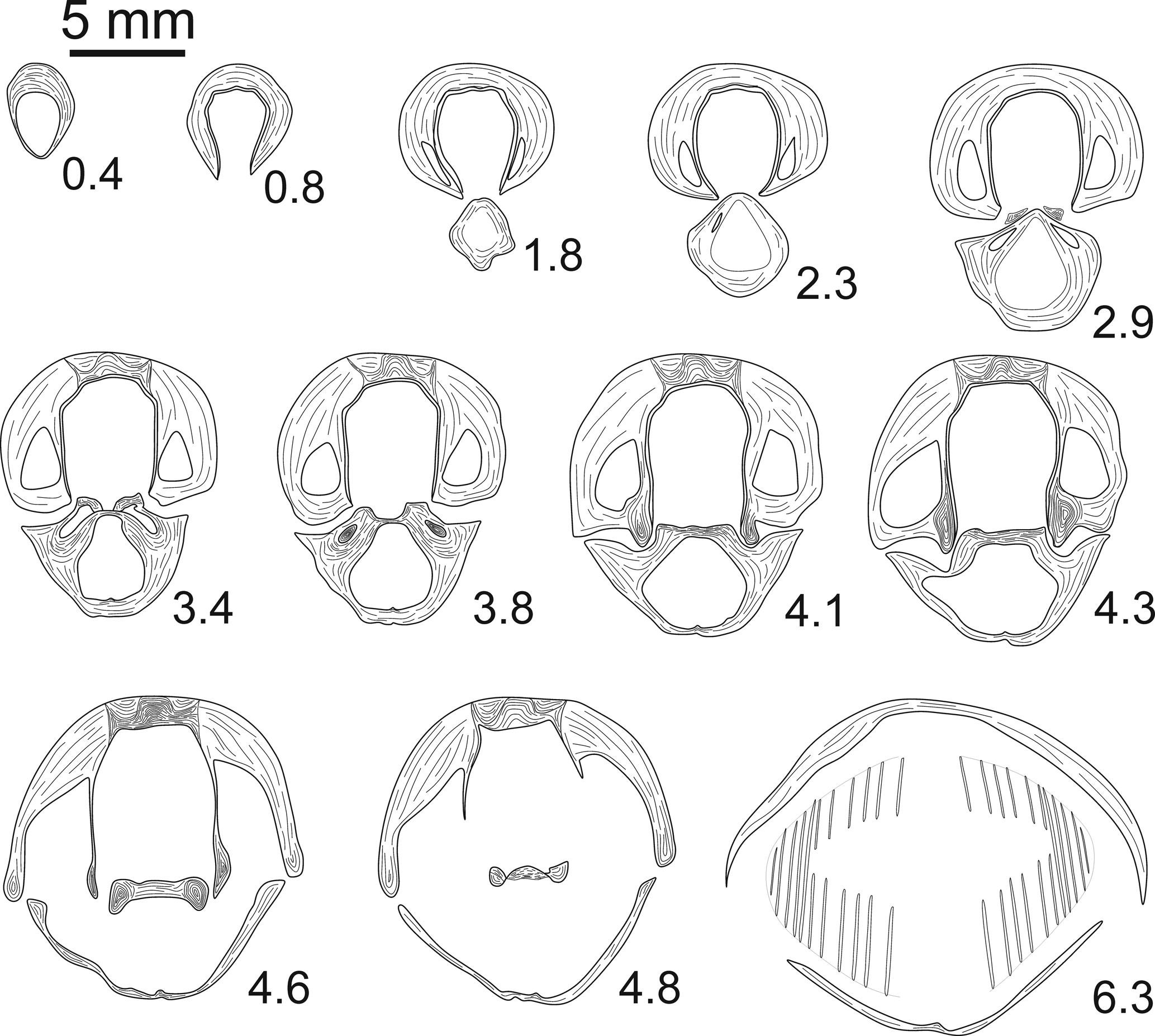
Ventral valve interior (Fig. 6) with thin, relatively long dental plates converging ventrally; teeth stout; central apical cavity wide.
Transverse serial sections of Sartenaerus charakensis (Brice, Reference Brice1967) (IGSNASRAGM 3910/PS 3016) from the lower Famennian ‘Cyrtospirifer’ orbelianus Zone in Armenia, Ertych section. Numbers refer to distances in mm from the tip of the ventral umbo.

Dorsal valve interior (Fig. 6) with long and high septum, extending up to ~20% of the valve's length; septalium narrow, relatively long, moderately deep, covered anteriorly by stout connectivum bearing a median crest; hinge plates long, connected; crural bases nearly horizontal.
Materials
Twenty-two articulated specimens and four ventral valves, eight articulated specimens and two ventral valves come from the Ertych section, five articulated specimens and two ventral valves from the Djravank section, and nine articulated specimens from the Noravank section.
Ontogeny
The size distributions of Sartenaerus charakensis during the growth represented by the scatter plots of length versus width, thickness versus width, and width of sulcus versus width show a continuous and progressive growth with no distinct stages (Figs. 16–18). The relative proportions of S. charakensis represented by more or less sufficient material remain constant (Figs. 16–18), as in S. baitalensis (linear regression: y = ax + b; significant probability value: p < 0.01). To complete the scatter plots, the measurements (in mm) of numerous individuals of S. charakensis are also presented in Table 2.
Remarks
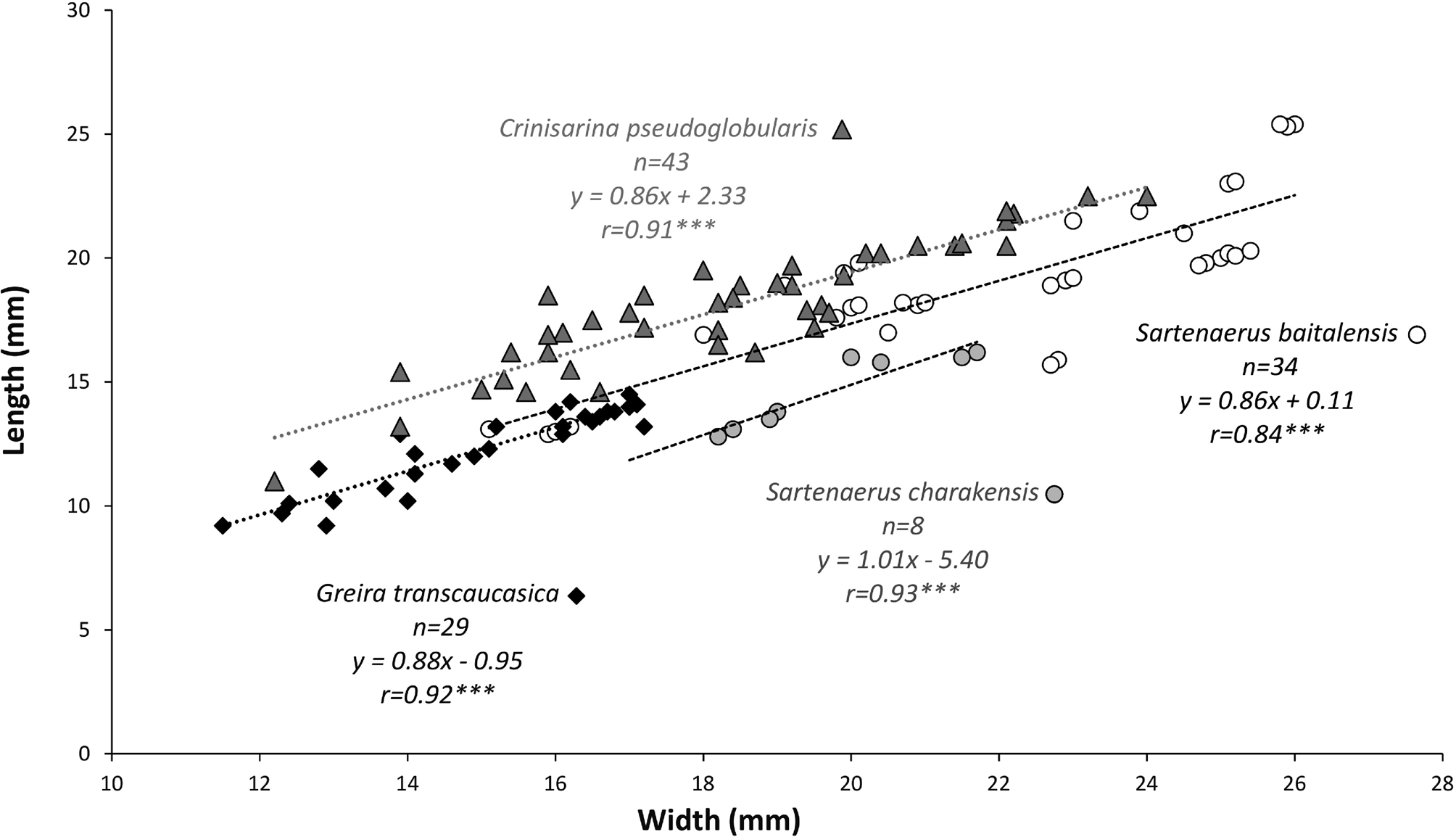
Our material differs slightly from the S. charakensis from Afghanistan (Brice, Reference Brice1967, Reference Brice1971) by its Y-shaped, moderately deep and thin septalium. Jafarian (Reference Jafarian2000) reported S. charakensis from the upper Famennian of Iran, but his illustrated specimens appear to be closer to Sinotectirostrum delicatacostatum (Abrahamyan, Reference Abrahamyan1957) rather than Brice's species because they clearly have a different shell shape, strongly emarginate anterior commissure, and markedly inflated ventral umbo. Moreover, the size distributions between these two studied species (S. charakensis and S. baitalenis) have been compared and tested. For the width/length ratios (Fig. 16), if equal adjusted means (between-groups effect) in the two studied species can be rejected at p < 0.05 (p = 0.00069***), equality of slopes cannot be rejected (p = 0.7493). Thus, if Sartenaerus charakensis and Sartenaerus baitalensis show similar slope for their width/length ratios, it is noteworthy that Sartenaerus baitalensis is longer relative to its width than Sartenaerus charakensis. Similarly, for their width/thickness ratios (Fig. 17), if equal adjusted means in Sartenaerus baitalensis and S. charakensis can be rejected at p < 0.05 (p = 0.00028***), equality of slopes cannot be rejected (p = 0.9728). Thus, Sartenaerus baitalensis is thicker relative to its width than S. charakensis.
Genus Porthmorhynchus Sartenaer, Reference Sartenaer2001
Type species
Rhynchonella ferquensis Gosselet, Reference Gosselet1887.
Porthmorhynchus? sp.
Figures 5.11–5.15, 7
Occurrence
The specimens were collected from the lower Famennian orbelianus Zone of Abrahamyan (Reference Abrahamyan1957) at the Noravank and Djravank sections (Fig. 1).
Description
Shell small-sized (~14.9 mm in width, 13.4 mm in length, and 10 mm in thickness), dorsibiconvex, widest and highest more anteriorly to mid-length, rounded subpentagonal in outline; anterior margin slightly rounded; anterior commissure uniplicate.
Ventral valve slightly convex in posterior and lateral profile views, with flanks sloping gently towards the lateral commissures; highest in the posterior third of the valve; umbo inflated, prominent; beak and foramen unobserved (due to poor preservation); sulcus relatively wide, shallow to moderately deep, originating at about mid-length, flat-bottomed at the front; tongue moderately high, wide, subtrapezoidal in outline, perpendicular to commissural plane or slightly bent dorsally.
Dorsal valve inflated, strongly curved in posterior and lateral profile views, with convex flanks sloping steeply towards the lateral commissures; highest slightly posteriorly to the front, then progressively decreasing towards the posterior margin; fold starting at some distance from the umbo, low, wide, well defined, becoming relatively higher anteriorly.
Ornamentation of coarse, angular, low to moderately high ribs, radiating at beaks, becoming fainter towards the lateral cardinal extremities; ribs in sulcus and on fold slightly wider than those present on the flanks; two ribs in sulcus, three ribs on fold, 6–7 ribs on flanks, parietal ribs unobserved; ribs wider than interspaces.
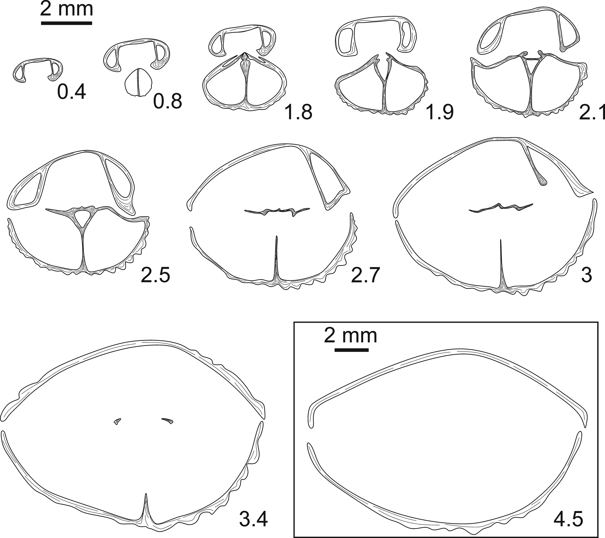
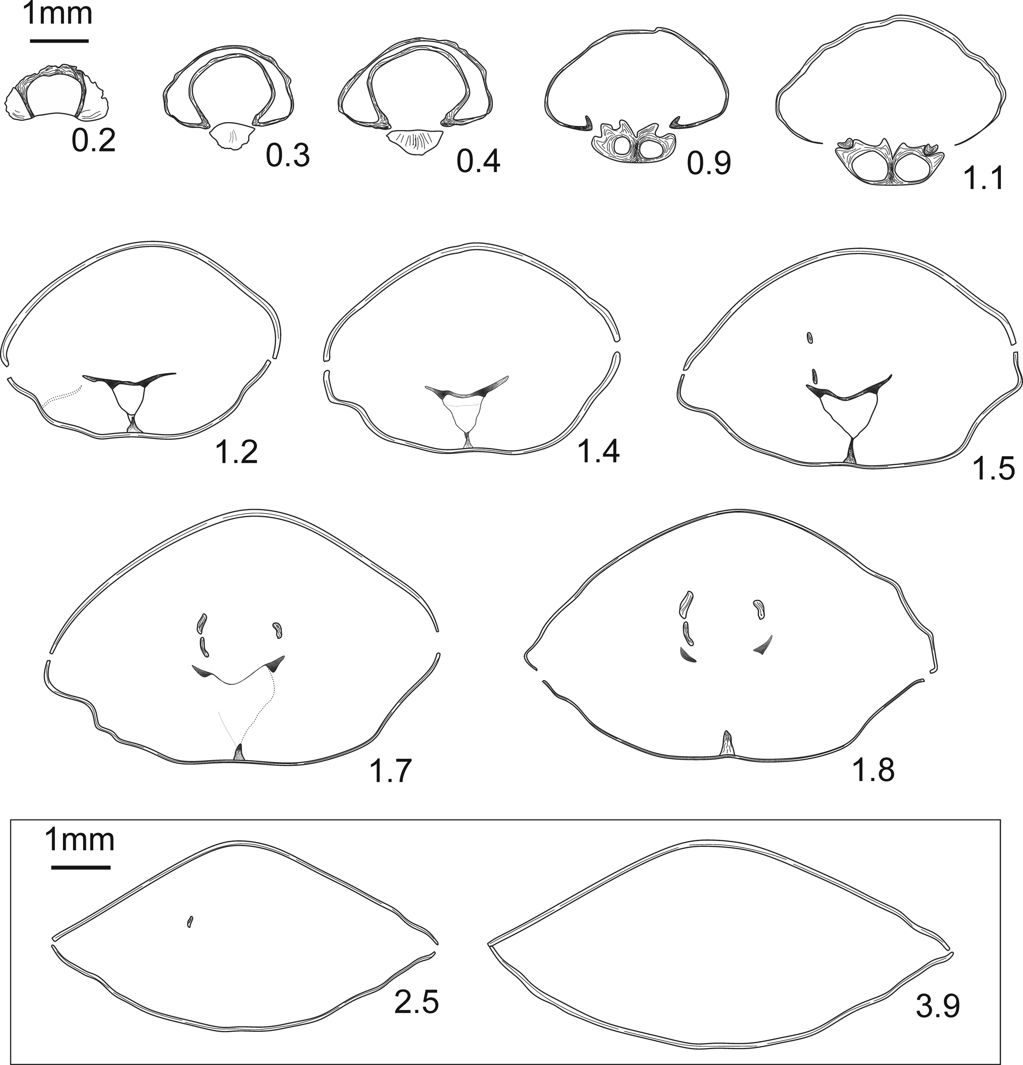
Ventral valve interior (Fig. 7) with relatively long and robust dental plates converging ventrally; teeth massive; central apical cavity wide; lateral apical cavities poorly infilled by callosities.
Transverse serial sections of Porthmorhynchus? sp. (IGSNASRAGM 3911/PS 3017) from the lower Famennian ‘Cyrtospirifer’ orbelianus Zone in Armenia, Noravank section. Numbers refer to distances in mm from the tip of the ventral umbo.

Dorsal valve interior with relatively high and long septum (Fig. 7, serial sections 1–2.7); septalium wide, relatively deep; connectivum not observed; hinge plates divided; dental sockets large; crural bases stout.
Materials
Two partly exfoliated articulated specimens.
Remarks
Porthmorhynchus Sartenaer is mainly known from middle-upper Frasnian strata of the Boulonnais region in Northern France (Brice in Brice and Meats, Reference Brice and Meats1972; Sartenaer, Reference Sartenaer2001). Our material is closely related to the genus Porthmorhynchus based on its rounded subpentagonal outline, dorsibiconvex profile, moderately high and subtrapezoidal tongue, massive teeth, and the development of septalium. However, it is doubtfully assigned to this genus because the development of its fold and sulcus (originating far from the beaks) and the orientation of its dental plates (converging ventrally) are in contradiction with the diagnosis of the genus. Furthermore, it is worth noting that Sartenaer (Reference Sartenaer2001) mentioned that the septalium of Porthmorhynchus is covered by a connectivum, which was not observed in our ground specimen. However, it should be stressed that this delicate structure is not always preserved. Additional material is needed to reach a more confident identification.
Subfamily Ripidiorhynchinae Savage, Reference Savage, Copper and Jin1996
Genus Gesoriacorostrum Sartenaer, Reference Sartenaer2003
Type species
Atrypa boloniensis d’Orbigny, Reference d'Orbigny1850.
Occurrence
The specimens were collected from the lower Famennian orbelianus Zone of Abrahamyan (Reference Abrahamyan1957) from the Djravank section (Fig. 1).
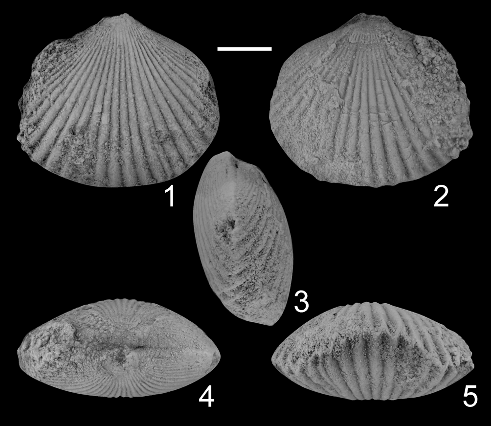
Gesoriacorostrum? sp. from the lower Famennian ‘Cyrtospirifer’ orbelianus Zone in Armenia, Djravank section. (1–5) IGSNASRAGM 3912/PS 3018, partly exfoliated specimen in ventral, dorsal, lateral, posterior, and anterior views. Scale bar = 5 mm for all.

Transverse serial sections of Gesoriacorostrum? sp. (IGSNASRAGM 3913/PS 3019) from the lower Famennian ‘Cyrtospirifer’ orbelianus Zone in Armenia, Djravank section. Numbers refer to distances in mm from the tip of the ventral umbo.

Description
Shell small-sized (~19 mm in width, 16.5 mm in length, 8.3 mm in thickness), dorsibiconvex, wider than long, widest and highest at about mid-length, transversally elliptic in outline; anterior margin slightly rounded; anterior commissure uniplicate.
Ventral valve slightly convex in posterior and lateral profile views, with flanks sloping gently towards lateral commissures; umbo small; beak and foramen unobserved (due to poor preservation); sulcus only perceptible close to anterior margin, very shallow, wide, round-bottomed at front; tongue low, wide, subtrapezoidal in outline, not perpendicular to commissural plane.
Dorsal valve highest at mid-length or slightly anteriorly to it, then decreasing progressively towards anterior margin; fold close to anterior margin, very low, wide, round-topped at front.
Ornamentation of up to 13 low, flattened, simple ribs on each flank originating at beaks and becoming progressively fainter towards posterolateral margins; in sulcus and on fold, 7–8 ribs, infrequently increasing by bifurcation; ribs wider than interspaces on the entire shell.
Ventral valve interior (Fig. 9) with thin, long, and subparallel dental plates; teeth strong; central apical cavity wide; lateral apical cavities poorly infilled by callosities.
Dorsal valve interior with relatively thick and low septum (Fig. 9, serial sections 1.9–2.1); septalium wide, relatively deep, covered anteriorly by stout connectivum bearing a median crest; dental sockets large.
Materials
Two partly exfoliated articulated specimens.
Remarks
The external and internal features observed (e.g., transversally elliptic outline, dorsibiconvex profile, weakly developed fold and sulcus, long and subparallel dental plates, relatively thick and low septum, relatively deep septalium, and connectivum bearing a median crest) in the two studied specimens indicate affinities to the genus Gesoriacorostrum Sartenaer, Reference Sartenaer2003, the presence of which in the SAB was recently documented by Pakhnevich in Alekseeva et al. (Reference Alekseeva, Afanasjeva, Grechishnikova, Oleneva and Pakhnevich2018a). However, the fold and sulcus are perceptible only anteriorly on our specimens, while the original material from the Boulonnais region in France displays fold and sulcus that start from mid-length. Furthermore parietal ribs are absent on our specimens.
Superfamily Rhynchoporoidea Muir-Wood, Reference Muir-Wood1955
Family Rhynchoporidae Muir-Wood, Reference Muir-Wood1955
Subfamily Greirinae Erlanger, Reference Erlanger1993
Genus Greira Erlanger, Reference Erlanger1993
Type species
Greira transcaucasica Erlanger, Reference Erlanger1993.
Greira transcaucasica Erlanger, Reference Erlanger1993
Figures 10, 11
- Reference Erlanger1993
Greira transcaucasica Erlanger, p. 120, text-figs. 1, 2.
- Reference Savage and Kaesler2002
Greira transcaucasica; Savage, p. 1233, fig. 840, 2a–o (copy of Erlanger, Reference Erlanger1993, fig. 1, 2a–d; fig. 2).
- Reference Alekseeva, Afanasjeva, Grechishnikova, Oleneva and Pakhnevich2018a
Greira transcaucasica; Pakhnevich in Alekseeva et al., p. 855, pl. 8, fig. 4; text-fig. 58.
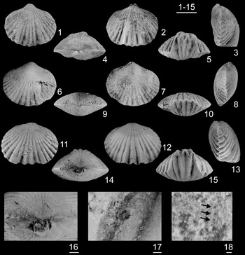
Greira transcaucasica Erlanger, Reference Erlanger1993 from the lower Famennian ‘Cyrtospirifer’ orbelianus Zone in Armenia. (1–5) IGSNASRAGM 3914/PS 3020 (Ertych section), almost complete specimen in ventral, dorsal, lateral, posterior, and anterior views; (6–10, 18) IGSNASRAGM 3915/PS 3021 (Ertych section), partly corroded specimen in ventral, dorsal, lateral, posterior, and anterior views, and close-up (18) of punctae (indicated by black arrows) observed in the sulcus (in interspace), near the anterior margin; (11–17) IGSNASRAGM 3916/PS 3022 (Djravank section), almost complete specimen in ventral, dorsal, lateral, posterior, and anterior views (11–15); close-up of the ventral umbo (thin dental plates are exposed because the ventral beak is broken (16); and close-up of a drill hole, perhaps of predatory origin, in the sulcus (17). Scale bars = 5 mm (1–15), 1 mm (16), 200 μm (17), 100 μm (18).

Transverse serial sections of Greira transcaucasica Erlanger, Reference Erlanger1993 (IGSNASRAGM 3917/PS 3023) from the lower Famennian ‘Cyrtospirifer’ orbelianus Zone in Armenia, Djravank section. Numbers refer to distances in mm from the tip of the ventral umbo.

Holotype
An articulated specimen (PIN 4127/1042) from the lower Famennian ‘Mesoplica’ meisteri Zone of Grechishnikova et al. (Reference Grechishnikova, Aristov, Reitlinger and Chizhova1982), left bank of the Arpa River, between Geran-Kalasy and Kabakhal mountains, Nakhichevan (Erlanger, Reference Erlanger1993, text-fig. 1a–d).
Occurrence
This species was considered by Erlanger (Reference Erlanger1993) as one of the oldest punctate rhynchonellides, which are particularly abundant and diverse with four species and subspecies (Abrahamyan, Reference Abrahamyan1954, Reference Abrahamyan1957; Sartenaer and Plodowski, Reference Sartenaer and Plodowski2003), known from the middle–upper Famennian succession of the SAB. Initially G. transcaucasica was described by Erlanger (Reference Erlanger1993) from the lower Famennian ‘Mesoplica’ meisteri Zone of Grechishnikova et al. (Reference Grechishnikova, Aristov, Reitlinger and Chizhova1982), equivalent to the ‘Mesoplica’ meisteri-Cyrtospirifer asiaticus Zone of Rzhonsnitskaya and Mamedov (Reference Rzhonsnitskaya and Mamedov2000) in Nakhichevan (Paknevich in Aleksseva et al., Reference Alekseeva, Afanasjeva, Grechishnikova, Oleneva and Pakhnevich2018a). This is the first time that this species is recorded within the Armenian sections, where it occurs in slightly younger strata, in the lower Famennian ‘Cyrtospirifer’ orbelianus Zone of Abrahamyan (Reference Abrahamyan1957).
Description
Shell medium-sized (reaching 17.1 mm in width, 14.5 mm in length, and 9.9 mm in thickness) (Table 3), equibiconvex to dorsibiconvex, wider than long, widest at about mid-length, transversally elliptic in outline; anterior margin slightly emarginate to straight; anterior commissure uniplicate and serrate; shell punctate (Fig. 10.18).
Measurements in mm and ratios of Greira transcaucasica Erlanger, Reference Erlanger1993. Abbreviations: W = width of the shell, L = length of the shell, T = thickness of the shell, Ws = width of the sulcus.

Ventral valve convex in posterior and lateral profile views, with flanks sloping gently towards the lateral commissures; umbo small but prominent; beak small, inclined to straight; foramen unobserved; sulcus wide, shallow to moderately deep, inconspicuously originating from mid-length or more anteriorly, flat- to round-bottomed at front; tongue variable in its height, low to moderately high, not perpendicular to the commissural plane, subtrapezoidal in outline.
Dorsal valve slightly inflated with flanks sloping gently to moderately towards the lateral commissures; fold wide, generally low, inconspicuously originating from mid-length or more anteriorly, becoming more or less prominent at the anterior margin, flat-topped at the front.
Ornamentation of low, angular ribs, running from beak, becoming weaker towards the posterolateral margins; 9–13 ribs on each flank, 4–5 ribs in sulcus, 4–6 ribs on fold; 5–6 ribs per 5 mm at the anterior margin near the sulcus and fold; ribs in sulcus and on fold somewhat wider than those present on the flanks; ribs wider than interspaces.
Ventral valve interior (Fig. 11) with thin, long, short, and divergent dental plates; lateral apical cavities poorly infilled by callus; teeth small, rounded in transverse section.
Dorsal valve interior (Fig. 11) with wide and Y-shaped septalium, covered anteriorly; sockets smooth; dorsal median septum low but long, extending one-quarter of valve's length, crural bases proximally triangle-shaped in section, dorsally convergent.
Materials
Twenty-six articulated specimens and two ventral valves, 14 articulated specimens and two ventral valves come from the Ertych section and 12 articulated specimens from the Noravank section.
Ontogeny
The size distributions of Greira transcaucasica during growth, as represented by the scatter plots of length versus width, thickness versus width and width of sulcus versus width, show a continuous and progressive growth with no distinct grouping (Figs. 16–18). The relative proportions of G. transcaucasica represented by sufficient material remain constant (linear regression: y = ax + b; significant probability value: p < 0.01, whatever the degree of development of individuals (Figs. 16–18). Moreover, the correlation is positive with width varying proportionally with length, thickness, and width of sulcus. To complete the scatter plots, the measurements (in mm) of numerous individuals of G. transcaucasica are also presented in Table 3.
Remarks
Our material slightly differs from the holotype of Greira transcaucasica; according to Erlanger (Reference Erlanger1993) the interspaces of this species are wider than ribs, but the specimens from Armenia display ribs that are wider than the interspaces. Our observation is consistent with the description of Pakhnevich in Alekseeva et al. (Reference Alekseeva, Afanasjeva, Grechishnikova, Oleneva and Pakhnevich2018a) for this species found in the lower Famennian of Nakhichevan. Thus, these differences in the width of ribs and interspaces are considered as expression of the intraspecific variability.
The size distributions of two previously described species assigned to the genus Sartenaerus have been compared with Greira transcaucasica (Figs. 16–18). For the width/length ratios (Fig. 16), if equal adjusted means in Greira transcaucasica, Sartenaerus baitalensis, and S. charakensis can be rejected at p < 0.05 (p = 9.521 x 10⁻⁵***), equality of slopes cannot be rejected (p = 0.9201). It is interesting to underline that Sartenaerus baitalensis and Greira transcaucasica show similar length/width ratios with Sartenaerus baitalensis being represented by rather larger individuals, while Greira transcaucasica is represented by smaller individuals. For the width/thickness ratios (Fig. 17), if equal adjusted means can be rejected at p < 0.05 (p = 2.201 x 10–09***), equality of slopes cannot be rejected (p = 0.9216). Thus, Sartenaerus baitalensis is thicker relative to its width than Greira transcaucasica. Finally, for the width/width of sulcus ratios (Fig. 18), equal adjusted means in the three studied taxa and equality of slopes cannot be rejected (p = 0.5363 and p = 0.4579, respectively). As for the width/length ratios, it is interesting to underline that Sartenaerus baitalensis and Greira transcaucasica show similar width/width of sulcus ratios with Sartenaerus baitalensis being represented by rather larger individuals, while Greira transcaucasica is represented by smaller individuals. Interestingly, Sartenaerus charakensis seems to be represented as an intermediate form with rather similar width/width of sulcus ratios.
Genus Sharovaella Pakhnevich, Reference Pakhnevich2012
Type species
Sharovaella mirabilis Pakhnevich, Reference Pakhnevich2012.
Sharovaella? sp.
Figures 12, 13
Occurrence
The specimens were collected from the lower Famennian orbelianus Zone of Abrahamyan (Reference Abrahamyan1957) at the Ertych section (Fig. 1).
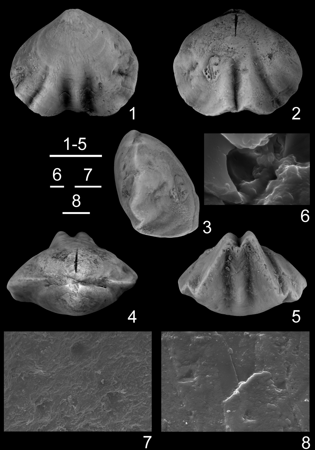
Sharovaella? sp. from the lower Famennian ‘Cyrtospirifer’ orbelianus Zone of Armenia, Ertych section. (1–8) IGSNASRAGM 3918/PS 3024, almost complete specimen in ventral, dorsal, lateral, posterior, and anterior views (1–5), and close-up of shell surface showing punctae on ventral valve, near umbo (6–8). Scale bars = 5 mm (1–5), 100 μm (8), 10 μm (7), 2 μm (6).

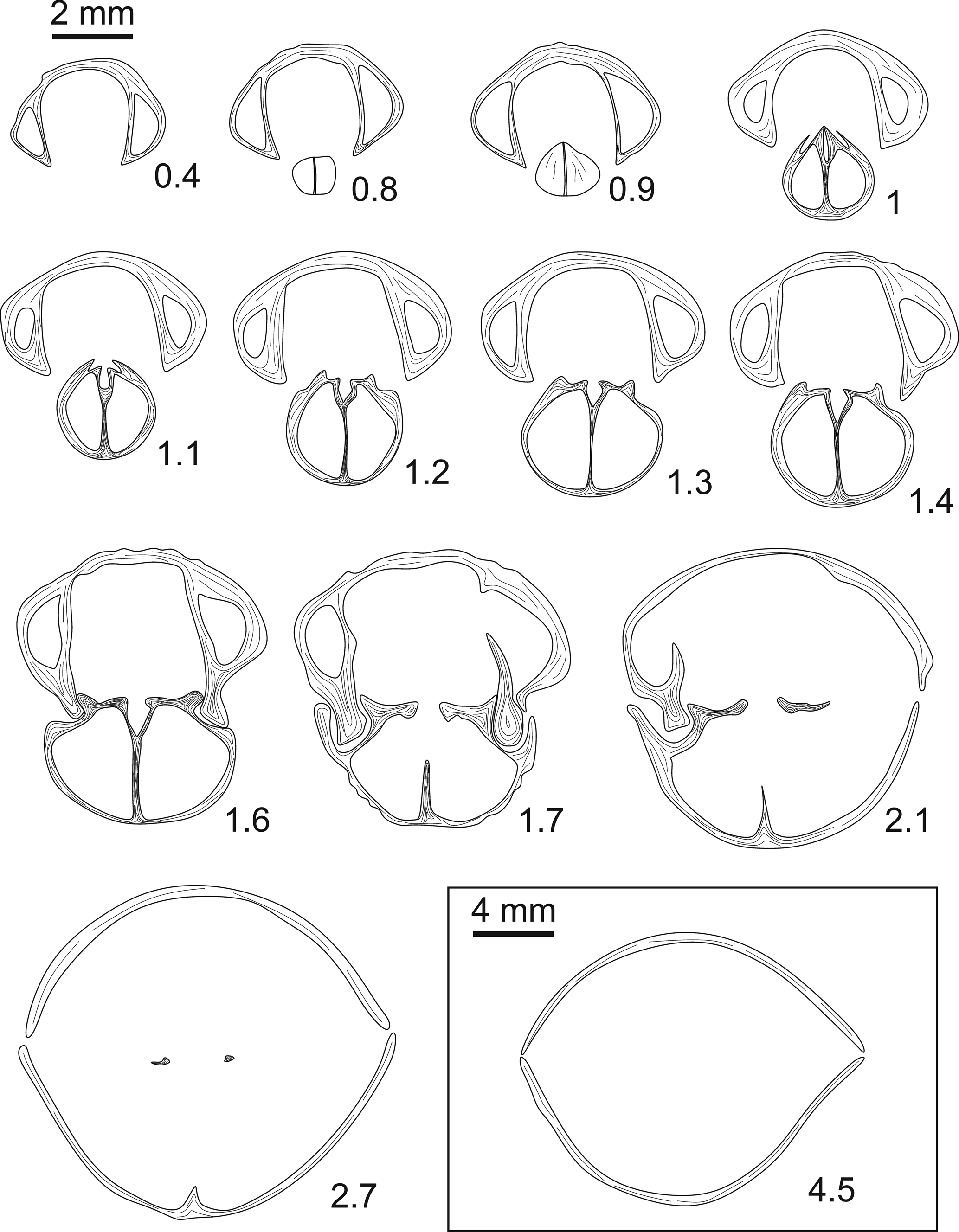
Transverse serial sections of Sharovaella? sp. (IGSNASRAGM 3919/PS 3025) from the lower Famennian ‘Cyrtospirifer’ orbelianus Zone of Armenia, Ertych section. Numbers refer to distances in mm from the tip of the ventral umbo.

Description
Shell small-sized (12.7 mm in width, 11 mm in length, and 8.4 mm in thickness), wider than long, strongly dorsibiconvex, rounded subpentagonal in outline, widest at mid-length, highest at mid-length or slightly anteriorly; anterior margin slightly emarginate; anterior commissure uniplicate.
Ventral valve gently inflated in posterior and lateral profile views, with slightly convex flanks sloping gently towards the lateral commissures; highest at mid-length, then progressively decreasing towards the anterior margin; umbo prominent, somewhat curved; beak small, straight to inclined; foramen unobserved; sulcus well defined by bounding ribs, wide, deep, originating from mid-length or slightly posteriorly, flat- to round-bottomed at the front; tongue wider than high, perpendicular to the commissural plane or slightly bent dorsally, high, subtrapezoidal in outline.
Dorsal valve strongly inflated with markedly convex flanks sloping moderately to steeply towards the lateral commissures; highest at mid-length or more anteriorly to it, then decreasing progressively towards the posterior margin; umbo flattened, not prominent; beak small, overhanging the hinge line; fold high, well delimited, wide, originating at mid-length or slightly posteriorly, then becoming higher anteriorly, round-topped at front.
Ornamentation of 3–4 coarse, strong ribs on flanks originating at mid-length by intercalations, becoming shorter and weaker towards the posterolateral margins; in sulcus and on fold, 1–2 ribs originating near the umbo or more anteriorly to it; ribs wider than interspaces, having rounded apices and being triangular in cross section; growth lamellae densely crowded, relatively prominent (2–3 lamellae per 1 mm) and penetrated by irregularly arranged punctae situated near the umbo on ventral valve. Ventral valve interior (Fig. 13) with thin intrasinal and slightly divergent dental plates, central and lateral apical cavities poorly filled in by callosities.
Dorsal valve interior with low but long septum (Figs. 12, serial sections 2, 12; 4; 13, serial section 1.15); septalium covered and narrow; hinge plates short; crural bases stout.
Materials
Three articulated specimens.
Remarks
Pakhnevich (Reference Pakhnevich2012) examined several specimens collected by Grechishnikova from different sections of Armenia and Nakhichevan that were identified as Eoparaphorhynchus triaequalis (Gosselet, Reference Gosselet1877). He noticed that this material differs from Gosselet's species by the presence of punctae on the shell walls. Consequently, he erected the new monospecific genus Sharovaella with its type species Sharovaella mirabilis. Although the external and internal morphology of our material is a reminiscent of S. mirabilis in terms of size, general shape, well-developed fold and sulcus, short dental plates, covered, narrow septalium, and low septum, this limited material is tentatively assigned to Sharovaella because it lacks strong ribs running from the umbo on flanks and in sulcus and on fold, contrary to what it is observed in Pakhnevich's material. Moreover, Pakhnevich observed irregularly spaced punctae along the whole shell, whereas in our material puncta-like structures are displayed only near the umbo (Fig. 12, serial sections 6–8). These structures may be an artefact of preservation, reflecting dissolution caused by meteoric water.
Order Athyrida Boucot, Johnson, and Staton, Reference Boucot, Johnson and Staton1964
Suborder Athyridina Boucot, Johnson, and Staton, Reference Boucot, Johnson and Staton1964
Superfamily Athyroidea Davidson, Reference Davidson1881
Family Athyridae Davidson, Reference Davidson1881
Subfamily Cleiothyridininae Alvarez, Rong, and Boucot, Reference Alvarez, Rong and Boucot1998
Genus Crinisarina Cooper and Dutro, Reference Cooper and Dutro1982
Type species
Crinisarina stainbrooki Mottequin, Reference Mottequin2008 (pro Cleiothyridina reticulata Stainbrook, Reference Stainbrook1947).
Crinisarina pseudoglobularis new species
Figures 14, 15
- Reference Arakelyan1952
Athyris globularis; Arakelyan, p. 32.
- Reference Abrahamyan1957
Athyris globularis; Abrahamyan, p. 9, table 2.
- Reference Abrahamyan, Mkrtchian, Vardaniants, Gabrielian, Maghakian and Paffenholz1964
Athyris globularis; Arakelyan, p. 75.
- Reference Abrahamyan, Arakelyan, Azizbekov, Rzhonsnitskaya, Nalivkin, Rzhonsnitskaya and Markovski1973
Athyris globularis; Abrahamyan et al., p. 218.
- Reference Akopian and Akopian1974
Athyris globularis; Akopian, p. 9.
- Reference Rzhonsnitskaya and Mamedov2000
Athyris ex gr. globularis; Rzhonsnitskaya and Mamedov, p. 330.
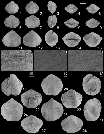
Crinisarina pseudoglobularis n. sp. from the lower Famennian ‘Cyrtospirifer’ orbelianus Zone in Armenia. (1–5) IGSNASRAGM 3920/PS 3026 (Ertych section), almost complete juvenile specimen in ventral, dorsal, lateral, posterior, and anterior views; (6–10) IGSNASRAGM 3921/PS 3027 (Noravank), partly exfoliated juvenile specimen in ventral, dorsal, lateral, posterior, and anterior views.; (11–18) IGSNASRAGM 3922/PS 3028 (Djravank section), almost complete specimen in ventral, dorsal, lateral, posterior, and anterior views (11–15); close-up of the ornamentation (radially aligned solid spines) on ventral valve near the anterior margin (16–18); (19–23) IGSNASRAGM 3923/PS 3029 (Noravank section), partly exfoliated specimen in ventral, dorsal, lateral, posterior, and anterior views.; (24–28) IGSNASRAGM 3924/PS 3030 (Ertych section), partly exfoliated specimen in ventral, dorsal, lateral, posterior, and anterior views. Scale bars = 5 mm (1–15, 19–28), 500 μm (16), 100 μm (17), 40 μm (18).

Transverse serial sections of Crinisarina pseudoglobularis n. sp. (IGSNASRAGM 3925/PS 3031) from the lower Famennian ‘Cyrtospirifer’ orbelianus Zone of Armenia, Noravank section. Numbers refer to distances in mm from the tip of the ventral umbo.

Holotype
An almost complete, articulated specimen (IGSNASRAGM 3918/PS 3024; Fig. 14.24–14.28) from the lower Famennian orbelianus Zone of Abrahamyan (Reference Abrahamyan1957) Ertych section, which is correlated with the Ertych horizon (Fig. 2); Ertych section (6 km southeast from Areni, Vayots Dzor Province, central Armenia).
Diagnosis
Shell medium-sized (up to 24 mm in width, 22.5 mm in length, 21.2 mm in thickness), equibiconvex, relatively equidimensional, rounded pentagonal in outline; anterior margin straight to poorly emarginate; anterior commissure paraplicate; fold and sulcus very shallow, wide at the front, more developed anteriorly; tongue moderately high to high, subtrapezoidal to semioval in outline; micro-ornamentation of imbricate, densely crowded growth lines projecting anteriorly and anterolaterally as flat, solid, spine-like outgrowths; dental plates thin, relatively long and arched supporting cyrtomatodont teeth, lateral apical cavities strongly filled in by callosities; dorsal interior with cardinal plate pierced apically by a dorsal foramen; inner hinge plates conjunct, weakly concave ventrally to nearly flat, depending on specimens; outer hinge plates flat; myophragm very short.
Occurrence
This species is only found in the SAB. More particularly, according to Abrahamyan (Reference Abrahamyan1957), it is restricted to the lower Famennian ‘Cyrtospirifer’ orbelianus Zone of Abrahamyan (Reference Abrahamyan1957) and according to Rzhonsnitskaya and Mamedov (Reference Rzhonsnitskaya and Mamedov2000) in the ‘Cyrtiopsis’ orbelianus-‘Cyrtiopsis’ armenicus Zone of Nakhichevan.
Description
Shell medium-sized (Table 4), relatively equidimensional, rounded subpentagonal in outline, equibiconvex, widest at about mid-length or slightly posteriorly; highest in the posterior third of the valve; anterior margin straight to poorly emarginate; anterior commissure paraplicate.
Measurements in mm and ratios of Crinisarina pseudoglobularis n. sp. Abbreviations: W = width of the shell, L = length of the shell, T = thickness of the shell, Ws = width of the sulcus.

Ventral valve inflated with flanks sloping moderately to strongly towards the lateral commissures; highest in the posterior third of the valve, then decreasing progressively towards the anterior margin; umbo strongly inflated, prominent; beak suberect to erect, pierced by a permesothyrid foramen; sulcus poorly defined posteriorly, more defined anteriorly, wide at the front, very shallow, flat- to round-bottomed at the front, subtrapezoidal to semioval in outline, perpendicular to the commissural plane, with distal part sometimes bent dorsally.
Dorsal valve with upper surface dome-shaped (median part sometimes raised by the fold), and strongly curved in posterior and lateral profile views with flanks sloping moderately towards the lateral commissures; highest in the anterior third of the valve (near umbo in juveniles) then progressively decreasing towards the anterior margin; umbo prominent; fold low, inconspicuously originating close to umbo, wide at the front, poorly defined, round-topped at the front.
Ornamentation of imbricate, densely crowded growth lines projecting anteriorly and anterolaterally as flat, solid spine-like outgrowths (Fig. 14, serial sections 16–18). Growth lines less densely crowded posteriorly.
Ventral valve interior with thin, relatively long, and arched dental plates, supporting cyrtomatodont teeth that are subrectangular in transverse section (Fig. 15); thick lens of prismatic calcite is developed in the middle part of the valve below the well-developed umbonal cavity; lateral apical cavities moderately filled in by callosities.
Dorsal valve interior (Fig. 15) with conjunct, weakly concave ventrally to nearly flat inner hinge plates; outer hinge plates flat; myophragm low but long; spiral cones with at least 11 whorls laterally oriented.
Etymology
In reference to the confusion made with the athyride species Composita globularis (Phillips, Reference Phillips1836).
Materials
Forty-three articulated specimens and five ventral valves, 15 articulated specimens and three ventral valves come from the Ertych section, 16 articulated specimens and two ventral valves from the Djravank section, and 12 articulated specimens from the Noravank section.
Ontogeny
Juveniles display a rectimarginate to slightly paraplicate anterior commissure, semi-elliptic tongue, and sharp lateral commissures. Although not visible on juveniles, sulcus and fold are more prominent in older shells. Beak inclination angle does not change with age because juveniles also have a suberect to erect beak. The maximum height of the dorsal valve is near the umbo in juveniles, then moves towards the anterior margin. Shell growth follows a linear model, which is especially reflected by the rather high coefficient of correlation obtained for the ratios of length versus width, thickness versus width, and width of sulcus versus width (Figs. 16–18). The relative proportions of Crinisarina pseudoglobularis n. sp. represented by sufficient material remain constant (linear regression: y = ax + b; significant probability value: p < 0.01, whatever the degree of development of individuals). Moreover, the correlation is positive with width varying proportionally with length, thickness, and width of sulcus. To complete the scatter plots, the measurements (in mm) of numerous individuals of Crinisarina pseudoglobularis n. sp. are also presented in Table 3.
Scatter diagrams exhibiting the relation between shell width and length (in mm) of Sartenaerus baitalensis (Reed, Reference Reed1922), Sartenaerus charakensis (Brice, Reference Brice1967), Greira transcaucasica Erlanger, Reference Erlanger1993, and Crinisarina pseudoglobularis n. sp. Abbreviations: n, number of specimens measured; y = ax + b, linear model; r, coefficient of correlation.

Scatter diagrams exhibiting the relation between shell width and thickness (in mm) of Sartenaerus baitalensis (Reed, Reference Reed1922), Sartenaerus charakensis (Brice, Reference Brice1967), Greira transcaucasica Erlanger, Reference Erlanger1993, and Crinisarina pseudoglobularis n. sp. Abbreviations: n, number of specimens measured; y = ax + b, linear model; r, coefficient of correlation.

Scatter diagram exhibiting the relation between shell width (in mm) and width of sulcus (in mm) of Sartenaerus baitalensis (Reed, Reference Reed1922), Sartenaerus charakensis (Brice, Reference Brice1967), Greira transcaucasica Erlanger, Reference Erlanger1993, and Crinisarina pseudoglobularis n. sp. Abbreviations: n, number of specimens measured; y = ax + b, linear model; r, coefficient of correlation.

Remarks
Reported initially by Arakelyan (Reference Arakelyan1952) as Athyris globularis, this species was found both in Armenia and Nakhichevan, but it was never described or illustrated. It was considered by Abrahamyan et al. (Reference Abrahamyan, Arakelyan, Azizbekov, Rzhonsnitskaya, Nalivkin, Rzhonsnitskaya and Markovski1973) and Arakelyan et al. (Reference Arakelyan, Malxasyan, Mkrtchyan and Paffenholz1975) as a biostratigraphically important species for the lower Famennian orbelianus Zone. Moreover, until now it was confused with Spirifera globularis Phillips, Reference Phillips1836, which is a poorly known species from the Viséan of Bolland (Yorkshire, UK) that currently is assigned to the genus Composita (see Brunton and Tilsley, Reference Brunton and Tilsley1991). This species is assigned herein to Crinisarina Cooper and Dutro, Reference Cooper and Dutro1982 rather than to Cleiothyridina Buckman, Reference Buckman1906 because of its markedly inflated shell, rounded pentagonal outline, paraplicate commissure, and highly inflated umbones. Crinisarina pseudoglobularis n. sp. is very close to C. stainbrooki Mottequin, Reference Mottequin2008, known from the lower Famennian of southern Belgium and the USA (Nevada, New Mexico) (see references in Mottequin, Reference Mottequin2008); however, C. pseudoglobularis n. sp. differs externally by its more inflated shell, equibiconvex lateral profile, narrower tongue, and by its ornamentation consisting of growth lines that bear solid spine-like outgrowths. Internally, the new species differs by the presence of a thick lens of prismatic calcite in the middle part of the ventral valve and a long myophragm. Crinisarina pseudoglobularis n. sp. displays strong similarities to Crinisarina angelicoides (Merriam, Reference Merriam1940), a species known from the lower Famennian to ?middle Famennian of Nevada, southern Belgium, Cracow area and Holy Cross Mountains (Poland), and doubtfully reported from Alberta (Canada) and the Harz Mountains (Germany) (see references in Baranov et al., Reference Baranov, Sokiran and Blodgett2016); however C. pseudoglobularis is nearly as wide as long and it thus displays a different outline. Moreover, C. angelicoides has a sharply parasulcate anterior commissure, a markedly higher tongue, and more developed sulcus and fold. The most significant internal difference is the presence of strong callosities in lateral apical cavities of C. angelicoides, whereas the lateral apical cavities of C. pseudoglobularis n. sp. are moderately filled in. The new species may be distinguished from C. reticulata (Gosselet, Reference Gosselet1877), as revised by Mottequin (Reference Mottequin2008), by its more inflated and less transverse shell, less-developed sulcus and fold, and subtrapezoidal to semioval tongue. Crinisarina pseudoglobularis n. sp. differs externally from C. shashishanensis Mottequin, Reference Mottequin2008, from the middle Famennian of South Tianshan (China), by its larger and thicker shell, poorly emarginate to straight anterior margin, less-developed fold and sulcus, and relatively lower subtrapezoidal to semioval tongue. Internally, the new species is distinguished by the presence of a thick lens of prismatic calcite developed in the middle part of the ventral valve, well-developed umbonal callus, and the absence of a stout dorsal myophragm. Crinisarina pseudoglobularis n. sp. differs superficially from C. neutra Modzalevskaya and Pushkin in Modzalevskaya, Reference Modzalevskaya2012, occurring in the lower Famennian of Belarus, by its shell size, general outline, and poorly developed sulcus. Furthermore, our material is also distinguished internally by its relatively stout dental plates, the presence of a thick lens of prismatic calcite developed in the middle part of the ventral valve, a well-developed umbonal callus, and comparably long dorsal myophragm. Crinisarina pseudoglobularis n. sp. may be distinguished from C. ligularis Modzalevskaya and Pushkin in Modzalevskaya, Reference Modzalevskaya2012, a middle Famennian species from Belarus, by its larger shell with width exceeding length and more or less equidimensional lateral profile. It is difficult to compare the internal morphologies of both because Modzalevskaya and Pushkin in Modzalevskaya (Reference Modzalevskaya2012) did not illustrate serial sections of their material. Crinisarina pseudoglobularis n. sp. seems to be similar to C. reticulatiformis Modzalevskaya and Pushkin in Modzalevskaya, Reference Modzalevskaya2012 from the middle Famennian of Belarus, but the latter differs by its always longer than wide shell and by its more solid spines covering the shell. The most significant difference lies in the ventral internal morphology. In C. reticulatiformis, the umbonal callus is divided into two parts by the median groove and lateral apical cavities are strongly filled in, whereas the lateral apical cavities of our material are only moderately filled in and the umbonal callus is not divided, instead it is supported by a thick lens of prismatic calcite developed in the middle part of the valve.
Discussion
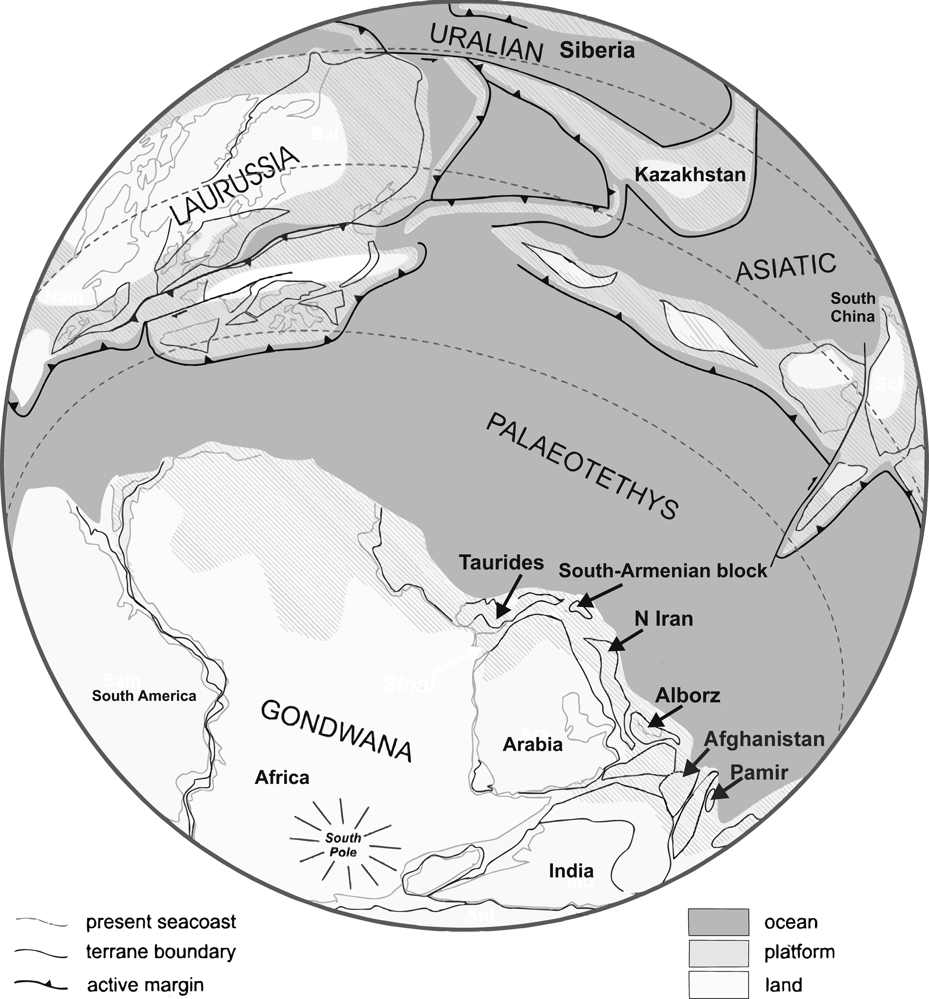
Although a precise paleolatitudinal evaluation of the Late Devonian position of the SAB is still hampered due to the remagnetization of upper Paleozoic rocks following the Late Cretaceous obduction of ophiolites (Meijers et al., Reference Meijers, Smith, Kirscher, Mensink, Sosson, Rolland, Grigoryan, Sahakyan, Avagyan, Langereis and Müller2015), existing paleogeographic reconstructions place the SAB at the junction between the Iranian terranes (to the east) and the Anatolide-Tauride microplate (to the west). It was thus an integral part of the northern passive margin of Gondwana, facing the Paleotethys ocean to the north (Fig. 19). Thus, during the Late Devonian, it was part of a huge platform that was positioned within the southern hemisphere tropical carbonate development zone (Brock and Yazdi, Reference Brock and Yazdi2000).
Late Devonian paleogeographic reconstruction of the Paleotethys ocean and its surrounding continents, including the position of the South Armenian Block along the northern margin of the Gondwana megacontinent (redrawn and modified after Denayer and Hoşgör, Reference Denayer and Hoşgör2014, based on the maps of Stampfli et al., Reference Stampfli, Von Raumer, Borel, Martinez Catalan, Hatcher, Arenas and Diaz Garcia2002).

The taxonomic assessment of brachiopods collected from the lower Famennian ‘Cyrtospirifer’ orbelianus Zone of Abrahamyan (Reference Abrahamyan1957) cropping out in the Ertych, Djravank, and Noravank sections of central Armenia allows us to recognize the presence of six rhynchonellide and one athyride species. Because rhynchonelliform brachiopods have a benthic mode of life and produce lecithotrophic larvae, their paleobiogeographic patterns have the potential to reveal past geographic boundaries and oceanographic connections, although caution should be exercised on questions of taxonomic consistency, comparison of assemblages of similar age, and biofacies (Brock and Yazdi, Reference Brock and Yazdi2000). In that respect the two species of the Famennian genus Sartenaerus found in the ‘Cyrtospirifer’ orbelianus Zone of Armenia (S. baitalensis and S. charakensis) are of particular paleobiogeographic significance because they point to a likely wide lower Famennian brachiopod bioprovince developed along the northern Gondwana margin, from the SAB to Afghanistan and Pamir (Fig. 19). However, it should be stressed that in spite of the well-constrained age of the occurrence of S. baitalensis in the Lesser Caucasus (the species occurs only in the ‘Cyrtospirifer’ orbelianus Zone), this is not the case for the occurrence of S. baitalensis in Pamir (Reed, Reference Reed1922). Finally, the apparent absence of both species in other parts of the northern Gondwanan margin, especially in Morocco, may be due to the presence of deeper (cephalopod-bearing) facies for this interval, and therefore it may reflect a paleoenvironmental rather than paleobiogeographical signal.
This is the first time that the genus Porthmorhynchus is recognized (although doubtfully) from the Armenian sections. It is also worth stressing that the genus is known so far only from the Frasnian—its type species, P. ferquensis, is known from the middle-upper Frasnian of northern France (Boulonnais region) and Nakhichevan (Pakhnevich in Alekseeva et al., Reference Alekseeva, Afanasjeva, Grechishnikova, Oleneva and Pakhnevich2018a). The other species assigned to this genus (P. elburzensis) is known from the upper Frasnian of the Alborz Mountains (northern Iran; Gaetani, Reference Gaetani1965). Thus, the doubtful report of this genus from the lower Famennian ‘Cyrtospirifer’ orbelianus Zone of Armenia is significant, although it requires further study to confirm its presence. Greira transcaucasica is one of the oldest punctate rhynchonellide species and, because it is only known from the SAB, may represent an interesting case of endemicity.
The single athyride species recognized (Crinisarina pseudoglobularis n. sp.) also represents the first report of the genus Crinisarina in the SAB, and indeed in the entire northern margin of Gondwana, although it cannot be excluded that Crinisarina representatives are present in the Famennian of North Africa (Algeria and Morocco), where taxonomical studies have been mostly dedicated to the rhynchonellides, productidines, and spiriferides, whereas the other elements of the brachiopod faunas received scant attention (see references in Mottequin et al., Reference Mottequin, Malti, Benyoucef, Crônier, Samar, Randon and Brice2015). Similar to other species of Crinisarina (see discussion in Mottequin, Reference Mottequin2008), it is likely that C. pseudoglobularis n. sp. had a preference for soft substrates that were comprised of argillaceous sediments on which it was attached with a thin pedicle. This corresponds well to our own observations, given that this species was found mainly in shaly intervals.
Conclusions
Taxonomic reassessment of the rhynchonellide brachiopods collected from the lower Famennian ‘Cyrtospirifer’ orbelianus brachiopod Zone of Abrahamyan (Reference Abrahamyan1957) from three distinct localities of central Armenia confirm the presence of a number of species known previously (e.g., Sartenaerus baitalensis, S. charakensis, and Greira transcaucasica). However, this is the first detailed description of the internal structure of Sartenaerus baitalensis provided, which is of particular significance because it is the type species of a biostratigraphically important genus. Thus, the brachiopod record from Armenia firmly establishes the presence of the Famennian genus Sartenaerus since the lower Famennian and not since the middle Famennian, as was previously thought (Mottequin et al., Reference Mottequin, Brice and Legrand-Blain2014). It is also the first documentation of S. charakensis in the Armenian sections; moreover, its stratigraphic range is extended here down to the ‘Cyrtospirifer’ orbelianus Zone (it was previously known from the ghorensis-pamiricus Zone). The intraspecific variability of Greira transcaucasica is documented and its stratigraphic range revised to include its presence up to the orbelianus Zone. The new athyride species, Crinisarina pseudoglobularis n. sp., represents the first report of the genus Crinisarina in the SAB and, indeed, in the entire north Gondwanan realm. The paleobiogeographic distribution of the rhynchonellides and athyrides within the lower Famennian of Armenia suggests that the brachiopod communities thriving on the shallow water carbonate platform of the SAB contained some apparently endemic species (G. transcaucasica and C. pseudoglobularis n. sp.), but also species that were common in other parts of the Gondwanan northern margin extending eastwards to Afghanistan and Pamir.
Acknowledgments
V. Serobyan is grateful to the French Embassy in Armenia and the MOBLILEX International Mobility Grant Programme of the University of Lille for funding his studies in France. T. Danelian and A. Grigoryan acknowledge the financial support of the Erasmus + International Credit Mobility Programme for their staff mobility in 2018 and 2019, respectively. C. Crônier acknowledges the financial support of the Lille Institute on Environmental Sciences (IRePSE). Fieldwork was also facilitated by logistic support of the Institute of Geological Sciences (Armenian Academy of Sciences). The authors thank the Région Hauts-de-France, and the Ministère de l'Enseignement Supérieur et de la Recherche (CPER Climibio), and the European Fund for Regional Economic Development for their financial support. S. Régnier (UMR Evo-Evo-Paleo, ULille) is gratefully acknowledged for the high-quality SEM pictures obtained on specimens deposited at IGNASRAGM. The manuscript benefited from the reviews of A. Balinski and an anonymous reviewer, and from the editorial help of J. Jin and C. Sproat, to whom all we are much indebted.