Implications
The importance of selenium (Se) in influencing biological processes, including regulation of redox/immune functions, makes its supplementation in dairy cattle diets a routine practice. Se can be supplemented in quantities up to 0.3 or 0.5 mg/kg dry matter in North America and the European Union, respectively. We evaluated the effects of these two levels of supplementations, in organic or inorganic forms, on neutrophil immune functions of high-producing dairy cows. Our results showed that the higher levels of supplementation significantly increased the respiratory burst activity of neutrophils. Also, neutrophil apoptosis decreased when cows were fed organic Se as compared to inorganic Se. Our data suggest further benefits at a higher level of supplementation.
Introduction
The transition from pregnancy to lactation is accompanied by immune incompetence and occurrence of early lactation-related infectious diseases (Mehrzad et al., Reference Mehrzad, Duchateau, Pyorala and Burvenich2002). Leukocytes are prone to be apoptotic around calving (Van Oostveldt et al., Reference Van Oostveldt, Vangroenweghe, Dosogne and Burvenich2002). Consequently, the number and function of leukocytes in blood and milk are generally low at this time (Mehrzad et al., Reference Mehrzad, Duchateau, Pyorala and Burvenich2002). Furthermore, it is known that the survival rate of milk neutrophils can affect the outcome of mastitis (Mehrzad et al., Reference Mehrzad, Duchateau and Burvenich2004). Therefore, the periparturient period is characterized by compromised immunity and a surge in the prevalence of infectious diseases in high-producing dairy cows.
Selenium (Se), an essential micronutrient, serves several biological purposes as an integral component of a variety of enzymes or selenoproteins. It also regulates redox and immune functions, supports thyroid hormone metabolism and has anticarcinogenic properties (Spears, Reference Spears2000; Ibeagha et al., Reference Ibeagha, Ibeagha-Awemu, Mehrzad and Zhao2007). For dairy cows, Se influences both the acquired and adaptive immune systems including antibody production, cell proliferation, cytokine production and neutrophil function (Larsen, Reference Larsen1993; Ndiweni and Finch, Reference Ndiweni and Finch1995). In cows suffering from Se deficiency, Boyne and Arthur (Reference Boyne and Arthur1986) noted a decrease in the ability of neutrophils to kill phagocytosed Candida albicans. Also, a lack of Se lowered the production of leucotrienes by polymorphonuclear leukocytes, which led to lowered chemotaxis of neutrophils (Aziz and Klesius, Reference Aziz and Klesius1986). Several reports associated increased concentration of blood Se with decreased infection rates such as subclinical and clinical mastitis in cows (Jukola et al., Reference Jukola, Hakkarainen, Saloniemi and Sankari1996; Ranjan et al., Reference Ranjan, Swarup, Naresh and Partra2005). Furthermore, several studies have revealed positive associations of Se supplementation with health conditions of the cow (Weiss et al., Reference Weiss, Hogan, Smith and Hoblet1990; Malbe et al., Reference Malbe, Klaassen, Fang, Myllys, Vikerpuur, Nyholm, Sankari, Suoranta and Sandholm1995; Kommisrud et al., Reference Kommisrud, Østeras and Vatn2005). In addition, Se status of the mammary gland is a vital regulator of selenoprotein expression and activities in the gland (Bruzelius et al., Reference Bruzelius, Hoac, Sundler, Önning and Äkesson2007). Thus, bioavailable Se is essential for an optimum immunological response following pathogen attack in cattle, but the exact mechanism of action is not yet clear. The positive action of Se in biological systems is thought to be supported by vitamin E. Several reports in the past decades examined the role of vitamin E and Se on bovine health (Hogan et al., Reference Hogan, Smith, Weiss, Todhunter and Schockey1990; Ndiweni and Finch, Reference Ndiweni and Finch1995; Jukola et al., Reference Jukola, Hakkarainen, Saloniemi and Sankari1996; Sivertsen et al., Reference Sivertsen, Øvernes, Østerås, Nymoen and Lunder2005) with the general consensus that adequate amounts of these nutrients may enhance cow health.
Dietary Se supplementation is either in the organic (selenized yeast) or inorganic (sodium selenite or sodium selenate) forms. Several studies have shown variability between organic and inorganic Se in terms of Se bioavailability and concentration in blood and milk (Knowles et al., Reference Knowles, Grace, Wurms and Lee1999; Weiss and Hogan, Reference Weiss and Hogan2005; Juniper et al., Reference Juniper, Phipps, Jones and Bertin2006). In general, when compared to the inorganic form of Se, organic Se possesses higher capacity of assimilation and bioavailability, and increases blood and milk Se concentrations (Ortman and Pehrson, Reference Ortman and Pehrson1999; Knowles et al., Reference Knowles, Grace, Wurms and Lee1999; Weiss and Hogan, Reference Weiss and Hogan2005; Juniper et al., Reference Juniper, Phipps, Jones and Bertin2006). Furthermore, organic Se had a higher tendency to lower milk somatic cell counts (SCC) than inorganic Se (Knowles et al., Reference Knowles, Grace, Wurms and Lee1999). However, less is known about the effects of organic Se supplementation on the functionality of neutrophils around the periparturient period. We hypothesized that supplementation of organic Se around calving would improve neutrophil functions of periparturient dairy cows, which may have the effect of improving cellular immunity and reducing the risk of early lactation-related infections. Thus, the purpose of this study was to determine the effects of supplemented organic and inorganic Se on neutrophil functions of high-producing dairy cows during the periparturient period, as well as on plasma Se concentrations in cows and calves.
Material and methods
Animals and experimental diets
Holstein cows (n = 25) were grouped according to calving dates and allocated to five dietary treatments (five cows per treatment). The cows were in their first to fourth parities. Cows calved within ±3 days of the expected dates. The farm records (individual cows) indicated that the animals were free from pathogens responsible for intramammary infections. The cows were individually housed, during the entire experimental period, in identical stalls with sawdust as bedding and had ad libitum access to drinking water. The study was approved by the McGill University Animal Care Committee.
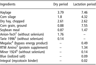
Animals were routinely fed diets containing approximately 0.3 mg/kg of dry matter (DM) of inorganic Se (sodium selenite) on the farm but this was discontinued for at least 4 months prior to the start of the trial. The five dietary treatments included: control, no Se supplementation; IN 0.3, low level of inorganic Se supplementation (0.3 mg/kg DM); IN 0.5, high level of inorganic Se supplementation (0.5 mg/kg DM); OR 0.3, low level of organic Se supplementation (0.3 mg/kg DM) and OR 0.5, high level of organic Se supplementation (0.5 mg/kg DM). All diets had similar composition and included a basal Se concentration of 0.016 mg/kg DM (Table 1), except for variations due to the level of Se supplementation. The organic Se used in this experiment was Sel-Plex® (Alltech, Inc., Nicholasville, KY, USA) and the inorganic one was sodium selenite. Se was premixed in the concentrate feed and fed to cows in the morning and evening rations throughout the experimental period (4 weeks prior to expected calving dates and 4 weeks after calving).
Ingredient composition of dietsFootnote 1 fed during the experiment (% of dry matter (DM))
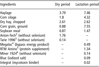
1 The selenium content of the total mixed ration was 0.016 mg/kg DM (analysis by Valacta). The feed was made by Agribrands Purina (Agribrands Purina Canada Inc., St-Hubert, Quebec, Canada). Apart from the control diet, diets were further supplemented with 0.3 or 0.5 mg/kg DM organic or inorganic selenium. The form of organic selenium used was Sel-Plex® (Alltech, Inc., Nicholasville, KY, USA) while inorganic was sodium selenite.
2 Anionic supplement to prevent milk fever.
3 Dry cow supplement.
4 Rumen bypass fat.
5 Protein supplement (contains amino acids that are not degradable in the rumen) for lactating cows.
6 Mineral supplement in milking to maintain bone health and reproductive system.
Blood sampling
Blood samples were either collected from the caudal vein of cows or jugular vein of calves, by venipuncture, into vacutainer tubes coated with heparin anticoagulant (BD Biosciences, Franklin Lakes, NJ, USA). Blood sampling was performed once weekly prior to calving and after calving (until post-partum week 4), and used for the determination of plasma Se concentration. After collection, samples were stored on ice and analyzed within 4 h. On the day of calving (day 0) however, blood samples were collected 8 h after calving from both dams and calves. The first blood samples from calves were taken after consumption of colostrum. Neutrophil functions were measured using blood samples collected during week 4 post partum.
Neutrophil phagocytosis
Phagocytosis was measured using the phagocytosis assay, PHAGOTEST® (Orpegen Pharma, Heidelberg, Germany). The assay is based on the ingestion of fluorescent-labeled bacteria and detection by flow cytometry (FC). PHAGOTEST® allows the quantitative determination of neutrophil phagocytosis. Analysis was done following the PHAGOTEST® protocol with slight modifications. Briefly, 100 μl of heparinized whole blood was incubated with fluorescein isothiocyanate (FITC)-labeled Escherichia coli bacteria in a 12 × 75 mm FC tube at 38.5°C for 10 min, according to manufacturer’s specification and as modified by Kampen et al. (Reference Kampen, Tollersrud, Larsen, Roth, Frank and Lund2004). A negative control sample was incubated on ice. The reaction was stopped by placing the samples on ice and adding a quenching solution. This solution discriminates attached and internalized bacteria by quenching the FITC fluorescence of surface bound bacteria, leaving the fluorescence of internalized particles unaltered. After two wash steps (centrifugation at 250 × g, 4°C for 5 min with a washing solution and subsequent aspirations), erythrocytes were removed by addition of a lysing buffer and fixed for acquisition by FC.
Respiratory burst of neutrophils
The BURSTTEST Kit (PHAGOBURST® Kit; Orpegen Pharma) was used for measurement of the respiratory burst of neutrophils. This test allows the quantitative determination of neutrophil respiratory burst (oxygen-dependent intracellular killing of bacteria) in heparinized whole blood. Dihydrorhodamine (DHR) 123 was used as the fluorogenic substrate for determination of the respiratory burst. In total, a volume of 100 μl heparinized whole blood was incubated with unlabeled opsonized E. coli bacteria provided in the kit for 10 min, following the manufacturer’s protocol. During optimization steps, the best results were obtained from the bacterial stimulation rather than stimulation with phorbol myristate acetate (also provided in the kit as a stimulant), which appeared to be damaging to the neutrophils, introducing false positive apoptotic and necrotic expressions, variations and experimental errors. For consistency, therefore, the unlabeled opsonized bacteria were used throughout the respiratory burst experiment. Formation of reactive oxidants during the respiratory burst was monitored by the addition and oxidation of DHR 123. The ROS (reactive oxygen species) produced convert the colorless DHR 123 to green fluorescent rhodamine, which was detected by the FC. The reaction was stopped by the addition of the lysing solution, which removes erythrocytes and results in partial fixation of neutrophils. The cells were later washed as described above and analyzed by FC.
Apoptosis and necrosis of neutrophils
Apoptosis is a form of programmed cell death that is characterized by a variety of morphological features, including changes in plasma membrane such as loss of membrane asymmetry and attachment, cell shrinkage, chromatin condensation and chromosomal DNA fragmentation. Annexin-V is a sensitive probe for identifying neutrophils that are undergoing apoptosis because phospholipid phosphatidylserine (PS) exposure occurs early in the apoptotic process (Koopman et al., Reference Koopman, Reutelingsperger, Kuitten, Keehnen, Pals and van Oers1994). During the process of apoptosis, PS is translocated from the inner to the outer leaflet of the plasma membrane, thereby exposing it to the external cellular environment. Annexin-V is a Ca2+-dependent phopholipid binding protein that has affinity for PS and binds to cells exposed to PS. Therefore, 100 μl of blood was first suspended in 1 ml of 1 × binding buffer (BioVision Inc., Mountain Vew, CA, USA) for 10 min. Then, 5 μl of Annexin-V–FITC and 10 μl of propidium iodide (PI) (50 μg/ml) were added, and the tube gently vortexed and incubated for 20 min at room temperature in the dark on an Orbitron rotator (Boekel Industries Inc., Feasterville Trevose, PA, USA). PI was used to distinguish apoptotic cells (Annexin V–FITC positive, PI negative) from necrotic cells (Annexin V–FITC positive, PI positive). After incubation, the samples were centrifuged at 250 × g for 5 min at 4°C to spin down the neutrophils. The supernatant was aspirated, leaving approximately 400 μl in the FC tube. The cells were washed twice by adding Dulbecco’s phosphate buffered saline, and centrifuged at 250 × g for 5 min at 4°C. Erythrocytes were lysed with the lysis buffer, according to Van Oostveldt et al. (Reference Van Oostveldt, Vangroenweghe, Dosogne and Burvenich2001), and analyzed within 30 min by FC. Blood initially incubated with actinomycin D (Sigma-Aldrich, Oakville, ON, Canada) was used as a positive control. Actinomycin D is an RNA synthesis inhibitor. This molecule has been shown to induce apoptosis to varying degrees in bovine neutrophils (Van Oostveldt et al., Reference Van Oostveldt, Dosogne, Burvenich, Paape, Brochez and Van den Eeckhout1999).
Using FC in these assays has the advantage of evaluating phagocytosis, respiratory burst and apoptosis in whole blood samples, thus circumventing the stimulatory effects often encountered in lengthy purification procedures with the classical methods (Smits et al., Reference Smits, Burvenich and Heyneman1997; Kampen et al., Reference Kampen, Tollersrud, Larsen, Roth, Frank and Lund2004). Furthermore, only small volumes of whole blood are required for evaluation.
The flow cytometer used was BD FACSAria™ (Becton Dickinson Immunocytochemistry Systems, San José, CA, USA). Cells were selectively gated for analysis and data were collected from 10 000 events per sample. Analysis was performed in duplicates. The percent or mean channel fluorescence was used as a quantitative index of neutrophil phagocytosis, respiratory burst, apoptosis and necrosis. Data were analyzed using the BD FACSDiva® software (Becton Dickinson Immunocytochemistry Systems).
Se concentrations in cow plasma and calf plasma
Se concentrations in cow plasma and calf plasma were measured by HPLC, using a modified method of Hawkes and Kutnink (Reference Hawkes and Kutnink1996), and by the University of Montreal Veterinary Laboratory, St Hyacinthe, Quebec. Briefly, a sample at 250 μl volume was mixed with 2.5 ml double-distilled nitric acid (70%) and 1 ml perchloric acid (70%). The samples were heated at 140°C for 90 min and then at 200°C for 75 min. After cooling to room temperature, 1 ml of 4 M HCl was added to the digested samples and the tubes were block heated for 15 min at 150°C to reduce Se VI to Se IV. In all, 1 ml of 2 M glycine base, 0.09 M Na4EDTA (ethylenediaminetetraacetic acid) and 4 drops of 0.02% cresol red were added, and the pH was adjusted to 1.5–2.0 with 7 M NH4OH. Glycine (2 M, pH 1.75), in the volume 1.5 ml, was added and the samples were diluted to 8 ml with distilled water, to minimize the tube-to-tube pH variation. A volume of 1 ml 0.1% (w/v) 2,3-diaminonaphthalene hydrochloride in 0.1 M HCl (extracted once with cyclohexane just before use) was added, and the mixture was heated for 45 min at 50°C. After cooling, 3 ml cyclohexane was added to the tubes, sealed with polyethylene stoppers and mechanically shaken for 15 min to extract the fluorescent piazselenol. The cyclohexane extracts were placed directly into 2-ml autosampler vials and analyzed by a HPLC system consisting of a HP 1100 (Hewlett–Packard Co., Palo Alto, CA, USA) and a Water-Xterra column (Waters Corporation, Milford, MA, USA). Elution was isocratic with a mobile phase consisting of 90% cyclohexane and 10% ethyl acetate, at a flow rate of 0.5 ml/min. The fluorescent derivative, naphtho-2-selena-1,3-diazole (4,5-benzopiazselenol), eluted at 2 min, was detected by a HP 1046A fluorescence detector (Scientific Support Inc., Hayward, CA, USA) at an excitation wavelength of 378 nm and emission wavelength of 530 nm.
Statistical analysis
Data were analyzed using the MIXED procedure of SAS (SAS, 2003). A one-way ANOVA was used for data parameters within a time interval (phagocytosis, respiratory burst, apoptosis, necrosis and calf plasma Se concentrations) with cows serving as experimental unit. Data for parameters at different time intervals (cow plasma) were analyzed by a two-way ANOVA with repeated measurements and cows nested within treatments. The model included the main effects of treatment (TRT), time (week), and treatment × time interaction, and the random effect of cow. Treatment means were separated using the least square means option of SAS. Differences between treatment means were tested using Scheffe’s Multiple Comparison test.

Results
Cow and calf plasma Se concentrations
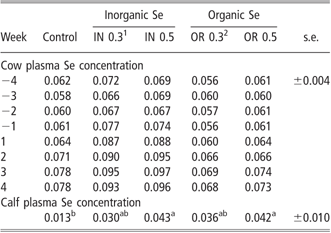
The cow and calf plasma Se concentrations are given in Table 2. Cow plasma Se concentrations were not different at any time interval whether diets contained organic or inorganic Se. Calf plasma Se concentrations were highest in OR 0.5 (0.042 μg Se/ml) and IN 0.5 (0.043 μg Se/ml) groups. However, feeding dairy cows the IN 0.3 and OR 0.3 diets did not significantly alter calf plasma Se concentrations in comparison with the control diet.
Effect of organic and inorganic selenium (Se) supplementation on cow plasma and calf plasma selenium concentrations (μg/ml ± standard error) during the trial
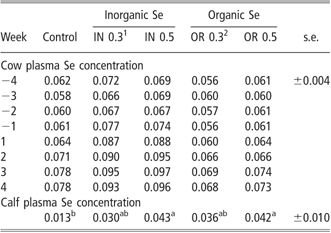
1IN 0.3, inorganic selenium (sodium selenite) at 0.3 mg/kg dry matter (DM).
2OR 0.3, organic selenium (Sel-Plex®) at 0.3 mg/kg DM.
a,bValues with different superscripts within each row differ significantly (Scheffe t-test, P < 0.05).
Neutrophil functions
Whole blood samples were gated for neutrophils for measurement of phagocytosis, respiratory burst, apoptosis and necrosis. The percentage of neutrophils that phagocytosed E. coli was not affected by dietary treatments and averaged about 91.6% (Figure 1). Conversely, neutrophils from cows fed organic Se showed higher respiratory burst activity than those fed the control or inorganic Se supplemented diets (P < 0.01) (Figure 2). Apoptosis of neutrophils was significantly lower (P < 0.01) in control cows and cows fed organic Se diets (Figure 3) than those fed inorganic Se diets. Necrosis was not significantly different among dietary treatments (0.43% ± 0.61 for OR 0.5, 0.68% ± 0.61 for OR 0.3, 1.53% ± 0.61 for IN 0.5, 2.03% ± 0.61 for IN 0.3 and 0.85% ± 0.61 for the control). Live cell percentages were significantly higher (P < 0.01) in control cows (93.88% ± 1.46) and in cows fed organic Se (91.58% ± 1.46 for OR 0.3 and 95.80% ± 1.46 for OR 0.5) than inorganic Se fed cows (81.28% ± 1.46 for IN 0.3 and 79.73% ± 1.46 for IN 0.5).
The effects of inorganic (sodium selenite) and organic selenium (Sel-Plex®) supplementation on bovine neutrophil phagocytosis measured by phagocytosis assay, PHAGOTEST® and detection by flow cytometry. There was no effect of selenium supplementation on neutrophil phagocytosis. Data presented are mean values (bar) of five animal replicates within each treatment.

The effect of inorganic (sodium selenite) and organic selenium (Sel-Plex®) supplementation on respiratory burst of bovine neutrophils as measured by BURSTTEST assay. Selenium supplementation significantly increased respiratory burst when compared to the control diet, but was highest in organic selenium fed cows. Data presented are mean values (bar) of five animal replicates within each treatment. a,bValues with different superscript differ significantly (Scheffe t-test, P < 0.01).

The effects of inorganic (sodium selenite) and organic selenium (Sel-Plex®) supplementation on apoptosis of bovine blood neutrophils as measured by the BD FACSAria™ flow cytometer. In comparison with inorganic selenium, apoptosis was significantly reduced when cows were fed organic selenium in a dose-dependent manner. Data presented are mean values (bar) of five animal replicates within each treatment. a,bValues with different superscript differ significantly (Scheffe t-test, P < 0.01).

Discussion
The periparturient period is critical in dairy cow productivity, due to immunosuppression and the occurrence of early lactation-related diseases during this period. The immune functions of circulating neutrophils are consequently markedly compromised during this period. Our study demonstrates that Se supplementation, especially in the organic form and at the higher level, enhances respiratory burst activities of neutrophils during the periparturient period and this may be beneficial for cows’ health and productivity.
In this study, neutrophils from cows fed organic Se showed significantly higher respiratory bust activities than their inorganic and control counterparts even though their phagocytic abilities were not different. Se as an antioxidant protects cells from oxidative damage from free radicals and peroxides by catalyzing reduction of peroxides that can damage cells and tissues (Spears, Reference Spears2000). Higher respiratory burst activity of neutrophils could lead to higher intracellular kill of bacteria. Our findings are in agreement with several authors who demonstrated that neutrophils from cows with supplemental Se (inorganic) increased intracellular kill of bacteria, enhanced viability and reduced extracellular hydrogen peroxide concentration as compared to non supplemented cows (Gyang et al., Reference Gyang, Stevens, Olsen, Tsitsamis and Usenik1984; Grasso et al., Reference Grasso, Scholz, Erskine and Eberhart1990; Hogan et al., Reference Hogan, Smith, Weiss, Todhunter and Schockey1990). Malbe et al. (Reference Malbe, Attila and Atroshi2006) also demonstrated possible involvement of organic Se supplementation (4 mg/day) with Staphylococcus aureus inhibition in cow’s whey. In our study, these effects were more pronounced with organic Se at both levels of supplementation. Moreover, Se adequacy has been associated with increased glutathione peroxidase (GSH-Px) activity (Ortman and Pehrson, Reference Ortman and Pehrson1999; Malbe et al., Reference Malbe, Klaassen, Kaartinen, Attila and Atroshi2003 and Reference Malbe, Attila and Atroshi2006), which enabled the continued production of radicals and bacterial clearance. In a recent study, Bruzelius et al. (Reference Bruzelius, Hoac, Sundler, Önning and Äkesson2007) demonstrated the activity of GSH-Px 1, 3 and 4, thioredoxin reductase (TrxR) 1 and selenoprotein P in bovine mammary tissue, which was enhanced by adequate Se status. However, significant respiratory burst activities of organic Se supplemented cows in our study were not accompanied by increased levels of plasma Se. This contradicts most of the earlier reports, which showed an increase in plasma Se concentrations with Se supplementation (Knowles et al., Reference Knowles, Grace, Wurms and Lee1999; Ortman and Pehrson, Reference Ortman and Pehrson1999; Weiss and Hogan, Reference Weiss and Hogan2005; Juniper et al., Reference Juniper, Phipps, Jones and Bertin2006). The exact reason for no significant increase in plasma Se concentration after supplementation is unknown. Relatively high Se concentrations in the control group, even after withdrawal of Se supplementation before the experiment, might be responsible for lack of significant increases after supplementation. According to Weiss et al. (Reference Weiss, Hogan, Smith and Hoblet1990), plasma Se concentrations were correlated positively with intakes of Se at a certain limit but were independent of Se intakes above the limit. Further, retention of absorbed Se is influenced both by animal status and the chemical form of Se administrated. Se retention is dependent on tissue demands. It will be interesting to determine whether retention of Se in cells such as neutrophils or tissues such as mammary glands is increased or not after Se supplementation.
Phagocytosis by neutrophils constitutes an essential arm of host defense against bacterial infections. In our study, all cows had the same ability to phagocytose pathogens irrespective of dietary treatments. Similar findings have been reported in several studies with cows fed diets supplemented with Se (Hogan et al., Reference Hogan, Smith, Weiss, Todhunter and Schockey1990; Smits et al., Reference Smits, Burvenich and Heyneman1997; Weiss and Hogan, Reference Weiss and Hogan2005). Neutrophils are short-lived cells that play a vital role in inflammatory responses. It is reported that the number and function of leukocytes in blood are low around calving time and are prone to be apoptotic (Mehrzad et al., Reference Mehrzad, Duchateau, Pyorala and Burvenich2002; Van Oostveldt et al., Reference Van Oostveldt, Vangroenweghe, Dosogne and Burvenich2002). Thus, a longer neutrophil lifespan around parturition time would be desirable for enhanced cow health. The present study shows that organic Se greatly reduced apoptotic neutrophils than inorganic Se fed cows, suggesting the beneficial effect of organic Se. Despite the lack of improvement in the phagocytic ability of neutrophils due to Se supplementation, a possible increase in the life span of neutrophils may prolong the clearance of pathogens at sites of infection. This finding may be attributed to the fact that the Se in the organic Se treated groups was more available and could have enhanced the production of selenoproteins and selenocompounds (Gyang et al., Reference Gyang, Stevens, Olsen, Tsitsamis and Usenik1984; Musik et al., Reference Musik, Koziol-Montewka, Tos-Luty, Pasternak, Latuszynska, Tokarska and Kielczykowska1999). This might have, in turn, supported longevity of neutrophils thereby making them available during a critical period. It has also been reported that the onset of apoptosis down-regulates neutrophil respiratory burst and phagocytic activity (Van Oostveldt et al., Reference Van Oostveldt, Vangroenweghe, Dosogne and Burvenich2002). Similarly, our study demonstrates that decreased apoptosis by organic Se supplementation was accompanied by increased respiratory burst activity. However, why the control cows also had lowered incidence of apoptotic neutrophils in comparison with inorganic Se fed cows is not clear.
Cow Se status in our study may have directly influenced the Se status of calf plasma. Calf plasma Se concentrations were significantly higher in cows supplemented with a higher Se level (0.5 mg/kg DM) irrespective of source as compared to the control, even though Se supplementation did not significantly affect cow plasma Se concentration. The exact reason for that apparent discrepancy is unknown. It might be related to the Se retention in tissues and the mammary gland transfer. The mammary barrier is selective, as higher Se in cow milk results from supplemental organic Se than from inorganic Se (Ortman and Pehrson, Reference Ortman and Pehrson1999). We hypothesize that the retention of Se in the tissue such as mammary glands could be higher for supplemented groups and subsequently led to higher calf plasma Se concentrations. In support of our findings, several authors have confirmed that Se supplementation of cows improved the Se status of their calves (Pehrson et al., Reference Pehrson, Ortman, Madjid and Trafikowska1999; Gunter et al., Reference Gunter, Beck and Phillips2003; Weiss and Hogan, Reference Weiss and Hogan2005). Furthermore, Gunter et al. (Reference Gunter, Beck and Phillips2003) have reported high GSH-Px activities in calves from cows fed supplemental Se. In a study conducted with sows, Se supplementation reduced the number of stillbirths per litter and increased piglet serum Se concentration (Yoon and McMillan, Reference Yoon and McMillan2006). Therefore, Se supplementation is necessary to enhance the Se status of cows during parturition, which in turn may confer higher Se transfer to calves for a healthy start in life.
This study also distinguishes between the organic and inorganic forms of Se and dosage (0.3 or 0.5 mg/kg DM) and their possible health benefits to dairy cows. Our findings indicate that the higher level of organic Se (OR 0.5) enhanced respiratory burst activities of neutrophils and could extend further benefits to neutrophil immune functions. In contrast to inorganic Se, the organic form of Se has been reported to show higher capacities of assimilation and bioavailability (Knowles et al., Reference Knowles, Grace, Wurms and Lee1999; Ortman and Pehrson, Reference Ortman and Pehrson1999; Weiss and Hogan, Reference Weiss and Hogan2005; Juniper et al., Reference Juniper, Phipps, Jones and Bertin2006). Furthermore, poor absorption of inorganic Se is likely due to the activities of ruminal microbes which oxidized them to insoluble, and hence, unavailable elemental Se that is excreted via the feces (Van Saun, Reference Van Saun1990). However, the assimilation of organic Se into selenoproteins, and their effects on neutrophil immune functions and bovine cellular immunity remains to be further investigated.
Acknowledgements
This research was supported by a grant from Alltech, Inc., Nicholasville, KY, USA. Special thanks to the staff of Macdonald Campus Farm (McGill University) for their support throughout the trial, Benjamin Olaniyan for assistance with blood collection and Jaime Sanchez-Dardon for technical support in flow cytometry.