
Introduction
Plasmodium coatneyi is an agent of malaria that infects a variety of old-world monkeys in South East Asia. The parasite was first identified in 1961 in an Anopheles mosquito from Malaya (Eyles et al., Reference Eyles, Fong, Warren, Guinn, Sandosham and Wharton1962). Subsequently, its complete lifecycle was described following experimental inoculation into a rhesus macaque. The species has been reported as part of a survey of blood parasites in cynomolgus macaques (Macaca fascicularis) which was conducted in the Philippines and resulted in significant interest in the use of the parasite as a model for severe human malaria associated with Plasmodium falciparum (Eyles et al., Reference Eyles, Fong, Warren, Guinn, Sandosham and Wharton1962, Reference Eyles, Dunn, Warren and Guinn1963; Aikawa et al., Reference Aikawa, Brown, Smith, Tegoshi, Howard, Hasler, Ito, Collins and Webster1992; Kawai et al., Reference Kawai, Aikawa, Kano and Suzuki1993; Moreno et al., Reference Moreno, Cabrera-Mora, Garcia, Orkin, Strobert, Barnwell and Galinski2013; Lombardini et al., Reference Lombardini, Gettayacamin, Turner and Brown2015).
The plasmodial parasites of Cercopithecidae monkeys from Asia are comprised of 10 species including P. coatneyi and the closely related Plasmodium knowlsei. This group also includes Plasmodium gonderi, infectious to Cercopithecidae from Africa; Plasmodium hylobati, which parasitizes Hylobatidae monkeys from Asia; and Plasmodium vivax (Escalante et al., Reference Escalante, Barrio and Ayala1995; Leclerc et al., Reference Leclerc, Hugot, Durand and Renaud2004; Hayakawa et al., Reference Hayakawa, Culleton, Otani, Horii and Tanabe2008; Lombardini et al., Reference Lombardini, Gettayacamin, Turner and Brown2015).
While not closely related to P. falciparum, P. coatneyi has a range of biological features that it shares with P. falciparum as well as several important distinguishing phenotypic traits: Both parasites have an erythrocytic stage with a 48-hour cycle (Eyles et al., Reference Eyles, Fong, Warren, Guinn, Sandosham, Wharton, Coatney, Collins, Warren and Contacos1971), there is evidence of microvascular cytoadherence, with the formation of rosettes between infected erythrocytes and uninfected red blood cells and several early reports have described the development of electron-dense knobs on the cell surface of infected erythrocytes which have been purported to be involved in cytoadhesion (Kilejian et al., Reference Kilejian, Abati and Trager1977; Aikawa et al., Reference Aikawa, Brown, Smith, Tegoshi, Howard, Hasler, Ito, Collins and Webster1992; Lombardini et al., Reference Lombardini, Gettayacamin, Turner and Brown2015). Furthermore, both P. falciparum and P. coatneyi undergo deep vascular sequestration of red blood cells infected with trophozoites and schizonts (Desowitz et al., Reference Desowitz, Miller, Buchanan and Permpanich1969; MacPherson et al., Reference MacPherson, Warrell, White, Looareesuwan and Warrell1985; Pongponratn et al., Reference Pongponratn, Riganti, Punpoowong and Aikawa1991; Sein et al., Reference Sein, Brown, Maeno, Smith, Corcoran, Hansukjariya, Webster and Aikawa1993; Smith et al., Reference Smith, Brown, Nakazawa, Fujioka and Aikawa1996).
Plasmodium coatneyi has been reported as causing both natural and experimentally induced disease in a variety of non-human primate species (Kawai et al., Reference Kawai, Aikawa, Kano and Suzuki1993, Reference Kawai, Aikawa, Suzuki and Matsuda1998; Sein et al., Reference Sein, Brown, Maeno, Smith, Corcoran, Hansukjariya, Webster and Aikawa1993; Smith et al., Reference Smith, Brown, Nakazawa, Fujioka and Aikawa1996; Moreno et al., Reference Moreno, Cabrera-Mora, Garcia, Orkin, Strobert, Barnwell and Galinski2013; Lombardini et al., Reference Lombardini, Gettayacamin, Turner and Brown2015). The cynomolgus macaque is described as being one of the natural hosts of the parasite and it is theorized that the parasite may have coevolved with the cynomolgus, resulting in reduced clinical disease in comparison with experimental infections in the rhesus or the Japanese macaque (Eyles et al., Reference Eyles, Fong, Warren, Guinn, Sandosham, Wharton, Coatney, Collins, Warren and Contacos1971; Kawai et al., Reference Kawai, Aikawa, Kano and Suzuki1993, Reference Kawai, Kano and Suzuki1995, Reference Kawai, Aikawa, Suzuki and Matsuda1998; Moreno et al., Reference Moreno, Cabrera-Mora, Garcia, Orkin, Strobert, Barnwell and Galinski2013; Lombardini et al., Reference Lombardini, Gettayacamin, Turner and Brown2015). Tissue pathology associated with P. coatneyi infection in the rhesus macaque is variable and likely is dependent on the genetic heterogenicity of the infected population, degree of blood parasitaemia, coinfection with other agents, immune status, age and overall health of the animals (Lombardini et al., Reference Lombardini, Gettayacamin, Turner and Brown2015, Reference Lombardini, Turner, Brown, Inamnuay, Kaewamatawong, Sunyakumthorn and Ferguson2021).
A large project has been undertaken in order to evaluate many aspects of the pathology of P. coatneyi in rhesus macaques and to evaluate its potential as a model for human falciparum malaria. In the present study, the ultrastructural features of P. coatneyi infected red blood cells (iRBC) and their in-situ interaction with host tissue are described. The features identified will be directly compared to those of P. falciparum infections in humans as were previously examined using identical techniques (Pongponratn et al., Reference Pongponratn, Turner, Day, Phu, Simpson, Stepniewska, Mai, Viriyavejakul, Looareesuwan, Hien, Ferguson and White2003).
Materials and methods
Six splenectomized Indian origin rhesus macaque monkeys were used in two studies in 2013 and 2014 at the Armed Forces Research Institute of Medical Sciences (AFRIMS) (Lombardini et al., Reference Lombardini, Gettayacamin, Turner and Brown2015, Reference Lombardini, Turner, Brown, Inamnuay, Kaewamatawong, Sunyakumthorn and Ferguson2021), in addition to the inclusion of two spleen intact animals in 2015. The research was conducted in compliance with the Animal Welfare Act and other federal statutes and regulations relating to animals and experiments involving animals and adhered to principles stated in the Guide for the Care and Use of Laboratory Animals, NRC Publication, 2011 edition. All animal use was approved by the AFRIMS Institutional Animal Care and Use Committee Protocol Number 12-08. The splenectomized experimental animals were comprised of four adult males and two adult females, while the spleen intact animals were both females. The mean age of the male animals was 11.7 years, and the mean weight was 11 pounds. The mean age of the female animals was 10 years and the mean weight was 12 pounds. The first six animals were enrolled in pathophysiology and model refinement study in which parasitaemia levels and parasite staging were conducted every other hour for the 3 days associated with peak parasitaemia (Lombardini et al., Reference Lombardini, Gettayacamin, Turner and Brown2015). In all instances, the experimental animals were inoculated from one donor rhesus to another through a blood challenge of P. coatneyi infected erythrocytes. Inoculation began in an infected donor by intravenous inoculation of freshly thawed cryopreserved P. coatneyi strain infected blood. The stabilate stock used was the Hackeri strain of P. coatneyi archived at AFRIMS, thereby ensuring that the stain of plasmodium was homogenous and standardized across the experimental animals. Once the parasitaemia in donor animals had reached >50 000 parasitized RBCs per μL, 10–20 mL of blood was drawn into a heparinized tube, diluted with sterile PBS solution to a concentration of 5 × 10⁶ infected erythrocytes per 1 mL suspension, and divided into equal 1 mL aliquots. These 1 mL aliquots were subsequently used to infect the study animals via intravenous inoculation. In accordance with the approved protocol, animals were euthanized based on the combined criteria of parasitaemia levels of 20% or greater, blood values and clinical signs. In these animals, comprehensive autopsies were performed with the collection of all tissues immediately post-mortem. All tissues were processed for histopathological, immunohistochemical and ultrastructural analyses using a standardized tissue sampling protocol (Lombardini et al., Reference Lombardini, Gettayacamin, Turner and Brown2015). Tissues sampled included multiple areas of the central nervous system (thalamus, hippocampus, forebrain, cerebellum, brain stem, spinal cord), kidneys and heart in all six experimental animals. At autopsy, tissue samples were collected from target organs, trimmed into ~1 mm3 blocks which were immersed within 1 min of collection in 2.5% glutaraldehyde in 0.1 M phosphate buffer and subsequently processed for routine transmission electron microscopy (TEM). In summary, samples were post-fixed in osmium tetroxide, dehydrated and embedded in Spurr's epoxy resin (TABB Limited UK). Thin sections of areas of interest were cut to a thickness of approximately 70 nm, and stained with uranyl acetate and lead citrate, and subsequently, grids were examined in a JEOL 1200EX electron microscope (JEOL UK). This process was similar to that described previously for a human autopsy study of fatal P. falciparum malaria (Pongponratn et al., Reference Pongponratn, Turner, Day, Phu, Simpson, Stepniewska, Mai, Viriyavejakul, Looareesuwan, Hien, Ferguson and White2003).
In addition, the blood from two spleen intact animals infected with P. coatneyi was obtained once parasitaemia was achieved and processed for both transmission and scanning electron microscopy. These animals underwent curative treatment. For TEM, the cells were centrifuged and the pellet fixed in 2.5% glutaraldehyde in 0.1 M phosphate buffer and processed for routine TEM as described above. Scanning electron microscopy (SEM) was conducted on P. coatneyi infected red cells after magnetic enrichment, using standard SEM protocols as in Malleret et al. (Reference Malleret, Xu, Mohandas, Suwanarusk, Chu, Leite, Low, Turner, Sriprawat, Zhang, Bertrand, Colin, Costa, Ong, Ng, Lim, Nosten, Renia and Russell2013). Briefly, sorted cells were allowed to settle on a poly-lysine (Sigma) pretreated glass coverslip for 15 min at room temperature (RT) prior to fixing in gluteraldehyde and post-fixing in osmium tetroxide (as described above). After washing in deionized water, cells were dehydrated and critical point dried. Samples were sputter-coated with gold prior to examination with a field emission scanning electron microscope (JSM-6701F, JEOL).
Microscopic enumeration of IRBCs was performed using thin blood smears stained with Giemsa. A minimum of 4000 RBCs were counted (20 fields at 100× magnification). Live cell subvital staining of reticulocytes and of parasites was done using Giemsa (Lee et al., Reference Lee, Russell, Lau, Fong, Chu, Sriprawat, Suwanarusk, Nosten and Renia2013).
Results
Tissue samples
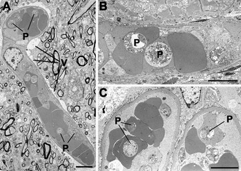
Ultrastructural examination of grids from the three selected tissues (brain, kidney and heart) from the six experimental animals, allowed for evaluation of the distribution of iRBC within the deep microvasculature. Counting the percentage of iRBCs to uninfected RBCs in each tissue gave 41% in the brain, 46% in the heart muscle and 38% in the kidney (based on a minimum of 12 blood vessels and over 120 erythrocytes for each tissue). There was a very strong synchrony of parasite stage of maturation and as such, the vast majority of the observed parasites were trophozoites characterized by their spherical appearance with cytoplasm containing a nucleus and food vacuole (Figs 1, 2A, B, 3B and C). Only a relatively few mature schizonts (Fig. 3A) and ring forms were observed. In all three tissues, iRBCs were identified in both large and small blood vessels with no obvious difference in the number or appearances of iRBCs between tissues (Figs 1–3).
(A–C) Low power transmission electron micrographs of tissue from Plasmodium coatneyi infected splenectomized animals showing the distribution of iRBCs within the blood vessels of the brain (A) heart (B) and the glomerulus of a kidney (C). Note the cytoplasmic vacuolation (V) around the cerebral blood vessel. P – Plasmodium coatneyi parasite. Bars are 10 μm.

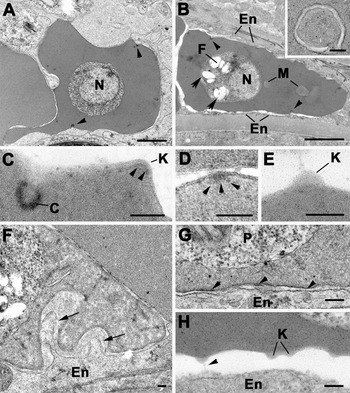
Transmission electron micrographs showing the structure of Plasmodium coatneyi iRBCs and their relationship to host endothelial cells. (A) Section through an iRBC showing a trophozoite with a centrally located nucleus (N). Note that the surface of the iRBC exhibits caveoloa formation (arrowheads). Bar is 1 μm. (B) iRBC with a trophozoite displaying a nucleus (N) and hemazoin crystals (arrows) in the food vacuole (F). There are also membrane structures (M) in the cytoplasm and caveoloa (arrowheads) at the plasmalemma of the iRBC. Note the tight apposition of the iRBC plasmalemma with the endothelial cells (En) lining the vessel. Bar is 1 μm. Inset: Detail of part of the cytoplasm of a similarly infected RBC's cytoplasm showing the double membrane-bound tubular structure. Bar is 100 nm. (C) Detail from the surface of an iRBC showing an invagination of the plasmalemma to form a caveoloa (C) with underlying electron-dense material and a knob (K) with underlying dense material (arrowheads). Bar is 100 nm. (D) Detail from the surface of an iRBC in which electron-dense material (arrowheads) is associated with the plasmalemma of an iRBC but in the absence of any protrusion. Bar is 100 nm. (E) Detail from the surface of an iRBC showing a knob that appears to lack underlying dense material. Bar is 100 nm. (F) Section through the junction between an iRBC and an endothelial cell (En) showing two microvilli from the endothelial cell protruding into folds in the iRBC (arrows). Bar is 1 μm. (G) Enlargement of the junction between iRBC and endothelial cell (En) with no evidence of knob formation but proteinaceous-like material appearing to connect the plasma membranes (arrowheads). P – parasite. Bar is 100 nm. (H) Detail of junction between iRBC and endothelial cell (En) where the iRBC appears to form knobs (K). Note the proteinaceous-like material (arrowhead) connecting the knob to the endothelial cell (En). Bar is 100 nm.

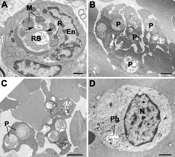
Transmission electron micrographs of Plasmodium coatneyi infected rhesus macaque tissues. (A) Cross-section through a small blood vessel showing an iRBC containing a mature schizont. Note the merozoites (M) with electron-dense rhoptries (R) around a residual cytoplasmic mass (RB), as well as hemazoin crystals (arrows). En – endothelial cell. Bar is 1 μm. (B) Low power from a large vessel showing a number of iRBCs interacting to form an auto-agglutinate-like structure. P – parasite. Bar is 1 μm. (C) Part of a vessel in which iRBCs are interacting with RBCs to form rosette-like structures. P – parasites. Bar is 1 μm. (D) Section through a vessel showing macrophage that had phagocytise a packet of pigment (Pg). N-Nucleus. Bar is 1 μm.

The smaller blood vessels were typically tightly packed with a mixture of uninfected and infected erythrocytes (Figs 1–3). While the majority of infected cells had a single parasite, a number had multiple parasites (Figs 1A and 3C). The parasites were located within a parasitophorous vacuole limited by a unit membrane (Fig. 2G). Trophozoites appeared to increase in size, developing from stages with a single nucleus (Fig. 2A) to stages with multiple nuclei and finally into mature schizonts (Fig. 3A). Food vacuoles were initially small but increased in size into large vacuoles containing a number of hemozoin crystals (Figs 2B and 3A). The few mature schizonts were comprised of multiple merozoites, with apical rhoptries and a posterior nucleus, centred around a body of residual cytoplasm containing food vacuoles filled with hemazoin crystals (Fig. 3A). The erythrocytic cytoplasm frequently contained numbers of circular or curved bilayered channels interpreted as tubular vesicular structures with a narrow electron-lucent lumen (Fig. 2B insert). However, Maurer's clefts running parallel to the RBC plasmalemma, characteristic for P. falciparum iRBCs, were not observed. The majority of iRBCs within small blood vessels were more spherical in shape with a relatively regular outline (Figs 1A and 2B). However, others show marked deformation of shape consisting of irregular cytoplasmic protrusions giving the infected cells a more flattened, crenated appearance (Figs 2A and 3B). A feature of all iRBCs was the presence of a number of caveolae, which measured approximately 90 nm in diameter described as small spherical invaginations with an electron-dense internal coating but with no evidence of associated vesicles (Figs 2A–C). In addition, a variable number of blebs or excrescences consistent with knob formation were observed. These knob-like structures were 70–90 nm in diameter (Fig. 2C, E and H). However, detailed examination showed that while a few possessed some electron-dense material beneath the plasmalemma (Fig. 2C), the majority (>60%) appeared to lack the clearly defined sub-plasma membrane electron-dense material (Fig. 2G). In addition, it was also possible to identify rare (<10%) plaques of electron-dense material associated with the iRBC plasmalemma but lacking protuberance (Fig. 2D). The majority of iRBCs appeared to have relatively few knobs irrespective of the stage of parasite development, and those that were noted appeared to be randomly distributed. In addition, examination of numerous (50+) iRBCs did not appear to demonstrate knob number as being related to the stage of parasite development.
In small vessels, there was often a tight association between the iRBC and endothelial cells (Fig. 2B and G), which resulted in evidence of margination of iRBCs along the luminal wall (Fig. 2A). When examined in detail there appeared to be close apposition between the iRBC plasmalemma and the plasmalemma of the endothelial cells with areas showing possible connection via proteinaceous material (Fig. 2B and G). While in the majority of cases, knob-like protuberances were absent from such areas, they were observed on occasion. In these cases, proteinaceous-like material connecting the knob to the endothelial surface was observed (Fig. 2H). Also, there was evidence of limited microvillus formation by the endothelial cells with a cytoplasmic process protruding into invaginations of the iRBC surface (Fig. 2F). In the larger vessels, where the RBCs were more loosely packed, it was often possible to observe clumping of iRBCs consistent with possible auto-agglutination (Fig. 3B) and interactions between iRBCs and RBCs possibly related to rosette formation (Fig. 3C). The iRBCs in this situation showed more severe shape changes and there was less evidence of cytoadherence between iRBCs and the endothelium in such vessels. No example of vessel rupture was observed by electron microscopy, although several endothelial cells showed some degenerative changes. There did appear to be lucent and vacuolated areas around certain cerebral vessels, which were interpreted as oedema (Fig. 1A). Within the vessels, a number of macrophages and neutrophils were observed and certain macrophages appear to have phagocytosed packets of malarial pigment (Fig. 3D).
Blood samples
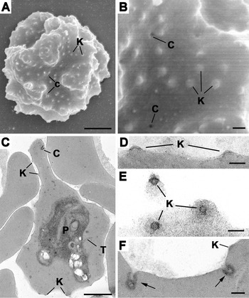
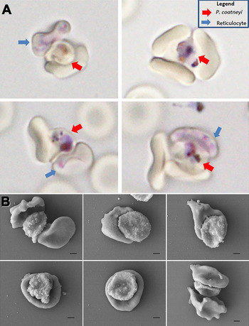
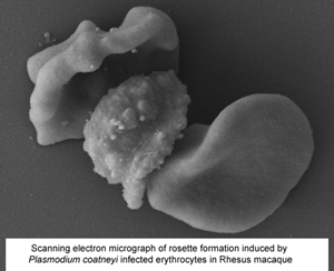
The observation of anomalies on the structure of the knob described above in splenectomized animals was unexpected. Therefore, to see if this was a phenomenon arising from the specific use of splenectomized animals, blood was obtained from femoral arteries in two spleen-intact animals infected with P. coatneyi and was examined by both TEM and SEM. In these blood samples, it was observed that the iRBCs could be identified by a markedly irregular outline (Fig. 4A and B). By SEM and TEM, the surface of the iRBCs showed the formation of variable numbers of caveolae and knob-like structures (Fig. 4A–F). The structure of the caveolae with the invagination of the plasmalemma with underlying electron-dense material could be seen by TEM (Fig. 4F) and appeared as surface pores by SEM (Fig. 4B). Detailed examination of the knob-like structures revealed circular electron-dense plaques beneath the plasmalemma shown in the longitudinal section (Fig. 4D) and tangential section (Fig. 2E). The knobs could be identified in SEM as slightly circular electron-lucent structures, which appeared to be random distribution (Fig. 4A and B). Spontaneous rosette formation with normocytes and reticulocytes were observed in wet Giemsa preparations (Fig. 5A) and by SEM (Fig. 5B).
Scanning electron (A, B) and transmission electron micrographs (C–F) of blood from Plasmodium coatneyi infected, spleen intact rhesus macaques. (A) Low power scanning electron micrograph of an iRBC showing the irregular (lumpy) surface appearance in which randomly distributed knobs (K) and caveolae (C) can be identified. Bar is 1 μm. (B) Detail of the surface of the iRBC showing the circular slightly raised electron-lucent knobs (K) and the openings of the caveolae (C). Bar is 100 nm. (C) Low power image showing an iRBC containing a mid-stage trophozoite (P) in which tubular structures (T), caveolae (C) and knobs (K) can be identified. Bar is 1 μm. (D) Longitudinal section through two knobs showing the underlying dense material (arrows). Bar is 100 nm. (E) Tangential section through the surface of an iRBC showing knobs (K) with the circular appearance of the underlying dense material. Bar is 100 nm. (F) Longitudinal section showing the caveolae formed by the invagination of the iRBC plasmalemma with underlying dense material (arrows). K – knob. Bar is 100 nm.

Plasmodium coatneyi rosettes. (A) Giemsa-stained blood from P. coatneyi infected, spleen intact rhesus macaque rosettes. P. coatneyi rosettes with both normocytes and reticulocytes. Live-cell staining of rosettes under fluid conditions (Note P. vivax rosettes with normocytes only). (B) Scanning electron micrographs of blood from P. coatneyi infected, spleen-intact rhesus macaques (scale bar is 1 μm).

Discussion
To date the majority of studies on P. coatneyi have concentrated on the use of the rhesus macaque model as a platform for cerebral malaria studies but with limited morphological evaluation (Kilejian et al., Reference Kilejian, Abati and Trager1977; Aikawa et al., Reference Aikawa, Brown, Smith, Tegoshi, Howard, Hasler, Ito, Collins and Webster1992; Kawai et al., Reference Kawai, Aikawa, Kano and Suzuki1993; Maeno et al., Reference Maeno, Brown, Smith, Tegoshi, Toyoshima, Ockenhouse, Corcoran, Ngampochjana, Kyle and Webster1993; Tegoshi et al., Reference Tegoshi, Udomsangpetch, Brown, Nakazawa, Webster and Aikawa1993; Moreno et al., Reference Moreno, Cabrera-Mora, Garcia, Orkin, Strobert, Barnwell and Galinski2013; Lombardini et al., Reference Lombardini, Gettayacamin, Turner and Brown2015). The present ultrastructural study was based on the examination of three tissues (brain, heart and kidney) in P. coatneyi infected splenectomized animals. Interestingly, quantitative comparisons of the level of sequestration in these three tissues, noted no tissue-specific differences. Prior studies have demonstrated that there is a significant amount of sequestration in the brain of macaques infected with P. coatneyi (Desowitz et al., Reference Desowitz, Miller, Buchanan and Permpanich1969; Sein et al., Reference Sein, Brown, Maeno, Smith, Corcoran, Hansukjariya, Webster and Aikawa1993; Smith et al., Reference Smith, Brown, Nakazawa, Fujioka and Aikawa1996; Lombardini et al., Reference Lombardini, Turner, Brown, Inamnuay, Kaewamatawong, Sunyakumthorn and Ferguson2021). However, the more diffuse tissue sequestration observed in the rhesus model differs from P. falciparum in humans where the disease is associated specifically with brain sequestration (MacPherson et al., Reference MacPherson, Warrell, White, Looareesuwan and Warrell1985; Pongponratn et al., Reference Pongponratn, Riganti, Punpoowong and Aikawa1991, Reference Pongponratn, Turner, Day, Phu, Simpson, Stepniewska, Mai, Viriyavejakul, Looareesuwan, Hien, Ferguson and White2003; Nguansangiam et al., Reference Nguansangiam, Day, Hien, Mai, Chaisri, Riganti, Dondorp, Lee, Phu, Turner, White, Ferguson and Pongponratn2007).
The changes to the host RBC associated with infection are the formation of tubular/vacuolar structures within cytoplasm and caveolae and knob-like protuberances at the red cell surface. The tubular/vesicular structures appear to be common to all species while caveolae and knobs appear species-specific and P. coatneyi is unusual in displaying both features. The caveolae observed for P. coatneyi are simple invaginations and lack the complex budding and associated vesicles that are characteristic of the caveola-vesicle complexes (CVC) associated with P. vivax and P. knowlsei iRBCs (Aikawa et al., Reference Aikawa, Miller and Rabbege1975; Aikawa, Reference Aikawa1988a, Reference Aikawa1988b; Barnwell et al., Reference Barnwell, Ingravallo, Galinski, Matsumoto and Aikawa1990; Akinyi et al., Reference Akinyi, Hanssen, Meyer, Jiang, Korir, Singh, Lapp, Barnwell, Tilley and Galinski2012; Liu et al., Reference Liu, Blanch, Namvar, Carmo, Tiash, Andrew, Hanssen, Rajagopal, Dixon and Tilley2019). Interestingly, both species are phylogenetically more closely related to P. coatneyi than is P. falciparum (LeClerc et al., Reference Leclerc, Hugot, Durand and Renaud2004; Hayakawa et al., Reference Hayakawa, Culleton, Otani, Horii and Tanabe2008). However, the role of these structures is still unclear although a number of proteins have been localized to the CVC (Udagama et al., Reference Udagama, Atkinson, Peiris, David, Mendis and Aikawa1988). As such their role in P. coatneyi is also uncertain. The presence of knobs and the cytoaherence of iRBCs to endothelial cells is similar that seen in P. falciparum iRBCs (Aikawa et al., Reference Aikawa, Rabbege, Udeinya and Miller1983, Reference Aikawa, Udeinya, Rabbege, Dayan, Leech, Howard and Miller1985; Aikawa, Reference Aikawa1988a, Reference Aikawa1988b; Atkinson and Aikawa, Reference Atkinson and Aikawa1990; Bannister et al., Reference Bannister, Hopkins, Fowler, Krishna and Mitchell2000; Pongponratn et al., Reference Pongponratn, Turner, Day, Phu, Simpson, Stepniewska, Mai, Viriyavejakul, Looareesuwan, Hien, Ferguson and White2003). However, the iRBCs in P. coatneyi differ from P. falciparum due to the absence of Maurer's clefts beneath the RBC plasmalemma (Bannister et al., Reference Bannister, Hopkins, Fowler, Krishna and Mitchell2000; Mundwiler-Pachlatko and Beck, Reference Mundwiler-Pachlatko and Beck2013). The presence of the characteristic features of both P. falciparum (knobs) and P. vivax (caveolae) iRBCs makes P. coatneyi a relatively unique phenotypic plasmodial parasite. The described comparative features between P. coatneyi, P. falciparum and P. vivax are highlighted in Table 1.
Comparison of the ultrastructural features present in the cytoplasm of malaria plasmodial infected erythrocytes

a More complex structures with associated vesicles.
The knob-like structures of P. falciparum have been demonstrated to be involved in sequestration and specific adherence ligands have been identified at the knob (Cooke et al., Reference Cooke, Mohandas and Coppel2004; Horrocks et al., Reference Horrocks, Pinches, Chakravorty, Papakrivos, Christodoulou, Kyes, Urban, Ferguson and Newbold2005; Subramani et al., Reference Subramani, Quadt, Jeppesen, Hempel, Petersen, Hassenkam, Hviid and Barfod2015; Watermeyer et al., Reference Watermeyer, Hale, Hackett, Clare, Cutts, Vakonakis, Fleck, Blackman and Saibil2016), and a similar association has been proposed in P. coatneyi infection (Kilejian et al., Reference Kilejian, Abati and Trager1977; Aikawa et al., Reference Aikawa, Brown, Smith, Tegoshi, Howard, Hasler, Ito, Collins and Webster1992; Kawai et al., Reference Kawai, Matsumoto, Aikawa and Matsuda2003). However, in the present study, the number and structure of the knobs appeared to be variable with some parasites having few knobs, which appeared to be independent of the stage of parasite development. The tissue samples examined in the present study were from splenectomized animals, which raises the question as to the effect of the absence of the spleen on the parasite. There has been renewed interest in the role of the spleen in both P. vivax and P. falciparum infections (Fernandez-Becerra et al., Reference Fernandez-Becerra, Bernabeu, Castellanos, Bruna, Correa, Obadia, Ramirez, Rui, Hentzschel, López-Montañés, Ayllon-Hermida, Martin-Jaular, Elizalde-Torrent, Siba, Vêncio, Arevalo-Herrera, Herrera, Alonso, Mueller and del Portillo2020; Kho et al., Reference Kho, Qotrunnada, Leonardo, Andries, Wardani, Fricot, Henry, Hardy, Margyaningsih, Apriyanti, Puspitasari, Prayoga, Trianty, Kenangalem, Chretien, Brousse, Safeukui, del Portillo, Fernandez-Becerra, Meibalan, Marti, Price, Woodberry, Ndour, Russell, Yeo, Minigo, Noviyanti, Poespoprodjo, Siregar, Buffet and Anstey2021) and a similar important role could be expected in P. coatneyi infections. In human P. falciparum infections, it is proposed that sequestration protects the parasite from clearance by avoiding passing through the spleen (Prommano et al., Reference Prommano, Chaisri, Turner, Wilairatana, Ferguson, Viriyavejakul, White and Pongponratn2005). It has also been noted that, in a splenectomized human infected with P. falciparum, the iRBC examined lacked knobs (Pongponratn et al., Reference Pongponratn, Viriyavejakul, Wilairatana, Ferguson, Chaisri, Turner and Looareesuwan2000) although others reported their retention (Ho et al., Reference Ho, Davis, Silamut, Bunnag and White1991). It has been shown that P. falciparum iRBCs lose their knobs in when cultured in vitro (Langreth et al., Reference Langreth, Reese, Motyl and Trager1979) and more recently it has been shown that culture conditions can have a marked effect on knob formation (Tilly et al., Reference Tilly, Thiede, Metwally, Lubiana, Bachmann, Roeder, Rockliffe, Lorenzen, Tannich, Gutsmann and Bruchhaus2015; Dörpinghaus et al., Reference Dörpinghaus, Fürstenwerth, Roth, Bouws, Rakotonirinalalao, Jordan, Sauer, Rehn, Pansegrau, Höhn, Mesén-Ramírez, Bachmann, Lorenzen, Roeder, Metwally and Bruchhaus2020). This would suggest that the host factor affects the expression of knobs and, in vivo, this could involve the spleen. Previous experimental studies have used both spleen-intact and splenectomized animals, however, the ultrastructure of iRBCs was not directly compared in those studies (Eyles et al., Reference Eyles, Fong, Warren, Guinn, Sandosham and Wharton1962; Desowitz et al., Reference Desowitz, Miller, Buchanan, Yuthasastrkosol and Permpanich1967). In the present study, there did appear to be some loss of the characteristic knob structures with variable loss of the underlying electron-dense material and/or the dome-shaped protuberance, which has not been reported previously. To investigate if such changes in the knobs could be related to the use of splenectomized animals, blood containing iRBCs of the same strain of P. coatneyi from spleen-intact animals was examined. It was found that iRBCs from spleen-intact animals possessed knobs with the characteristic sub-structure. This raises the possibility that the reduced selection pressure experienced in splenectomized animals could result in a progressive loss of intact knobs. The interaction of the iRBCs with endothelial cells with close apposition between the iRBC plasmalemma of the endothelial cell plasmalemma particularly at the site of knobs is similar to that seen with P. falciparum (MacPherson et al., Reference MacPherson, Warrell, White, Looareesuwan and Warrell1985; Pongponratn et al., Reference Pongponratn, Turner, Day, Phu, Simpson, Stepniewska, Mai, Viriyavejakul, Looareesuwan, Hien, Ferguson and White2003). The cytoadherance of knob-negative parasites in the present study has not been reported for P. falciparum in spleen-intact patients (Pongponratn et al., Reference Pongponratn, Turner, Day, Phu, Simpson, Stepniewska, Mai, Viriyavejakul, Looareesuwan, Hien, Ferguson and White2003). The structure of the knob involves multiple proteins both structural and adhesive ligands. In vitro studies have shown that certain strains of knob negative (loss of structural proteins) parasites can adhere to cultured endothelial cells and still express clumps of adhesive ligand at the surface in the absence of knobs (Horrocks et al., Reference Horrocks, Pinches, Chakravorty, Papakrivos, Christodoulou, Kyes, Urban, Ferguson and Newbold2005; Quadt et al., Reference Quadt, Barfod, Andersen, Bruun, Gyan, Hassenkam, Ofori and Hviid2012). Theoretically, it is possible that the variable changes observed in the knobs in the present study may represent a partial loss of knob proteins related to the lack of selection pressure associated with passage through the spleen, however additional investigation would be required to assess the further impact of parasite density and stage of parasite development on the development or loss of knobs in this model. The observation of rosettes between iRBCs and RBCs has been reported previously for P. coatneyi (Kilejian et al., Reference Kilejian, Abati and Trager1977; Udomsangpetch et al., Reference Udomsangpetch, Brown, Smith and Webster1991; Aikawa et al., Reference Aikawa, Brown, Smith, Tegoshi, Howard, Hasler, Ito, Collins and Webster1992; Kawai et al., Reference Kawai, Kano and Suzuki1995; Lombardini et al., Reference Lombardini, Gettayacamin, Turner and Brown2015) and is similar to that reported for certain isolates of P. falciparum (David et al., Reference David, Handunnetti, Leech, Gamage and Mendis1988; Handunnetti et al., Reference Handunnetti, David, Perera and Mendis1989; Udomsangpetch et al., Reference Udomsangpetch, Wåhlin, Carlson, Berzins, Torri, Aikawa, Perlmann and Wahlgren1989; Wahlgren et al., Reference Wahlgren, Carlson, Udomsangpetch and Perlmann1989; Carlson et al., Reference Carlson, Helmby, Hill, Brewster, Greenwood and Wahlgren1990; Ho et al., Reference Ho, Davis, Silamut, Bunnag and White1991; Carlson, Reference Carlson1993; Fernandez and Wahlgren, Reference Fernandez and Wahlgren2002). However, the formation of clumps of iRBCs (auto-agglutination) has not been reported before for P. coatneyi although this observation is similar to findings reported for certain isolates of P. falciparum (Hasler et al., Reference Hasler, Handunnetti, Aguiar, van Schravendijk, Greenwood, Lalliger, Cegielski and Howard1990; Roberts et al., Reference Roberts, Pain, Kai, Kortok and Marsh2000; Fernandez and Wahlgren, Reference Fernandez and Wahlgren2002; Watermeyer et al., Reference Watermeyer, Hale, Hackett, Clare, Cutts, Vakonakis, Fleck, Blackman and Saibil2016). Interestingly, P. coatneyi parasites seem to form rosettes with reticulocytes and normocytes when P. falciparum and Plasmodium vivax rosette primarily with normocytes (Lee et al., Reference Lee, Malleret, Lau, Mauduit, Fong, Cho, Suwanarusk, Zhang, Albrecht, Costa, Preiser, McGready, Renia, Nosten and Russell2014).
In conclusion, ultrastructural examination of P. coatneyi infection in the rhesus macaques demonstrated that the model replicates many of the host–parasite ultrastructural features associated with P. falciparum in humans and as such reinforces the potential suitability of this animal model for further studies of severe malaria. Those commonalities with P. falciparum, include the presence of knobs; tubular/vacuolar structures; close apposition to endothelial cells; interdigitating processes; similar parasite development; formation of rosettes and the formation of auto-agglutinates. There are also important differences including the presence of caveolae; lack of Maurer's clefts; no evidence of tissue-specific sequestration; and examples of incomplete knob formation.
It is important to note the possibility that the use of splenectomized animals in this and other P. coatneyi studies could compromise the observed similarities with falciparum malaria in infected humans and should be considered in the development of future studies and further refinement of this animal model.
Acknowledgements
We are extremely grateful to the generations of researchers and scientists who have laboured towards a greater understanding of P. coatneyi malaria, to the staff of the Department of Veterinary Medicine of AFRIMS, and to constructive discussions and advice of Professor Nick White, Mahidol Oxford Tropical Medicine Research Unit and the University of Oxford. Additionally, the authors acknowledge the Electron Microscopy Unit (EMU), the National University of Singapore, and particularly thank Miss Tan Suat Hoon.
Author contributions
EL, AB and GT conceived and designed the study; AR, NP and TK were critical in the technical performance and data gathering of the study; EL, GT, BR, BM and DF performed data analysis and EL, AB, GT, BM, BR and DF wrote the article.
Financial support
Grant support from the Wellcome Trust of Great Britain (grant 089275/Z/09/Z). Benoit Malleret was funded by NUHS Start-up grant (NUHSRO/2018/006/SU/01) and core funds to SIgN from A*STAR and Mister Lu Thong Beng.
Conflict of interest
E. D. Lombardini is a Colonel in the United States Army. The opinions or assertions herein are those of the authors and do not necessarily reflect the view of the Department of the Army or The Department of Defense. Otherwise, the authors declare there are no conflicts of interest.
Ethical standards
The research was conducted in strict compliance with the United States Animal Welfare Act and other federal statutes and regulations relating to animals and experiments involving animals and adhered to principles stated in the Guide for the Care and Use of Laboratory Animals, NRC Publication, 2011 edition. All animal use was approved by the AFRIMS Institutional Animal Care and Use Committee Protocol Number 12-08.