The skin has a crucial role as a barrier to the external environment and its associated bacterial flora. Therefore, efficient functioning of the immune system in the skin is critical to our well-being. However, evidence now implicates the immune system as an integral contributor to the pathogenesis of several skin diseases. In particular, T cells are believed to promote inappropriate and damaging dermal and epidermal inflammation in atopic dermatitis, allergic contact dermatitis and psoriasis. The mechanisms whereby these cells are recruited to the skin have been the focus of extensive investigation, using animal models of inflammatory skin disease. These studies have revealed the important roles of adhesion molecules and chemokines in promoting accumulation of T cells in the skin. Research using intravital microscopy, which enables visualisation of the intact dermal microvasculature and analysis of the leukocyte–endothelial cell interactions that are required for leukocytes to exit the vasculature, has provided a detailed understanding of the subtleties of this process. Intravital microscopy is essentially the imaging of tissues in living animals. It is used to study inflammatory events, such as leukocyte–endothelial cell interactions within the microvasculature and alterations in vascular permeability and vascular tone in situ. Intravital microscopy was initially applied to transparent tissues such as the frog tongue and rat mesentery. However, application of fluorescence-based imaging has allowed visualisation of solid tissues such as liver, brain and skin. In recent years, the capabilities of intravital microscopy have been greatly enhanced by the advent of multiphoton confocal microscopy.
The aim of this review is to summarise work examining the mechanisms of lymphocyte recruitment into inflamed skin, with a particular emphasis on the use of intravital microscopy to determine the mechanisms of dermal lymphocyte recruitment.
Inflammatory skin diseases associated with lymphocyte recruitment
The three most common inflammatory diseases of the skin that feature extensive lymphocyte infiltration are atopic dermatitis, allergic contact dermatitis and psoriasis. Dermatitis is an umbrella term that describes skin conditions characterised by itch, erythema, scale formation and leukocyte recruitment, including extensive infiltration of lymphocytes. CD4+ T helper cells control the local inflammatory response via the secretion of various cytokines. The Type 1 cytokines interferon-γ (IFN-γ) and interleukin-2 (IL-2) are produced by the Th1 lymphocyte subset, the Type 2 cytokines IL-4, IL-5 and IL-13 are produced by the Th2 subset, and the more recently described Th17 subset produce IL-17A, IL-17F, IL-22 and IL-21 (Ref. Reference Saint-Mezard1). Atopic dermatitis is a common condition, particularly in the paediatric population, of which 10–20% are affected (Ref. Reference Jin2). It is associated with immune-mediated sensitivity to environmental allergens in allergy-prone (atopic) individuals. Evidence implicates a central role for CD4+ T cell production of Th2 cytokines in the early phases of this disease, although production of Th1 cytokines has been found to increase in more chronic disease (Ref. Reference Hamid, Boguniewicz and Leung3). In addition, reduced skin barrier function resulting from mutations in barrier proteins such as profilaggrin or filaggrin is implicated in disease development, and is associated with an increased risk of atopic dermatitis (Ref. Reference Fallon4). Increased epidermal permeability allows allergens greater access to the skin, and results in elevated immune priming against foreign proteins. Allergic contact dermatitis is associated with exposure to environmental sensitising chemical agents (e.g. nickel, urushiol from poison ivy), which modify endogenous proteins, inducing an antigen-specific T cell response. Re-exposure of the skin to the inducing agent results in T-cell-mediated dermal inflammation. Psoriasis is a relatively common (1–2% of the Caucasian population) skin disease of complex and poorly understood aetiology (Ref. Reference Griffiths and Barker5). Data support a role for genetic factors, although recent evidence now also strongly suggests a role for dysregulated T cell recruitment in disease progression. T cell infiltration of affected skin is routinely observed in psoriasis. Moreover, the success of therapies targeting T cells demonstrates the role of T cells in promulgating disease (Ref. Reference Ellis6).
Animal models of T-cell-mediated skin diseases
Investigation of the mechanisms of T-cell-mediated dermal inflammation typically involves the use of delayed-type hypersensitivity (DTH) and contact sensitivity (CS) skin reactions in experimental animals. DTH and CS responses are caused by exposure to protein antigens or sensitising chemicals (haptens), and typically consist of a sensitisation phase followed by the induction of a challenge response. Common rodent models of skin hypersensitivity use the immunogenic proteins ovalbumin (OVA), and keyhole limpet haemocyanin, or haptens such as oxazolone, trinitrochlorobenzene (TNCB), picryl chloride (PCl), dinitrofluorobenzene (DNFB) and fluorescein isothiocyanate (FITC). Haptens are not immunogenic in their own right, but by modifying endogenous cutaneous carrier proteins, function as antigens and elicit responses in hapten-mediated hypersensitivity (Ref. Reference Martin7).
Sensitisation by a protein antigen or hapten primes the immune system and, upon subsequent exposure, results in the induction of an inflammatory response. The nature of the influx of inflammatory cells involved in the skin hypersensitivity challenge response can vary depending on the protein or hapten involved and the precise details of exposure. There are differences between the mechanisms of what is considered the classical DTH response to protein antigens, and the hypersensitivity response caused by haptens (Ref. Reference Gocinski and Tigelaar8). As such, the term DTH is often used in a strict sense to refer to protein-induced hypersensitivity, with the term contact sensitivity (CS) being reserved for hapten-induced hypersensitivity. This convention will be observed here.
The cellular immunology of delayed-type hypersensitivity and contact sensitivity
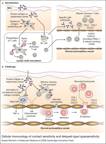
The sensitisation phase of skin DTH and CS (Fig. 1a) involves the initial exposure to antigen and its processing by antigen-presenting cells (APCs) that migrate to lymph nodes. The major APC populations in the skin are Langerhans cells in the epidermis, and dendritic cells in the dermis (Ref. Reference Roediger9). Sensitisation is usually initiated in the skin by direct application or intradermal or subcutaneous injection. The exposure of naive T cells to antigen presented in the lymph nodes gives rise to a population of antigen-specific memory T cells in lymphoid organs and the skin (Ref. Reference Robert and Kupper10). Sensitisation also induces B cells to produce antibodies that recognise the sensitising protein or haptenated peptide (Refs Reference Szczepanik11, Reference Campos12, Reference Tsuji13). The sensitisation process is complete after approximately 5 days in the mouse, but generally takes longer in humans.
Cellular immunology of contact sensitivity and delayed-type hypersensitivity. (a) In the sensitisation phase, after introduction of protein antigen or hapten into the skin, the antigen or hapten-modified protein is taken up by antigen-presenting cells in the skin, which migrate to the lymph node and initiate an antigen-specific immune response via antigen presentation to naive T cells. Some T cells develop into effector memory T (TEM) cells, altering their adhesion molecule and chemokine receptor expression by upregulating CCR4, CCR10 and a glycosylated form of P-selectin glycoprotein ligand-1 (PSGL-1), and increasing E-selectin-binding capacity. These alterations facilitate leukocyte migration into the skin, a process aided by local expression of the chemokines CCL1, CCL7 and CCL27. Similarly, B cells undergo activation and commence production of IgM antibodies that recognise the sensitising antigen. (b) Upon challenge with the sensitising antigen, IgM antibodies bind the antigen, resulting in immune complex formation, C5a generation and mast cell activation. Activated mast cells release inflammatory mediators including tumour necrosis factor (TNF) and serotonin. Similarly, skin-resident TEM cells can recognise the antigen and respond by releasing proinflammatory cytokines and chemokines. Together, the mediators released during this response activate the dermal endothelium, resulting in increased vascular permeability, which leads to skin swelling. In addition, activated endothelial cells in the dermal microvasculature support increased rolling and adhesion of leukocytes, and leukocyte emigration out of the vasculature. Expression of chemokines such as CCL1, CCL2, CCL5, CCL7 and CCL27 promote migration of lymphocytes into the skin.

The induction of a challenge response in DTH and CS (Fig. 1b) is immediate, and involves a massive inflammatory cell infiltrate and tissue swelling that peaks over the ensuing day or two. Events that occur in the first few hours following an antigen challenge are crucial for the development of a fully-fledged hypersensitivity response. Antigen-specific T effector memory (TEM) cells in the skin provide immediate effector function following challenge (Ref. Reference Sallusto14). Binding of hapten to antigen-specific IgM produced by B1 cells rapidly activates the complement system and mast cells, initiating the recruitment of the cell populations that mediate subsequent development of DTH and CS responses (Ref. Reference Askenase15). NKT cells have been implicated in stimulating this B1-cell-mediated response via the secretion of IL-4 (Ref. Reference Campos12). In CS, this early initiation phase involves the recruitment of a small population of pioneer CD4+ cells that are essential for the development of a hypersensitivity response (Ref. Reference Ptak, Herzog and Askenase16, Reference Tsuji17, Reference Hwang18, Reference Norman19). As the inflammatory response develops, T cells play a central role in the pathogenesis of skin hypersensitivity responses. CD4+ T cells are thought to be the primary effectors in DTH responses (Ref. Reference Doebis20). In the case of CS, various studies have demonstrated the importance of either CD4+ or CD8+ T cells as effectors (Refs Reference Hwang18, Reference Martin21, Reference Bour22, Reference Bouloc, Cavani and Katz23, Reference Yokozeki24, Reference Nakae25), but there is a growing consensus that both subsets contribute (Refs Reference Gocinski and Tigelaar8, Reference Wang26). In addition to effector T cells, neutrophils and macrophages are also recruited to the skin during hypersensitivity responses and might be involved in the development of inflammation (Refs Reference Tsuji17, Reference Doebis20, Reference Dilulio27).
Although T cells have a major role as effectors in skin hypersensitivity, they are also actively involved in the resolution of inflammation. Several studies have demonstrated the importance of a subpopulation of CD4+ cells with a regulatory phenotype (Tregs) in CS (Refs Reference Bour22, Reference Atarashi28, Reference Ring29, Reference Xu, DiIulio and Fairchild30) and DTH (Ref. Reference Siegmund31, Reference Mempel32, Reference Sather33). CD4+ Tregs are found in lymphoid organs and the periphery, and their depletion results in more severe or prolonged skin-hypersensitivity responses. Questions remain as to the relative immunosuppressive contributions of lymphoid versus peripheral Tregs and the precise mechanisms by which they fulfil their regulatory role.
Leukocyte-trafficking mechanisms
Leukocyte recruitment to the skin is central to the development of DTH and CS. However, much of our understanding of the mechanisms of leukocyte recruitment has come from analysis of leukocyte–endothelial cell interactions in postcapillary venules in transparent tissues (mesentery, cremaster muscle) that are amenable to examination by intravital microscopy. These studies have shown that for leukocytes in the blood to be recruited to sites of inflammation, they must first undergo a sequence of interactions with the endothelium lining blood vessels at the inflamed site (Refs Reference Griffiths and Barker5, Reference Ellis6). Leukocytes first tether and roll along the endothelial surface, before undergoing chemoattractant-dependent activation, leading to firm adhesion on the endothelium (Ref. Reference Gerszten34). Adherent leukocytes then migrate or ‘crawl’ across the endothelial surface in search of optimal sites for the final step in the sequence: transmigration out of the vasculature and into the inflamed site (Ref. Reference Phillipson35). The recently identified process of intravascular migration or ‘crawling’ has been shown to be important in allowing neutrophils to rapidly transmigrate in response to chemotactic stimulation, and is also the basis of a novel form of immune surveillance performed by monocytes in the dermal microvasculature and invariant NKT cells in the hepatic sinusoids (Refs Reference Phillipson35, Reference Hickey and Kubes36).
Distinct families of adhesion molecules mediate each of these steps. Leukocyte rolling is predominantly mediated by the selectins (P-selectin and E-selectin on endothelial cells, L-selectin on leukocytes) (Refs Reference Kanwar37, Reference Kanwar38), although α4-integrin can also mediate leukocyte rolling in the microvasculature (Refs Reference Johnston, Issekutz and Kubes39, Reference Hickey, Granger and Kubes40, Reference James, Bullard and Hickey41). Selectins mediate leukocyte rolling via interaction with glycosylated ligands, including the major P-selectin ligand, P-selectin glycoprotein ligand-1 (PSGL-1) (Ref. Reference Vachino42). Arrest and migration of rolling leukocytes is triggered by chemoattractants. A wide variety of molecules possess chemoattractant function for leukocytes, including lipid-based molecules such as leukotriene B4 (LTB4) and platelet-activating factor (PAF), products of the activated complement pathway such as C5a, and chemokines, which are small chemoattractant proteins. Chemoattractants interact with specific G-protein-coupled receptors on leukocytes, rapidly inducing signal transduction, which results in leukocyte arrest and subsequent migration (Refs Reference Ley43, Reference Bromley, Mempel and Luster44).
Leukocyte adhesion is typically mediated by leukocyte integrins. Adhesive function of this group of heterodimeric adhesion molecules is rapidly increased in response to chemokine receptor ligation. The major integrins used for interacting with endothelial cells are the β2 integrins LFA-1 (αLβ2, also known as CD11a/CD18) and Mac-1 (αMβ2, also known as CD11b/CD18) and the α4 integrins α4β1 (VLA4) and α4β7. They interact with members of the immunoglobulin superfamily expressed on endothelial cells. The major ligand for the β2 integrins is intercellular adhesion molecule-1 (ICAM-1) whereas the main ligand for α4β1 is vascular cell adhesion molecule-1 (VCAM-1) (Ref. Reference Springer45). Blockade of Mac-1 has been shown to inhibit intravascular migration of neutrophils and delay their transmigration, suggesting a key role for Mac-1 in migration across the endothelial surface.
Transmigration is arguably the most complex aspect of the leukocyte recruitment response, and it involves active contributions from both leukocytes and endothelial cells (Refs Reference Ley43, Reference Rao46, Reference Shulman47). The leukocyte integrins LFA-1, Mac-1 and VLA-4 are probably major contributors to this process. There are numerous molecules on the endothelial cell that are also potentially involved, including platelet–endothelial cell adhesion molecule-1 (PECAM-1/CD31, which is also expressed on leukocytes), ICAM-1, ICAM-2, junctional adhesion molecules (JAM-A, JAM-B, JAM-C), endothelial cell-selective adhesion molecule (ESAM), CD99 and CD47 (or integrin-associated protein) via interaction with the signal regulatory protein γ (SIRPγ) (Refs Reference Ley43, Reference Rao46, Reference Wegmann48, Reference Stefanidakis49). Much remains to be learnt regarding this step of the process, in part because the roles of several of these molecules appear to be stimulus- and cell-type-specific (Ref. Reference Ley43). For example, absence of PECAM-1 reduces leukocyte transmigration induced by IL-1β, but not by tumour necrosis factor (TNF), suggesting that PECAM-1 is involved in leukocyte recruitment induced by IL-1β but not TNF (Ref. Reference Thompson50, Reference Woodfin, Voisin and Nourshargh51). Similarly, under certain circumstances, leukocytes can transmigrate directly through endothelial cells, rather than in between them (Refs Reference Phillipson35, Reference Millan52). Together, these complexities mean that much further work is required to clarify the molecular basis of transmigration.
Studies of adhesion molecule and chemokine function in T-cell-mediated dermal inflammation
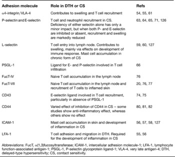
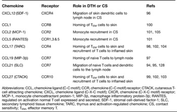
Numerous studies have examined the functional roles of adhesion molecules and chemokines in models of CS and DTH. These studies have used both antibody inhibition and gene deletion in models of acute DTH or CS, in response to either single or repeated hapten challenge (Tables 1 and 2).
Adhesion molecules involved in delayed-type hypersensitivity and contact sensitivity

Abbreviations: FucT, α(1,3)fucosyltransferase; ICAM-1, intercellular adhesion molecule-1; LFA-1, lymphocyte function-associated antigen-1; PSGL-1, P-selectin glycoprotein ligand-1; VLA-4, very late antigen-4; DTH, delayed-type hypersensitivity; CS, contact sensitivity.
Chemokines involved in delayed-type hypersensitivity and contact sensitivity

Abbreviations: CCL, chemokine ligand (C-C motif); CCR, chemokine (C-C motif) receptor; CTACK, cutaneous T-cell attracting chemokine; CXCL, chemokine ligand (C-X-C motif); CXCR, chemokine (C-X-C motif) receptor; MCP-1, monocyte chemoattractant protein-1; MIP-3β, macrophage inflammatory protein-3β; RANTES, regulated on activation normal T cell expressed and secreted; SDF-1, stromal cell-derived factor-1; SLC, secondary lymphoid tissue chemokine; TARC, thymus and activation-regulated chemokine; CS, contact sensitivity; TEM, effector memory T.
Adhesion molecules in the microvasculature
Early research focused on the roles of leukocyte integrins and their endothelial ligands, ICAM-1 and VCAM-1. These ligands have been found to be upregulated in CS, driven by the actions of the proinflammatory cytokine TNF (Ref. Reference McHale53). Use of function-blocking antibodies demonstrated roles for the β2-integrin pathway (LFA-1 and ICAM-1) and α4 integrin in mediating dermal leukocyte recruitment in sensitivity responses (Refs Reference Elices54, Reference Issekutz55, Reference Scheynius, Camp and Pure56), and these results are supported by similar findings in ICAM-1-deficient mice (Refs Reference Sligh57, Reference Xu58).
Subsequent studies focused on the role of the selectin family. Initial work with L-selectin-deficient mice reported dramatic reductions in leukocyte infiltration and swelling, suggesting a key role for L-selectin in mediating entry of leukocytes into the skin (Ref. Reference Tedder, Steeber and Pizcueta59). However, this defect was subsequently found to be due solely to inhibition of lymphocyte trafficking to lymph nodes, which resulted in impaired T cell activation and development of antigen-specific immunity, and consequently caused reduced hypersensitivity responses upon antigen challenge (Refs Reference Catalina60, Reference Catalina, Estess and Siegelman61).
Of the endothelial selectins, expression of both P- and E-selectin has been found to be increased for at least 48 h after CS challenge, suggesting an important role for these molecules (Ref. Reference Harari62). However, mice deficient in E-selectin have unaltered CS, whereas absence of P-selectin causes only a partial reduction in leukocyte recruitment, without altering the swelling response (Refs Reference Labow63, Reference Subramaniam64). These findings suggest overlapping roles for the selectins in dermal recruitment – a possibility supported by the significantly reduced CS in mice lacking both P-selectin and E-selectin (Ref. Reference Staite65). TNF and IL-1 are crucial for upregulation of these selectins, as demonstrated by studies in which inhibition of these cytokines results in reduced adhesion molecule expression (Ref. Reference Harari62).
The nature of the selectin ligands that contribute to DTH- or CS-associated leukocyte recruitment has been studied in detail. Absence of the major P-selectin ligand, PSGL-1, results in reduced T cell infiltration, but has no effect on dermal swelling (Ref. Reference Hirata66). Human memory T cells that migrate to the skin have been shown to express a unique skin-homing receptor termed cutaneous lymphocyte antigen (CLA). This is a carbohydrate epitope that is upregulated upon T cell activation and specifically binds to E-selectin (Ref. Reference Fuhlbrigge67). CLA expression is hypothesised to favour entry of T cells into the skin, via interaction with E-selectin constitutively expressed in dermal postcapillary venules (Ref. Reference Weninger68). Indeed, constitutive expression of E-selectin and E-selectin-dependent rolling in the absence of inflammatory stimulation is a specialisation of the dermal endothelium. In most other organs, E-selectin is not expressed in the absence of inflammation, as is also the case in isolated human umbilical vein endothelial cells (Refs Reference Eppihimer69, Reference Bevilacqua70).
A mouse equivalent of CLA has not been identified, although similar increases in E-selectin binding function do occur in mice upon T cell activation, particularly in Th1 cells, and these facilitate T cell entry into inflamed skin (Ref. Reference Austrup71). E-selectin binds to numerous ligands including PSGL-1, E-selectin ligand-1, CD43 and CD44 (Refs Reference Fuhlbrigge72, Reference Hidalgo73). Mice deficient in both PSGL-1 and CD43 have marked reductions in T cell selectin-binding function and reductions in swelling and recruitment that are greater than the effect associated with deficiency of either molecule alone (Refs Reference Alcaide74, Reference Matsumoto75). These studies support the view that PSGL-1 and CD43 are the major leukocyte ligands responsible for P- and E-selectin-dependent leukocyte recruitment in murine CS.
Optimal selectin-binding function is reliant on the presence of fucosyl transferases (FucTs), particularly FucT-IV and FucT-VII, which are necessary for the appropriate glycosylation of the selectin ligands. Deficiency in FucT-VII, and to a lesser extent, FucT-IV, either globally or specifically in antigen-specific T cells, significantly reduces leukocyte recruitment and swelling in CS and DTH (Refs Reference Doebis20, Reference Smithson76, Reference Erdmann77). Given that these deficiencies also affect L-selectin-mediated interactions, these effects could be due to reduced lymphocyte interactions in lymph node vessels, as well as in dermal vessels.
Finally, CD44 is a T-cell-expressed molecule that is capable of mediating rolling and leukocyte recruitment via interaction with hyaluronan (Refs Reference DeGrendele78, Reference Bonder79). Some studies have found that blockade of CD44 reduces leukocyte infiltration and swelling in CS (Refs Reference Camp80, Reference Gonda81), although others have observed a minimal role for CD44 (Refs Reference Catalina, Estess and Siegelman61, Reference Schmits82). The reasons for this discrepancy are unclear.
Adhesion molecules for extravascular migration
After leaving the vasculature, leukocytes also use adhesion molecules to migrate throughout the interstitium. Time-lapse intravital microscopy has demonstrated a crucial role for collagen-binding β1 integrins, particularly α2β1, in mediating neutrophil extravascular locomotion in the mesentery (Refs Reference Werr83, Reference Werr84). These molecules are expressed only minimally in nonactivated T cells, but their expression levels increase upon activation, and they are highly expressed on lymphocytes in inflamed skin (Ref. Reference de Fougerolles85). Furthermore, inhibition of either the α1 or α2 integrin reduces swelling and leukocyte infiltration in DTH and CS responses, and deficiency of α1 integrin results in reduced CS (Ref. Reference de Fougerolles85). Interestingly, these interventions did not reduce inflammation induced by a nonspecific irritant, suggesting that the role played by these integrins is specific to antigen-induced dermal inflammation. The mechanism for this is not clear, but may involve β1-integrin-mediated lymphocyte migration along interstitial collagen, as well as effects on lymphocyte activation and viability.
αE/CD103 is another T-cell-expressed adhesion molecule, which, in combination with β7 integrin, mediates T cell binding to E-cadherin expressed by epithelial cells (Ref. Reference Cepek86). αEβ7 has been suggested to be a marker of ‘effector memory’ Treg cells that can enter non-lymphoid tissues (Refs Reference Huehn87, Reference Lehmann88), and is believed to have a role in T cell localisation in the epidermis, and retention in the skin (Ref. Reference Suffia89). Expression of CD103 by Treg cells has been shown to define a population with the ability to migrate to the skin and inhibit a DTH response. In addition, mice lacking CD103 can be affected by spontaneous skin inflammation, supporting the contention that this integrin is important in migration and/or retention of T cells that dampen skin inflammation (Refs Reference Siegmund31, Reference Schon90).
Despite these findings, a recent study has raised the possibility that leukocyte integrins are dispensable for leukocyte migration (Ref. Reference Lammermann91). Leukocytes deficient in integrins of the β1, β2, β7 and αv families were found to migrate through three-dimensional substrates, and through the extravascular space, as efficiently as wild-type leukocytes, via a process of contraction and protrusion. These unexpected findings suggest that there remains much to be learnt regarding the mechanics of extravascular leukocyte migration.
Chemokines
Chemokines are diffusible chemoattractant proteins that are required for several steps in the development of a skin hypersensitivity response (Table 2). Various chemokines are produced by cells residing in the lymph nodes, in intrinsic skin cells and in infiltrating cells from the immune system. In addition, there is evidence that chemokines are retained on the luminal surface of endothelial cells in a manner that promotes leukocyte recruitment (Ref. Reference Middleton92). Chemokine receptors are a family of membrane-bound, G-protein-coupled receptors that display a degree of specificity for the chemokines that they are able to bind (Ref. Reference Zlotnik and Yoshie93). Thus, the chemokine receptor complement of a circulating or migrating cell influences its potential for tissue-specific recruitment.
The chemokines CCL21 (SLC) and CXCL12 (SDF-1) are involved in sensitisation and facilitate the migration of skin-localised APCs carrying antigen to the lymph node (Refs Reference Forster94, Reference Gunn95, Reference Kabashima96). The chemokines CCL21 and CCL19 (MIP-3β) also facilitate the homing of naive T cells to lymph nodes where they encounter these APCs. (Refs Reference Forster94, Reference Gunn95, Reference Baekkevold97). Following T cell activation, homing of antigen-specific TEM cells to the skin involves the chemokines CCL17 (TARC) and CCL27 (CTACK) and their respective receptors CCR4 and CCR10 (Fig. 1a) (Refs Reference Campbell98, Reference Morales99). Examination of healthy human skin has revealed that the majority of skin-resident T cells express CCR8, a receptor rarely found on T cells in other organs (Ref. Reference Schaerli100). These findings suggest a key role for the CCL1–CCR8 pathway in aiding homeostatic migration of TEM cells to the skin in humans.
The expression of several proinflammatory chemokines is upregulated during development of a hypersensitivity response (Refs Reference Tsuji17, Reference Goebeler101). As is the case with recruitment of TEM cells to the non-inflamed skin, CCL17 and CCL27 are required for skin-specific recruitment of T cells during the inflammatory phase of hypersensitivity (Refs Reference Reiss102, Reference Homey103, Reference Campbell, O'Connell and Wurbel104). CCL2 (MCP-1) and CCL5 (RANTES) expression is also increased in the inflamed skin, promoting the recruitment of monocytes (Fig. 1b) (Refs Reference Goebeler101, Reference Lu105).
In vivo imaging of leukocytes in the inflamed dermal microvasculature
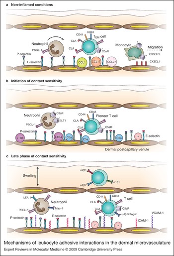
The critical barrier function of the skin is reflected in several aspects of the trafficking and functions of immune cells in this organ. The dermal microvasculature supports a high level of constitutive leukocyte rolling, which enables neutrophil recruitment to be induced rapidly upon inflammatory challenge (Fig. 2a). Constitutive rolling has been demonstrated in areas of skin such as the mouse ear, which can be examined using intravital microscopy without the requirement for surgical exteriorisation and its associated inflammatory response (Refs Reference Weninger68, Reference Mayrovitz106, Reference Janssen107). Ongoing rolling is dependent on constitutive expression of both P-selectin and E-selectin. More recent imaging studies have used in vivo confocal microscopy to examine nonperturbed dermal microvessels in transgenic mice in which green fluorescent protein is expressed exclusively by a subset of monocytes (Ref. Reference Auffray108). These leukocytes randomly migrate throughout the entire microvasculature, behaviour that has been interpreted to be a form of immune surveillance (Fig. 2a). Immediately upon inflammatory stimulation, the monocytes exit the vasculature and have a key role in driving subsequent immune responses in models such as local Listeria monocytogenes infection (Ref. Reference Auffray108). The relevance of these cells in dermal sensitivity responses remains to be determined, but it must be noted that this behaviour is not restricted to the skin or indeed organs exposed to the external environment – it is also seen in other microvascular beds such as the mesentery and brain (Refs Reference Auffray108, Reference Audoy-Remus109).
In vivo imaging has also revealed that the dermal microvasculature responds to inflammatory stimuli in a distinct fashion (Ref. Reference Hickey110). In an IgE-dependent model of antigen-induced leukocyte recruitment, we have previously shown that whereas antigen challenge in muscle induces upregulation of only P-selectin, the identical challenge in skin results in increased E-selectin expression but no change in P-selectin expression (Ref. Reference Hickey110). Within postcapillary venules, this results in different effects on leukocyte–endothelial cell interactions: in the skin the major effect was a reduction in leukocyte rolling velocity, whereas in the muscle, the number of rolling leukocytes increased in the absence of a change in rolling velocity. We have also observed key roles for P- and E-selectin in dermal leukocyte recruitment in vasculitis-prone MRL/faslpr mice (Ref. Reference Hickey111). These mice, which are affected by a systemic inflammatory stimulus, also undergo leukocyte recruitment to the cerebral microvasculature. The capacity of the dermal microvasculature to use tissue-specific combinations of adhesion molecules in response to a similar inflammatory stimulus is further illustrated by the finding that, in these mice, leukocyte adhesion in the skin occurs via different molecular mechanisms from those used in the cerebral microvasculature (Ref. Reference James, Bullard and Hickey41). Together, these imaging studies illustrate that leukocyte recruitment mechanisms in the skin are very different to those in many other organs, even in response to the same inflammatory stimulus.
Imaging the dermal microvasculature in contact sensitivity
More recent studies have examined the recruitment mechanisms at work in CS. As previously described, it is hypothesised that ‘pioneer’ T cells arriving early in the CS response are essential for the development of the subsequent antigen-specific inflammatory response. The recruitment mechanisms of this population have been difficult to examine. Hwang and co-workers addressed this question using intravital microscopy to examine leukocyte recruitment in dermal postcapillary venules during the initiation phase of a CS response in the mouse ear (Ref. Reference Hwang18). Several key points came out of this work. Following hapten challenge, two peaks of intravascular leukocyte adhesion were observed, a rapid one peaking at 2 h, and a more sustained one between 4 and 24 h (Fig. 2b,c). The initial phase of recruitment was dependent on both P- and E-selectin, whereas at 24 h, an additional role for the α4 integrin was observed. In addition, these studies examined the importance of the initial phase of leukocyte recruitment on the subsequent late-phase response. Several experiments generated findings consistent with the concept that early leukocyte recruitment acts to promote the subsequent inflammatory response. Inhibition of the initial phase of recruitment via combined blockade of P- and E-selectin completely eliminated rolling, adhesion and the dermal inflammatory response during the subsequent late (24 h) phase. However, if this selectin inhibition was applied 2 h after hapten challenge, the late phase developed as normal, indicating that the leukocyte recruitment during the initial 2 h period is sufficient to allow for full initiation of CS. This response was only marginally affected by neutrophil depletion, indicating that pioneer leukocytes other than neutrophils are responsible for this effect. To test the alternative hypothesis that this response was mediated by CD4+ T cells, an adoptive transfer approach was utilised. If CD4+ T cells from a sensitised mouse were transferred into a sensitised CD4 T-cell-deficient mouse prior to challenge, the late-phase response continued unabated. However, if the transfer was performed only 2 h after hapten challenge, the intravascular adhesion response at 24 h was eliminated. Together, these experiments provide direct evidence of the existence of a critical post-challenge period during which recruitment of effector T cells must occur to initiate a full CS response.
Mechanisms of leukocyte adhesive interactions in the dermal microvasculature. (a) Under resting conditions, endothelial cells in dermal postcapillary venules constitutively express P-selectin and E-selectin, which mediate basal leukocyte rolling, via interaction with leukocyte-expressed P-selectin glycoprotein ligand-1 (PSGL-1). Effector memory T (TEM) cells express CD43, which can promote rolling interactions with E-selectin ligand, and CD44 which can promote interactions with hyaluronan and potentially, E-selectin. Similarly, human skin-homing TEM cells express cutaneous lymphocyte antigen (CLA), an additional E-selectin ligand, and the receptor for the complement component, C5a. Constitutively expressed chemokines including CCL1, CCL17 and CCL27 facilitate migration of TEM cells into the skin, although the adhesion molecules required for this process are unknown. In addition, monocytes migrate or ‘crawl’ throughout the dermal vasculature in what is hypothesised to be a form of surveillance that allows them to mediate a rapid response upon the detection of inflammatory stimuli such as infection. This process is facilitated by fractalkine/CX3CL1 via interaction with monocyte-expressed CX3CR1. (b) In the initial phase of contact sensitivity (CS), neutrophils and ‘pioneer’ T cells undergo rolling interactions mediated by P- and E-selectin, PSGL-1 and potentially CLA and CD43. The rapidly generated chemoattractants C5a (attracts CD4+ T cells and neutrophils via interaction with the C5a receptor C5aR) and LTB4 (attracts neutrophils via interaction with the leukotriene B4 receptor, BLT1) promote arrest of specific leukocyte subsets within dermal venules, although other unidentified chemoattractants (designated by ‘?’) may also contribute to this process. (c) In the late phase of CS, in addition to the endothelial selectins, the α4 integrin has a role in mediating leukocyte–endothelial cell interactions, associated with upregulation of endothelial vascular cell adhesion molecule-1 (VCAM-1). Other adhesion molecules implicated include leukocyte-expressed LFA-1, which interacts with endothelial intercellular adhesion molecule-1 (ICAM-1), and CD44. The chemoattractants required for arrest (designated by ‘?’) of rolling cells have not been definitively identified. Upon entering the tissue, the collagen-binding integrins α1β1 and α2β1 may contribute to intradermal T cell migration, and the αE-integrin (CD103) is hypothesised to promote interaction of T cells with epidermal epithelial cells and retention in the skin. It is during this phase that skin swelling due to increased vascular permeability is most apparent.

Ring and colleagues used a dorsal skinfold chamber model to examine CS-induced leukocyte rolling and adhesion in non anaesthetised mice (Ref. Reference Ring29). As seen in the experiments discussed above, they observed a reduction in leukocyte rolling velocity and a very rapid (<30 min) increase in leukocyte adhesion in response to hapten challenge. This study showed that administration of CD4+ CD25+ Treg cells markedly inhibits the increased adhesive interactions in the dermal microvasculature, a response that is dependent on Treg-derived IL-10 (Ref. Reference Ring29). A similar approach has been used to examine the roles of CD44 and L-selectin in DTH-associated leukocyte rolling and adhesion (Ref. Reference Gonda81). Comparable levels of sensitisation were achieved in wild-type, CD44-deficient, L-selectin-deficient and double-knockout mice. However, the absence of CD44, but not L-selectin, abrogates the CS-associated reduction in leukocyte rolling velocity and also reduces leukocyte adhesion and DTH-associated swelling.
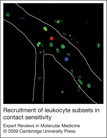
Despite the novelty of the findings from experiments using conventional intravital microscopy to examine leukocyte–endothelial cell interactions in real time in vivo, one weakness of these studies is that they do not differentiate between the various leukocyte subsets that may interact within the microvasculature. This is highly relevant in CS because of the potential functional roles of numerous cell types of varying frequencies, including CD4+ and CD8+ T cells, Tregs and neutrophils. To define adhesion mechanisms specific to each of these subsets, an alternative approach is required. The advent of high-speed in vivo multichannel intravital microscopy, a technique that allows several fluorochromes to be visualised simultaneously, has enabled this deficiency to be overcome (Ref. Reference Chiang112). Using small amounts of subset-specific antibodies (e.g. anti-CD4 for CD4+ T cells; anti-Gr-1 for neutrophils) conjugated with different fluorochromes, this technique now allows the online identification of leukocyte subsets and specific analysis of their recruitment mechanisms (Fig. 3).
Recruitment of leukocyte subsets in contact sensitivity. Confocal microscopy image of a dermal postcapillary venule during a contact sensitivity response, showing numerous interacting leukocytes. White lines denote the venular lumen. A CD4+ T cell (red, phycoerythrin-conjugated anti-CD4) and a CD8+ T cell (blue, allophycocyanin-conjugated anti-CD8) are visible interacting with the venular endothelium, and numerous neutrophils (green, Alexa Fluor 488-conjugated anti-Gr-1), are present within the vessel, and in the perivascular tissue. This imaging approach allows the mechanisms and kinetics of recruitment specific to each subset to be identified.

Norman and colleagues used in vivo spinning-disk confocal microscopy to examine the recruitment mechanisms of CD4+ T cells and neutrophils in the first 2 h of the CS response (Ref. Reference Norman19). They found marked differences in the frequency of interactions of these two populations: analysis of rolling cells showed that one CD4+ T cell was observed for every 10 to 18 neutrophils, and when examining leukocyte adhesion, there were six times fewer CD4+ T cells than neutrophils. This is noteworthy because despite the low frequency of CD4+ T cells, earlier studies have shown that these cells are essential contributors to the inflammatory response (Ref. Reference Hwang18). This study also demonstrated the roles of potential mediators involved in leukocyte arrest within the microvasculature during early CS, and showed that T cell adhesion is significantly reduced in C5aR-deficient mice (Fig. 2b), which is consistent with a role for complement activation in early CD4+ T cell recruitment in CS (Ref. Reference Norman19).
Use of in vivo imaging to examine T cell biology in the dermal interstitium
The final step in the process of T cell recruitment involves their exit from the vasculature and migration through the dermis. Recent advances in in vivo imaging, in particular the application of multiphoton microscopy, are increasing our understanding of the biology of T cells within the interstitium (Refs Reference Miller113, Reference Mempel, Henrickson and von Andrian114). This form of microscopy uses long-wavelength rapidly pulsed lasers to enable imaging of cells and structures up to 500 µm below the surface of tissues. A great deal of work has examined the actions of T cells, dendritic cells and other leukocyte populations within lymphoid organs, and demonstrated the active migration of T cells within lymph nodes and the dynamic nature of interactions between T cells and dendritic cells during the development of an antigen-specific immune response (Ref. Reference Mempel, Henrickson and von Andrian114). However, less is known of interstitial T cell migration in the periphery.
Multiphoton microscopy has been used to examine migration of passively transferred, activated myelin basic protein-specific CD4+ T cells in brain explants. In these studies, the majority of T cells were found to migrate randomly, although a small subset formed stable adhesions, actions that are thought to represent interactions with APCs (Ref. Reference Kawakami115). A similar approach was used to examine the behaviour of tumour-infiltrating lymphocytes in skin-resident tumours in mice, although in this case, a transgenic mouse in which the T cells expressed green fluorescent protein (GFP) was used. This technique provides the additional benefit of enabling the examination of migration of endogenous, rather than transferred, T cells (Ref. Reference Mrass116). As described for T cells in the brain, T cells recruited to subcutaneously implanted tumours randomly migrated throughout the tumour, but also interacted with antigen-bearing tumour cells for varying periods. In addition, these cells were observed to migrate along collagen fibres within tumours. Comparison of wild-type and CD44-deficient tumour-infiltrating lymphocytes in the same model demonstrated a role for CD44 in efficient migration through the tumour interstitium, via regulation of lymphocyte polarity (Ref. Reference Mrass117).
More recently, this technology has been applied specifically to imaging immune cells in the skin (Ref. Reference Roediger9). Dermal T cell migration has also been examined in a CD4+ T-cell-dependent model of DTH in the rat (Ref. Reference Matheu118). Activated ovalbumin-specific, CD4+ T cells were passively transferred to rats 48 h before intradermal challenge with ovalbumin. This delay allows the transferred T cells to migrate into the skin before introduction of the antigen. Three hours after antigen challenge, the majority of ovalbumin-specific T cells were not migrating, but rather maintained stable contact with an antigen-bearing cell, presumed to be an APC. By 24 h, the T cells had enlarged and had become highly motile and polarised, migrating extensively along dermal collagen fibres. This was thought to indicate that T cells undergo activation during the contact with APCs (Ref. Reference Matheu118). However, stronger evidence of the formation of an ‘immune synapse’ between a T cell and an APC could be provided via demonstration of the upregulation of T cell activation markers, or of T cell division. Alternatively, clear identification of the antigen-bearing cells as dermal dendritic cells or Langerhans cells would provide unequivocal evidence that the cells involved in these interactions are professional APCs Data of this nature are required before these interactions can be definitively described as interactions between T cells and APCs.
APC migration in mouse skin was examined in a recent study in which yellow fluorescent protein (YFP) was expressed under the control of the CD11c promoter, thereby labelling dendritic cells (Ref. Reference Ng119). In these animals, YFP-positive dermal dendritic cells were found to spontaneously migrate throughout the non-inflamed skin. However, after intradermal injection of Leishmania major, dermal dendritic cells ceased migration, enabling them to ingest the parasites and process them into intracellular compartments. These findings are thought to indicate that dermal dendritic cells undertake ongoing tissue surveillance while retaining the capacity to respond rapidly to pathogen invasion.
Clinical implications
Many existing therapies used to treat inflammatory diseases of the skin, such as corticosteroids, are nonspecific immune suppressants with multiple side effects. As such, there is a need for effective therapies of greater specificity with fewer side effects. Adhesion molecule blockade has great potential as a therapeutic strategy to treat skin inflammatory diseases. For example, Efalizumab, which is an inhibitory antibody against Mac-1/CD11a, has proven efficacious in psoriasis patients (Ref. Reference Pariser120). However, even these quite specific therapeutic agents are not without side effects. This is illustrated by the recent withdrawal of Efalizumab from clinical use, because of induction of the severe demyelinating disease progressive multifocal leukoencephalopathy (PML) in psoriasis patients (Ref. Reference Pugashetti and Koo121). It is conceivable that the complex nature of T-cell-mediated inflammation, involving contributions from several T cell subsets that are potentially recruited via different molecular mechanisms, will provide an opportunity for these therapeutic approaches to be further improved in the future. For example, existing strategies aimed at blocking effector T cell recruitment into the skin might also restrict entry of Tregs, thereby removing local regulation of inflammation. In addition, in chronic forms of skin inflammation, when many lymphocytes are already resident in the skin, targeting of ongoing leukocyte recruitment might be less effective than blocking the actions of extravascular leukocytes. Given these considerations, further research is required to completely understand the biology of leukocyte recruitment in the skin, and the potential for antiadhesive agents to be beneficial in a range of T-cell-mediated skin diseases.
Research in progress and outstanding research questions
The application of in vivo imaging to the field of leukocyte trafficking in T-cell-dependent dermal inflammation has greatly increased our understanding of this process. However, the immunology of this area is complex and much remains to be clarified, including the intravascular interactions of the various T cell subsets (Th1, Th2, Th17, Tregs, CD8+), as well as other cells involved in the response (mast cells, dendritic cells). In addition, imaging studies are only just beginning to examine the mechanisms of intradermal T cell migration and localisation. This is a crucial issue because T cell localisation within the skin is believed to be a key factor in the determination of T-cell-mediated responses.
Skin diseases mediated by sensitivity reactions often involve repeated or prolonged exposure to the inducing factor or antigen. As such, these diseases are commonly encountered during the chronic phase of inflammation. This complicates the immunological mechanisms further, because recruited T cells add to the already high number of skin-resident T cells (Ref. Reference Clark122). The role of the skin-resident T cell in CS and other T-cell-mediated dermal responses requires further examination. Finally, studies of chronic CS show that the peak of leukocyte infiltration and swelling occurs substantially more rapidly after hapten exposure than in acute CS (Refs Reference Kitagaki123, Reference Kitagaki124, Reference Man125), and that cytokine production becomes biased towards a Th2 pattern, with increased IL-4 and IL-10 expression and significantly elevated circulating IgE (Ref. Reference Kitagaki124). How these changes affect the process of dermal leukocyte recruitment has not been examined. Future application of in vivo imaging will provide an ideal approach for the investigation of these issues, and will continue to increase our understanding of the immunology of T-cell-mediated dermal inflammation.
Acknowledgements and funding
The authors would like to thank the reviewers for their helpful suggestions, which improved the quality of the manuscript. This work is supported by a Program Grant (334067) from the National Health & Medical Research Council of Australia. M.J.H. is a National Health & Medical Research Council Senior Research Fellow.