

Introduction
Grapevine (Vitis spp.) presents a great production of fruits with high economic value worldwide (Myles et al. Reference Myles, Boyko, Brown, Grassi, Owens, Aradhya, Prins, Reynolds, Chia, Ware, Bustamante and Buckler2011; This et al. Reference This, Lacombe and Thomas2006). Unfortunately, grapes have been susceptible to various pathogens, among them the dagger nematode, Xiphinema index (Hewitt et al. Reference Hewitt, Raski and Goheen1958). This migratory ectoparasitic nematode is considered a major pest in grape-growing countries in Mediterranean environments and temperate climates where grapevine grows (Hao et al. Reference Hao, Fayolle, van Tuinen, Chatagnier, Li, Gianinazzi and Gianinazzi-Pearson2012; M’rabet Samaali et al. Reference M’rabet Samaali, Mougou Hamdane, Toumi, Dhaouadi and Kallel2022). Xiphinema index is a nematode vector of nepovirus Grapevine fanleaf virus (GFLV), which is the causal agent of grapevine fanleaf degeneration disease (Belin et al. Reference Belin, Schmitt, Demangeat, Komar, Pinck and Fuchs2001; Hewitt et al. Reference Hewitt, Raski and Goheen1958). GFLV belongs to the genus Nepovirus of the family Comoviridae (Mayo & Robinson Reference Mayo and Robinson1996). It is one of the most destructive grapevine viruses worldwide (Nourinezhad Zarghani et al. Reference Nourinezhad Zarghani, Shams-Bakhsh, Sokhandan-Bashir and Pazhouhandeh2012). Upon infestation of host roots, the nematode perforates the cell wall with the stylet, followed by salivation and ingestion of the cell cytoplasm. This results in hypertrophy and necrosis at feeding sites. As a result, the root tips progressively swell and gradually transform into a terminal gall (Weischer & Wyss Reference Weischer and Wyss1976; Wyss Reference Wyss1977). The transmission of GFLV by X. index is non-circulative and semi-persistent (Brown & Weischer Reference Brown and Weischer1998; Mc Farlane Reference Mc Farlane2003; Taylor and Brown Reference Taylor and Brown1997). GFLV could be acquired from infested plants and transmitted to recipient plants within 1 to 10 min (Wyss Reference Wyss, Maramorosch and Mahmood2000). GFLV does not replicate within the nematode and has no negative impact on the reproduction of X. index (Das & Raski Reference Das and Raski1969).
Other Longidorus species, such as X. diversicaudatum Thorne, X. vuittenezi (Luc et al., Reference Luc, Lima, Weischer and Flegg1964), and X. italiae Meyl, are known or suspected of being vectors of nepoviruses (Wang et al. Reference Wang, Gergerich, Wickizer and Kim2002). Vector Xiphinema species could retain GFLV adhered to the surface of its cuticular lining in a specific region of the esophagus. Later, the nematode releases this nepovirus when its stylet is inserted into the parenchyma tissue of growing root tips (Demangeat et al. Reference Demangeat, Voisin, Minot, Bosselut, Fuchs and Esmenjaud2005; Wang et al. Reference Wang, Gergerich, Wickizer and Kim2002; Van Ghelder et al., Reference Van Ghelder, Reid, Kenyon, Esmenjaud and Lacomme2015). Several methods, including immunoassays, reverse transcriptase polymerase chain reaction (RT-PCR), and real-time RT-PCR, are used to detect viruses in nematodes (Deng et al., Reference Deng, Li and Tang2003; Osman et al. Reference Osman, Hodzic, Kwon, Wang and Vidalakis2015). Grapevine chrome mosaic virus and Arabis mosaic virus were vectored by X. vuittenezi and X. diversicaudatum, respectively (Andret-Link et al. Reference Andret-Link, Schmitt-Keichinger, Demangeat, Komar and Fuchs2004; Digiaro et al. Reference Digiaro, Elbeaino and Martelli2017; Van Ghelder et al. Reference Van Ghelder, Reid, Kenyon, Esmenjaud and Lacomme2015). Besides, X. italiae has also been reported as a vector of GFLV (Cohn et al. Reference Cohn, Tanne and Nitzani1970). Controversially, other studies have reported that X. italiae does not act as a specific vector of GFLV (Brown et al., Reference Brown, Robertson and Trudgill1995; Catalano et al., Reference Catalano, Savino and Lamberti1992; Martelli, Reference Martelli1975).
In this study, we aimed to detect GFLV from Xiphinema species. To this end, for each Xiphinema specimen, the posterior body half of the nematode was used for identification, and the anterior body half was used to detect GFLV. Furthermore, the ability of X. index and X. italiae to transmit GFLV when feeding on grapevine roots was studied.
Material and methods
Soil sampling and nematode isolation
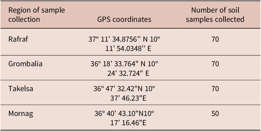
During the spring season, 260 rhizosphere soil samples were collected from four Tunisian grapevine-growing regions, including Rafraf, Grombalia, Takelsa, and Mornag (Table 1). Soil samples were collected at a depth of 30 – 80 cm from vineyards showing typical GFLV symptoms. Then, nematodes were extracted from each soil sample according to Cobb’s decanting and sieving methods (Brown & Boag Reference Brown and Boag1988 ; Flegg Reference Flegg1967). Nematode specimens were mounted on glass slides with sterile distilled water and visualized under a light microscope (Olympus C40, model SZX-ILLK200, Japan). The morphometric features reported by Luc and Dalmasso (Reference Luc and Dalmasso1975), Siddiqi (Reference Siddiqi1974), and Cohn (Reference Cohn1977) served to identify Xiphinema species. The built-in software (Nikon Eclipse 50i) was used to measure these morphometric characters.
Geographical location of soil sampling sites

Nematode molecular identification
Twenty specimens each of X. index and X. italiae were cut transversely into two fragments using a sterilized scalpel. The posterior body half was transferred to a sterilized tube (1.5 mL) containing 20 μL of RNase-free water and stored at –20°C for ulterior molecular analysis. The anterior part was placed in a sterilized Eppendorf tube (1.5 mL) containing 10 μL of RNA laterTM (Qiagen, Germany) and stored at –20°C for GFLV detection (Kulshrestha et al. Reference Kulshrestha, Hallan, Raikhy, Adekunle, Verma, Haq and Zaidi2005).
Total genomic DNA was extracted from each single nematode according to the modified protocol described by Wang et al. (Reference Wang, Bosselut, Castagnonen, Voisin, Abad and Esmenjaud2003). The posterior body half of the nematode was added to Eppendorf microtubes containing 2 μL of proteinase K (60 μg/mL) and 2 μL of Taq polymerase buffer 10X (Invitrogen). Then, the mixture was crushed gently using a sterilized cone and incubated for 1 h at 60°C and 10 min at 95°C. The obtained DNA lysate was stored at –20°C. For molecular identification of Xiphinima sp, the Internal Transcribed Spacer 1 (ITS1) sequences spanning the 18S and 5.8S ribosomal DNA were amplified using primer sets: S-ITS1 (5′-TGATTACGTCCCTGCCCTTTGTAC-3′) and A-ITS1 (5′-CGAGCCTAGTGATCCACCGCTTAG-3′). For the specific molecular identification of X. index and X. italiae species, specific PCR primers were used to amplify ITS1 gene sequences. To identify X. index, primers my-I27 (5′CGTTAGTACACACGGCGACGAA3′) and myA-ITS1 (5′CGAGCCTAGTGATCCACCGCTTAG3′) were used, whereas primer sets my-ITA26 (5′CCGTCGGTTTCGAAGGTCTG3′) and myA-ITS1 (5′CGAGCCTAGTGATCCACCGCTTAG3′) were used to identify X. italiae. These species-specific primers were newly designed.
Each PCR was performed in a 25 μL total volume, containing: 1X PCR buffer, 1.5 mM MgCl2, 0.2 mM dNTPs, 0.8 pmol for each primer, 0.5 units Taq polymerase (Bioron), 5 μL of the genomic DNA, and distilled water adjusted at 25 μL.
All PCR reactions were carried out using a thermocycler (Qiagen, Germany), programmed as follows: initial denaturation step at 94°C for 5 min, followed by 35 cycles of denaturation at 94°C for 30 s, annealing for 45 s at 56°C (for my-V18/myAITS1) or 58°C (for S-ITS1/A-ITS1, and my-ITA26/ myAITS1) or 60°C (for my-I27/myAITS1), and an elongation at 72°C for 60 s. The final extension was performed at 72°C for 10 min. PCR products were purified and sequenced by Genome Express services (Neylan, France). The Netprimer software was used to determine the annealing temperature of each primer set.
Phylogenetic analysis
The obtained sequences were deposited in the GenBank database using the basic local alignment search tool (BLASTn) of the National Centre for Biotechnology Information and were aligned using the ClustalW software implemented in MEGA 7 (Tamura et al. Reference Tamura, Peterson, Peterson, Stecher, Nei and Kumar2011). The likelihood method was used to calculate the trees using the Jones-Taylor-Thonnton evolutionary model in 1000 bootstrap replications (Jones et al. Reference Jones, Taylor and Thornton1992). The genetic distances among and within groups were determined by the same software. Meloidogyne Luci (LN713294) was used as the outgroup taxa.
Extraction of GFLV RNA from the nematode
Demangeat et al. (2005) reported that GFLV resides in the anterior region of the nematode. The GFLV-RNA extraction protocol from the anterior part of Xiphinema species was applied using the RNesay kit (Plant Mini Kit) and the QIAcube HT extractor (Qiagen, Germany), as described by Kulshrestha et al. (Reference Kulshrestha, Hallan, Raikhy, Adekunle, Verma, Haq and Zaidi2005).
Synthesis of complementary DNA
To linearize RNA, a mixture of 10 μL of TNA, 1 μg/μL random primers (Invitrogen Corporation, USA), and 1.5 μL RNase-free water was heated at 95°C for 5 min. Next, RT was carried out by adding to the prepared mixture: enzyme buffer Fs 5xcc (Invitrogen, USA), 0.1M DTT (Invitrogen, USA), 10 mM dNTPs (Promega, USA), and 200 U/μL reverse transcriptase enzyme Moloney Murine Leukemia Virus M-MLV (Invitrogen, USA). Then, the mixture was incubated at 39°C for 60 min, followed by 70°C for 10 min in a thermocycler (Qiagen, Germany). The obtained DNAc were stored at –20°C.
PCR
PCR amplification was performed using a mixture of 2.5 μL of DNAc, Taq polymerase buffer 10X, 1.5 mM MgCl2, 10 mM dNTPs, 10 μM of each primer, and 5 U/μL of Taq polymerase. GFLV-specific primers were used for RT-PCR, GT1076 (5′-CCAAGGATTGCCAGGCA-3′) and GT1826 (5′-TCCATAGTGTCCCGTTCC-3′) (Saamali M’rabet et al. Reference Saamali M’rabet, Toumi, Mougou-Hamdane, Sahbi and Kallel2018). PCR cycling conditions used were as follows: an initial denaturation step at 94°C for 4 min, followed by denaturation at 94°C for 30 s (35 cycles), annealing at 57°C for 45 s and elongation at 72°C for 60 s. A final extension was performed at 72°C for 7 min. PCR products were separated on 0.5% TAE (Tris–acetic acid–EDTA) buffered agarose gel stained with ethidium bromide.
Acquisition and transmission of GFLV by X. index and X. italiae under controlled condition
To evaluate the acquisition and the transmission of GFLV by X. index and X. italiae, for each nematode population, 20 Muscat d’Alexandrie grapevines (infected with GFLV), rooted in pots, were inoculated with nematode suspension (100 nematodes suspended in 10 mL of distilled water).
GFLV-carrying nematodes were obtained from grapevines showing GFLV symptoms. The presence of GFLV in grapevine samples was detected using DAS-ELISA (Double-Antibody Sandwich) according to the Bioreba protocol. DAS-ELISA was carried out using 1:1000 dilution of polyclonal antiserum anti-GFLV IgG. The optical density was measured at 405 nm using an automatic microplate reader (Multisacan Ascent, Labsystems USA). The positive signal threshold was set at twice the mean of healthy controls. A total of 20 healthy Muscat d’Alexandrie (free GFLV), taken from cuttings, were rooted in (2-L) pots and placed under greenhouse conditions.
The French population of X. index was reared on Ficus carica and used as a positive control. Fig plants were not found to be infected by X. index, which was not viruliferous (Esmenjaud et al. Reference Esmenjaud, Walter, Minot, Voisin and Cornuet1993). Fig cuttings were cultivated in black plastic bags (2 L) using sterilized soil as described by Demangeat et al. (Reference Demangeat, Komar, Cornuet, Esmenjaud and Fuchs2004) and maintained in greenhouse conditions. Healthy grapevine plants that were not inoculated with nematodes were used as a negative control.
Six weeks after inoculation, the infected grapevines were carefully removed from pots. Next, the soil in the pot of each grapevine-infected sample was used to retransplant the healthy Muscat d’Alexandrie (free GFLV). Six weeks after incubation, the transmission of the virus to these healthy plants by nematodes was checked using RT-PCR. The bioassay was performed under greenhouse conditions.
To evaluate the transmission of GFLV from nematodes to grapevine plants, the virus was detected in the root, leaf, and stem of each inoculated plant using RT-PCR. To evaluate the acquisition of GFLV, nematodes were extracted from each grapevine root, and re-identified molecularly using the PCR method as mentioned previously. Subsequently, for each nematode population (X. index-TN, X. italiae-TN, and X. index-FR), 20 specimens of nematode were used to detect the eventual presence of GFLV using RT-PCR.
Results
Molecular identification of nematodes
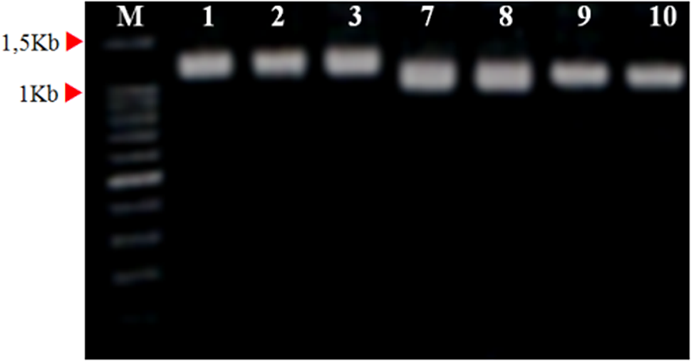
For DNA extracted from the posterior half of nematode, using primer pairs of my S-ITS1 and my A-ITS1, the approximate sizes of the amplified products obtained were 1.1 – 1.2 kbp. The findings indicated that the nematode populations used belong to the genus Xiphinema (Figure 1). The amplified PCR product, using specific primers (my-I27 and my-A-ITS), was 250 bp, corresponding to X. index (Figure 2A). Similarly, using specific primers (my ITA26 and my A-SIT), a PCR product of the expected size (900 bp) was obtained, corresponding to X. italiae (Figure 2B). The molecular identification was confirmed with morphological characterization.
PCR products obtained using primers my A-ITS1 and my S-ITS1. M: 100 bp DNA marker. Lanes 1, 2, and 3 correspond to the amplification of the genome of the posterior part of three individuals of X. index. Lanes 7, 8, and 9 correspond to the amplification of the genome of the posterior part of three individuals of X. italiae. Lane 10 corresponds to the amplification of the genome of X. index French population.

(A) Electrophoresis of the amplification product from DNA isolated from posterior parts of X. index of the three geographical regions (Rafraf, Grombalia, and Takelsa). M: 100 bp DNA marker. Lanes 1 – 2 and 3: X. Index from Rafraf. Lanes 4 – 5 X. index from Grombalia. Lanes 6 – 7: X. index from Takelsa. Lane 8: Negative control and Lane 9: positive control (X. index French population). (B) Electrophoresis of the amplification product from DNA isolated from posterior parts of X. italiae from the three geographical regions (Rafraf, Grombalia, and Takelsa). M: 100 bp DNA marker. Lanes 1 – 2: X. Italiae from Rafraf. Lanes 3 – 4 X. italiae from Grombalia. Lanes 5 – 6: X. italiae from Takelsa. Lane 7: Positive control.

Phylogenetic analyses
The phylogenetic analyses were performed in “MEGA7” to reconstruct the evolutionary history of gene sequences of the Tunisian populations of X. index as well as X. italiae. The Tunisian populations of X. index matched well with gene sequences deposited in GenBank, being 99% – 100% similar with the accessions AY430175, AJ437026, AY584243, HM921334, and JF37918, originating from Belgium, France, Italy, Spain, and Chile, respectively (Table 2). The phylogenetic tree showed that X. index isolates from different geographical regions are dispersed in the tree and do not form a distinct group, suggesting a low level of divergence among the different taxa (Figure 3). A clear separation between X. index isolates and X. italiae was observed, revealing that the two species of Xiphinema were genetically distinct. The Tunisian isolates of X. italiae showed 88% – 94% similarity to the Tunisian isolate (KX062698) (Guesmi-Mzoughi et al., Reference Guesmi-Mzoughi, Archidona-Yuste, Cantalapiedra-Navarrete, Palomares-Rius, Regaieg, Horrigue-Raouani and Castillo2017), the Spanish isolates (KX244936 and KX244937), and the French isolate (AJ437029) (Table 3). The 12 Tunisian populations of X. italiae formed a monophyletic clade with each other. However, the Tunisian isolate (KX062698) formed a separate clade with the Spanish and French isolates, which was distinct from the other 12 Tunisian populations (Figure 4).
Nucleotide identity (%) of the Tunisian sequences of X. index in comparison with the sequences deposited in GENBANK

Nucleotide identity (%) of the Tunisian sequences of X. italiae in comparison with the sequences deposited in GENBANK

Phylogenetic tree linking the different Tunisian populations of X. index (red rectangle) according to the likelihood method. The Tunisian populations of X. index are grouped together in the same clade, illustrated in red with the other foreign populations. The foreign populations of X. italiae are grouped together in another clade, illustrated in blue. The bootstrap values determined by the MEGA 7 over 100 replications are indicated near the nodes. The 0.05 bar represents the genetic distance.

Phylogenetic tree linking Tunisian populations of X. italiae according to the likelihood method. The Tunisian populations of X. italiae are grouped together in a single clade illustrated in green. The foreign populations of X. italiae are grouped together in another clade illustrated in red. The Tunisian population of X. italiae population of the olive tree illustrated in blue belongs to the clade which gathers the foreign populations. The bootstrap values determined by the MEGA 7 over 100 replications are indicated near the nodes.

Acquisition of GFLV by Xiphinema species under natural conditions
Total RNA of GFLV was extracted from all X. index populations and amplified based on RT-PCR, using virus-specific primers to detect the virus in its vector. The expected gene fragments (750 bp) were visualized and electrophoresed in a 1.5% agarose gel. The detection of GFVL was recorded at three localities in Tunisia: Rafraf, Grombalia, and Takelsa, with prevalences of 32%, 12%, and 12%, respectively. However, GFVL was not detected in X. italiae (Table 4).
Molecular characterization of GFLV in the anterior part of the nematode isolated directly from the rhizosphere of vines naturally infected with GFLV

Acquisition and transmission of GFLV by X. index and X. italiae under controlled conditions
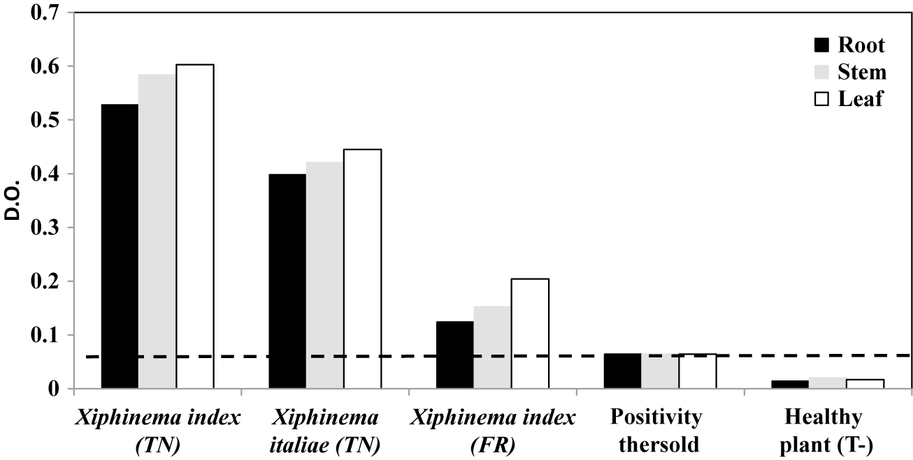
All grapevine plants inoculated with X. index, X. italiae, and the French population of X. index, were found to be 100% infected by GFLV. Both DAS-ELISA and RT-PCR successfully detected GFLV in root, leaf, and stem samples from each grapevine plant used in the experiment (Figure 5 and Figure 6). Thus, the acquisition and transmission of GFLV from healthy to infected grapevines were confirmed by both X. index and X. italiae, under controlled conditions (Figure 7).
GFLV titer in root, stem, and leaf samples measured by DAS-ELISA. Optic density values (nm) are shown.

Agarose gel analysis of GFLV obtained by RT-PCR (using GT1076/GT1826 primers) from grapevine leaf samples that were infested with X. index and X. italiae. M: 100pb Marker (S304105, Bioron). T-: negative control. T+: positive control

Visualization of PCR products obtained from Xiphinema index and X. italiae that were isolated from soil after transmission tests. M: marker size 100 bp (S304105, Bioron). Lanes 1 – 5 correspond to X. index Tunisian population. Lanes 6 – 7 correspond to the negative and positive controls, respectively. Lanes 1 –5 correspond to X. italiae Tunisian population. Lanes 6 – 7 correspond to the negative and positive controls, respectively. Molecular detection of GFLV from the anterior part of X. index was isolated from the soil after the transmission test. M: marker size 100 bp (S304105, Bioron). Lanes 1 – 5 correspond to the GFLV detected in the anterior part of the X. index. Lane 2 corresponds to the absence of GFLV in the anterior part of X. index. Lanes 6 – 7 correspond to the negative and positive controls, respectively (GFLV was isolated from the leaves). Lanes 1 – 2 and 5 correspond to the GFLV detected in the anterior part of X. italiae. Lanes 3 – 4 correspond to the absence of GFLV in the anterior part of X. italiae.

Discussion
Xiphinema is one of the most varied genera of plant ectoparasitic nematodes, with more than 280 species, belonging to the family Longidoridae (Cai et al. Reference Cai, Archidona-Yuste, Cantalapiedra-Navarrete, Palomares-Rius and Castillo2020; Archidona-Yuste et al. Reference Archidona-Yuste, Navas-Cortés, Cantalapiedra-Navarrete, Palomares-Rius and Castillo2016). Numerous species of Xiphinema are distributed worldwide, especially in different agricultural regions, such as X. index that is present in most if not all vineyards around the world, where it has been presumably introduced via grapevine plants from Mediterranean vineyards (Esmenjaud Reference Esmenjaud, Esmenjaud, Kreiter, Martinez, Sforza, Thièry, VonHelden and Yvon2008, Handoo et al. Reference Handoo, Carta, Skantar, Subbotin and Fraedrich2016). Also, X. italiae has been broadly found in grapevine plots (Van Ghelder al. 2015). In our study, these dagger nematodes were isolated from Tunisian vineyards. X. italiae was the most prevalent species in the surveyed vineyard plots. This is consistent with previous studies indicating that X. italiae is among the most widely distributed species in the Mediterranean region (Dalmasso Reference Dalmasso1970; Gutiérrez-Gutiérrez et al. Reference Gutiérrez-Gutiérrez, Palomares-Rius, Jiménez-Díaz and Castillo2011; Martelli et al. Reference Martelli, Cohn and Dalmasso1966). Xiphinema species are characterized by substantial intra- and interspecific homogeneity of the morphometric characters used for species discrimination (Cai et al. Reference Cai, Archidona-Yuste, Cantalapiedra-Navarrete, Palomares-Rius and Castillo2020; Archidona-Yuste et al., Reference Archidona-Yuste, Navas-Cortés, Cantalapiedra-Navarrete, Palomares-Rius and Castillo2016). The Tunisian population of X. italiae matched the morphological identification by Luc and Dalmasso (Reference Luc and Dalmasso1975) and Cohn (Reference Cohn1977). Similarly, the morphometric characteristics of the Tunisian population of X. index are consistent with those of Luc and Dalmasso (Reference Luc and Dalmasso1975) and Siddiqi (Reference Siddiqi1974). Raski et al. (Reference Raski, Goheen, Lider and Meredith1983) and Nguyen et al. (Reference Nguyen, Khallouk, Polidori, Truch, Portier, Lafargue and Esmenjaud2021) have reported that X. index males are rare and females reproduce parthenogenetically, which is consistent with our result. Similarly, for X. italiae, no male was found, suggesting a parthenogenetic reproduction of this species (Dalmasso & Younes Reference Dalmasso and Younes1969). The use of molecular tools based on PCR methods has been applied for Xiphinema diagnostics because of their specificity and sensitivity compared with traditional methods (Oliveira et al. Reference Oliveira, Fenton, Malloch, Brown and Neilson2005; Wang et al. Reference Wang, Bosselut, Castagnonen, Voisin, Abad and Esmenjaud2003). In our study, species-specific PCR primers, including my-I27/myAITS1, and my-ITA26/myAITS1 were designed for molecular identification of X. index and X. italiae, respectively. The phylogenetic analyses revealed that the Tunisian population of X. index is closely related to French, Spanish, and Italian populations.
On the other hand, X. index transmits GFLV, which is the most severe grapevine virus disease worldwide (Hewitt et al. Reference Hewitt, Raski and Goheen1958; Van Helden et al. Reference Van Helden, Villate, Laveau, Morin, Darrieutort and Van Leeuwen2011). The current study showed that the Tunisian populations of X. index are able to acquire and transmit the viral particles of GFLV. The transmission process is mediated by the ability of X. index to ingest GFLV particles from a virus source grapevine, retain virions at specific retention sites within its feeding apparatus, and subsequently infect a recipient vine by the release of virus particles from the retention sites (Demangeat et al. Reference Demangeat, Voisin, Minot, Bosselut, Fuchs and Esmenjaud2005; Schellenberger et al. Reference Schellenberger, Sauter, Lorber, Bron, Trapani, Bergdoll and Ritzenthaler2011). Esmenjaud et al. (Reference Esmenjaud, Demangeat, Van Helden and Ollat2013) reported that there was no variability in GFLV transmission between seven isofemal populations of X. index collected from five different countries with 87.5% – 96% efficiency, whereas other studies revealed differential transmission of nepoviruses associated with longidorid nematodes of different geographical locations (Brown & Trudgill Reference Brown and Trudgill1983; Brown Reference Brown1985, Reference Brown1986; Taylor & Brown Reference Taylor and Brown1997). GFVL was not detected from Tunisian populations of X. italiae under natural conditions. Thus, GFLV could not be naturally vectored by X. italiae. However, under controlled conditions, GFLV virus particles were successfully acquired and transmitted by X. index and X. italiae. In areas where grapevines are grown, X. italiae is frequently observed. Its potential as a GFLV vector is controversial because the transmission reported for a Middle East population was not experimentally reproduced with any other population of this species (Catalano Reference Catalano, Savino and Lamberti1992; Cohn et al. Reference Cohn, Tanne and Nitzani1970; Lamberti and Roca Reference Lamberti, Roca, Veech and Dickson1987). This association has never been confirmed by other studies. It is therefore very unlikely that it could be a specific vector of GFLV (Demangeat Reference Demangeat2007; Martelli & Taylor Reference Martelli, Taylor and Harris1990; Taylor & Brown Reference Taylor and Brown1997).
Brown and Weischer (Reference Brown and Weischer1998) demonstrated that specific associations between nematodes and viruses are constantly evolving, possibly resulting in some viruses losing their vector transmissibility or some vectors losing their ability to transmit viruses, while concurrently new virus and vector associations are becoming established.
Conclusion
Xiphinema species have caused a serious problem in viticulture worldwide, particularly X. index, which is the main vector of GFLV. In conclusion, this paper revealed the morphometric and molecular characteristics of Tunisian populations of X. index and X. italiae compared with foreign populations. Additionally, this study reported that the population of X. index was capable of acquiring and transmitting the viral particles of GFLV. This nepovirus was not found to be naturally retained by X. italiae. However, under controlled conditions, GFLV was successfully acquired and transmitted by this dagger nematode. The acquisition and transmission of GFLV by Xiphinema species still need further studies notably in Tunisian grapevines.
Acknowledgements
The authors thank the University of Carthage and the National Agronomic Institute of Tunisia for their support.
Financial support
This study was financed by the Ministry of Higher Education and Scientific Research, Tunisia.
Competing interest
The authors declare that they have no conflict of interest.
Ethical standard
This contains no studies with human participants or animals performed by any of the authors.