Introduction
Major depressive disorder (MDD) is characterized by emotional dysregulation with abnormalities in emotional processing as a core feature (Davidson, Reference Davidson2002). The fundamental mechanisms underlying the emotional dysregulation of MDD remain unclear; however, these mechanisms are likely to involve the neural system that subserves emotional processing, including its key components, the prefrontal cortex (PFC) and the amygdala. Convergent studies provide support for abnormalities in the structure and function of the PFC and the amygdala in MDD (Mayberg et al. Reference Mayberg, Liotti, Brannan, McGinnis, Mahurin, Jerabek, Silva, Tekell, Martin, Lancaster and Fox1999; Dougherty et al. Reference Dougherty, Rauch, Deckersbach, Marci, Loh, Shin, Alpert, Fischman and Fava2004; Johnstone et al. Reference Johnstone, van Reekum, Urry, Kalin and Davidson2007; Lee et al. Reference Lee, Seok, Lee, Cho, Yoon, Lee, Chae, Choi and Ham2008; MacQueen, Reference MacQueen2009; Frodl et al. Reference Frodl, Bokde, Scheuerecker, Lisiecka, Schoepf, Hampel, Moller, Bruckmann, Wiesmann and Meisenzahl2010). Positron emission tomography (PET) studies have shown increased metabolism in the amygdala (Drevets et al. Reference Drevets, Price, Bardgett, Reich, Todd and Raichle2002) and decreased metabolism in the PFC in MDD (Sackeim et al. Reference Sackeim, Prohovnik, Moeller, Brown, Apter, Prudic, Devanand and Mukherjee1990); similarly, functional magnetic resonance imaging (fMRI) studies have shown increased activation in the amygdala (Sheline et al. Reference Sheline, Barch, Donnelly, Ollinger, Snyder and Mintun2001; Anand et al. Reference Anand, Li, Wang, Wu, Gao, Bukhari, Mathews, Kalnin and Lowe2005a; Siegle et al. Reference Siegle, Thompson, Carter, Steinhauer and Thase2007) and decreased activation in the PFC in MDD (Siegle et al. Reference Siegle, Thompson, Carter, Steinhauer and Thase2007; Lee et al. Reference Lee, Seok, Lee, Cho, Yoon, Lee, Chae, Choi and Ham2008). As the amygdala and the PFC share reciprocal inhibitory connections, these findings implicate abnormalities in the PFC–amygdala connectivity in MDD.
Functional imbalance between the left and right PFC in emotion processing may also be involved in the neuropathophysiology of MDD (Davidson & Irwin, Reference Davidson and Irwin1999). Electroencephalographic (EEG) studies have shown that MDD is associated with less left than right PFC activity during preparation for a sad narrative (Nitschke et al. Reference Nitschke, Heller, Etienne and Miller2004), consistent with previous findings of an association between damage in the left PFC and secondary depressive syndromes (Sackeim et al. Reference Sackeim, Greenberg, Weiman, Gur, Hungerbuhler and Geschwind1982). Further evidence from PET (Martinot et al. Reference Martinot, Hardy, Feline, Huret, Mazoyer, Attar-Levy, Pappata and Syrota1990), transcranial magnetic stimulation (TMS; Bajwa et al. Reference Bajwa, Bermpohl, Rigonatti, Pascual-Leone, Boggio and Fregni2008) and fMRI (Keedwell et al. Reference Keedwell, Andrew, Williams, Brammer and Phillips2005; Johnstone et al. Reference Johnstone, van Reekum, Urry, Kalin and Davidson2007; Grimm et al. Reference Grimm, Beck, Schuepbach, Hell, Boesiger, Bermpohl, Niehaus, Boeker and Northoff2008) studies have also indicated that frontal functional asymmetry is probably important in the neuropathophysiology of MDD (Sackeim et al. Reference Sackeim, Greenberg, Weiman, Gur, Hungerbuhler and Geschwind1982), with possibly greater dysfunction in the left PFC versus the right PFC.
Several task-related fMRI studies have shown functional connectivity abnormalities in MDD (Johnstone et al. Reference Johnstone, van Reekum, Urry, Kalin and Davidson2007; Chen et al. Reference Chen, Suckling, Ooi, Fu, Williams, Walsh, Mitterschiffthaler, Pich and Bullmore2008; Frodl et al. Reference Frodl, Bokde, Scheuerecker, Lisiecka, Schoepf, Hampel, Moller, Bruckmann, Wiesmann and Meisenzahl2010), but their results are inconsistent. The conflicting findings may be due to differences in brain activation between various paradigms or the existence of high-functioning resting-state patients. Resting-state fMRI studies, in which subjects do not perform a task but instead rest quietly throughout the scan, are reported to be especially useful in the study of neuropsychiatric disorders as they may better reflect an individual's natural mental state at the time of scanning (Raichle et al. Reference Raichle, MacLeod, Snyder, Powers, Gusnard and Shulman2001). In resting-state fMRI, the magnitude of temporal correlation between low-frequency blood oxygen level-dependent (BOLD) signal fluctuations in spatially separated regions is measured as an index of the functional connectivity between the regions (Lowe et al. Reference Lowe, Dzemidzic, Lurito, Mathews and Phillips2000). Studies using this method have reported abnormal functional connectivity in neuropsychiatric disorders (Greicius, Reference Greicius2008), with a few specifically in MDD. For example, Anand et al. (Reference Anand, Li, Wang, Wu, Gao, Bukhari, Mathews, Kalnin and Lowe2005a) detected decreased connectivity between the anterior cingulate cortex (ACC) and the medial thalamus, the amygdala and the pallidostriatum in 15 unmedicated depressed patients using a region of interest (ROI) analysis. The ACC–medial thalamus connectivity was increased after 6 weeks of treatment with sertraline (Anand et al. Reference Anand, Li, Wang, Wu, Gao, Bukhari, Mathews, Kalnin and Lowe2005b). Greicius et al. (Reference Greicius, Flores, Menon, Glover, Solvason, Kenna, Reiss and Schatzberg2007) found increased connectivity of the subgenual ACC and the thalamus in 28 medication-free, depressed patients using independent component analysis (ICA), whereas Veer et al. (Reference Veer, Beckmann, van Tol, Ferrarini, Milles, Veltman, Aleman, van Buchem, van der Wee and Rombouts2010), also using ICA, found decreased connectivity of the amygdala and the left anterior insula in 19 medication-free, MDD patients. In addition to differences in methodology, differences in co-morbidities and medication exposure may account for the inconsistencies among these studies.
In the current study we used resting-state fMRI to examine the functional connectivity between the amygdala and other brain regions in treatment-naive subjects with MDD and matched healthy control (HC) subjects. We hypothesized that there would be decreased functional connectivity between the amygdala and cortical regions, especially the PFC, in MDD subjects.
Method
Subjects
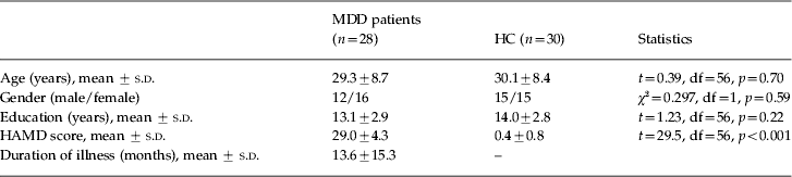
Twenty-eight treatment-naive MDD subjects [mean age 29.3 years (s.d. = 8.7), 57% female] were recruited from the out-patient clinic at the Department of Psychiatry, First Affiliated Hospital of China Medical University and the Mental Health Center of Shenyang. MDD subjects were diagnosed by two trained psychiatrists using the Structured Clinical Interview for DSM-IV Disorders (SCID) and met the following inclusion criteria: (1) fulfilled DSM-IV criteria for MDD; (2) did not have a co-morbid Axis I diagnosis; (3) had a score on the 17-item Hamilton Depression Rating Scale (HAMD-17) of ≥24 (so as to obtain participants with severe depression who were most likely to have prominent biological abnormalities); and (4) did not have a history of psychopharmacotherapy, electroconvulsive therapy or psychotherapy. The mean years of education was 13.1 (s.d. = 2.9), the mean duration of illness was 13.6 (s.d. = 15.3) months, and the mean HAMD score was 29.0 (s.d. = 4.3). The duration of illness was calculated as the difference between the participant's age of first onset of depressive symptoms (as reported by the participant and confirmed by other sources including prior medical records and close relatives) and their age at the time of scanning.
The HC group comprised 30 participants [mean age 30.1 years (s.d. = 8.4), 50% female] recruited from the community who, along with their first-degree family members, had no DSM-IV Axis I disorder. The absence of DSM-IV Axis I disorders in HC subjects were confirmed using the SCID by two independent psychiatrists. For both MDD and HC groups, individuals were excluded for the following: any MRI contraindications, history of head injury or neurological disorder, and any concomitant medical disorder. All subjects were right-handed, scanned within 24 h of initial contact, and rated on the HAMD at the time of scanning. The participants provided written informed consent after receiving a detailed description of the study as approved by the Institutional Review Board (IRB) of China Medical University.
MRI data acquisition
fMRI data were acquired using a 3-T GE MR scanner (General Electric, USA) at the First Affiliated Hospital, China Medical University, Shenyang, China. Head motion was minimized with restraining foam pads. A standard head coil was used for radiofrequency transmission and reception of the nuclear magnetic resonance signal. The subjects were asked to keep their eyes closed but remain awake throughout the resting-state scan. fMRI images were acquired using a spin echo planar imaging (EPI) sequence, parallel to the anterior–posterior commissure (AC–PC) plane with the following scan parameters: repetition time (TR) = 2000 ms; echo time (TE) = 40 ms; image matrix = 64 × 64; field of view (FOV) = 24 × 24 cm2; 35 contiguous slices of 3 mm and without gap; scan time 6 min 40 s.
Functional connectivity processing
The resting-state fMRI data preprocessing was carried out by using SPM8 (www.fil.ion.ucl.ac.uk/spm/software/spm8) and the Resting-State fMRI Data Analysis Toolkit (REST) V1.5_101101 (www.restfmri.net). The first 10 volumes were deleted, then data preprocessing included slice timing correction, head motion correction, spatial normalization and smoothing. Head motion parameters were computed by estimating translation in each direction and the angular rotation about each axis for each volume. Participants were excluded if their head motion was >2 mm maximum displacement in any of the x, y or z directions or 2° of any angular motion throughout the course of the scan (no participants were excluded). There are no group differences in head motion between the two groups. The spatial normalization was performed by using a standard EPI template from the Montreal Neurological Institute (MNI). The voxel size was resampled to 3 × 3×3 mm3. Spatial smoothing was performed with an 8-mm full-width at half-maximum (FWHM) Gaussian filter. Then, linear detrending and temporal bandpass (0.01–0.08 Hz) filtering were performed to remove low-frequency drifts and physiological high-frequency noise (Cordes et al. Reference Cordes, Haughton, Arfanakis, Carew, Turski, Moritz, Quigley and Meyerand2001). Linear regression of head motion parameters, global mean signal, white matter signal and cerebrospinal fluid signal was performed to remove the effects of the nuisance covariates (Liu et al. Reference Liu, Hu, Zhou, Shen and Wang2008; Fox et al. Reference Fox, Zhang, Snyder and Raichle2009).
The left and right amygdala ROIs were defined separately with the WFU PickAtlas Tool (www.fmri.wfubmc.edu/download.htm). For each subject, the mean time course for the amygdala ROI was calculated by averaging the time course for all voxels within the amygdala ROI. The time course of the amygdala ROI was then correlated with the time course of each pixel in the brain, resulting in a correlation map for each subject that contained the correlation coefficient for each voxel with that of the amygdala ROI. The resulting correlation coefficients were transformed into z scores by Fisher's z transform to create subject-specific maps of resting-state correlations to the amygdala ROI.
Statistical analyses
Independent-sample t tests and χ2 tests were used to compare demographic data and HAMD scores between the MDD and HC groups with SPSS version 13.0 (SPSS Inc., USA). The subject-specific maps of resting-state correlations from the amygdala to all brain voxels were combined across subjects within the MDD group and within the HC group using voxel-based one-sample t tests to produce group whole-brain composite maps. Contrast maps to assess between-group differences were then created using voxel-based two-sample (MDD versus HC) t tests. The contrast maps were corrected for multiple comparisons using Monte Carlo simulation [AlphaSim command line in Analysis of Functional NeuroImages (AFNI; Cox, 1996)] within the PFC, which was our hypothesized region. The PFC ROI was defined with the WFU PickAtlas Tool (www.fmri.wfubmc.edu/download.htm), including Brodmann areas (BAs) 9–12, 24/25/32 and 44–47. The spatial smoothness between the voxels modeled by the FWHM of a Gaussian kernel was estimated by the 3dFWHMx AFNI routine (http://afni.nimh.nih.gov/pub/dist/doc/manual/AlphaSim.pdf). Therefore, the combination criteria were determined by the AlphaSim program at a single voxel threshold of p < 0.001 and cluster size >18 voxels (486 mm3), corresponding to a corrected p < 0.05. Exploratory whole-brain analyses were conducted to explore other possible brain regions, not hypothesized a priori. Multiple comparisons correction was performed analogous to that described above. Findings in these regions were considered significant at a single voxel threshold of p < 0.001 and cluster size >31 voxels (837 mm3), corresponding to a corrected p < 0.05. Post-hoc exploratory Pearson correlation analyses were performed in MDD participants to assess the correlation of the HAMD scores and the duration of illness with z scores in the PFC regions showing significant differences between the HC and MDD groups.
Results
There were no significant differences in age (p = 0.7), gender (χ2 = 0.297) or education (p = 0.22) between the MDD group and the HC group. The MDD group had significantly higher HAMD scores than the HC group (p < 0.001, Table 1).
Demographic and clinical data of participants
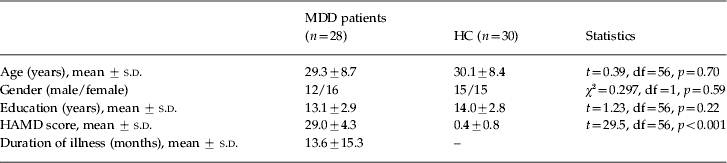
HAMD, Hamilton Depression Rating Scale; MDD, major depressive disorder; HC, healthy controls; s.d., standard deviation; df, degrees of freedom.
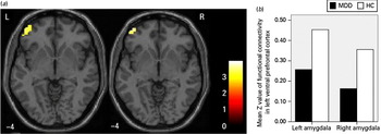
We observed significantly decreased left ventral prefrontal cortex (VPFC) functional connectivity in the MDD group, compared with the HC group [from the left amygdala: maximal MNI coordinates of the left VPFC region: x = –51, y = 42, z = –9, 100 voxels (2700 mm3), T = 4.00, p < 0.001 uncorrected; from the right amygdala, maximal MNI coordinates of the left VPFC region: x = –42, y = 57, z = 0, 24 voxels (648 mm3), T = 3.72, p < 0.001 uncorrected] (Fig. 1). These findings correspond to a corrected p < 0.05 by AlphaSim correction. Whole-brain analysis did not reveal additional group differences in decreased functional connectivity between the amygdala and other brain regions. In post-hoc correlation analyses, neither the HAMD score (from the left amygdala: r = 0.01, p = 0.959; from the right amygdala: r = –0.188, p = 0.337) nor the duration of illness (from the left amygdala: r = 0.108, p = 0.583; from the right amygdala: r = 0.059, p = 0.764) had any significant associations with VPFC functional connectivity in MDD participants.
(a) The axial-oblique images (MNI coordinate z = –4 mm) display the regions in the ventral prefrontal cortex (VPFC) that show reduced functional connectivity from the left amygdala (left) and the right amygdala (right) in participants with major depressive disorder (MDD), compared to healthy comparison participants (HC), at rest. The color bar represents the range of T values. L, left brain; R, right brain. (b) The graph depicts the mean z values of functional connectivity in the VPFC with the left and right amygdala.

Discussion
To our knowledge, this is the first study to investigate functional connectivity abnormalities between the amygdala and the PFC in treatment-naive MDD individuals using a resting-state fMRI method. We have demonstrated decreased functional connectivity between both the left and the right amygdala and the left VPFC in MDD participants compared with HC participants.
The VPFC and the amygdala are key components of the cortico-limbic circuit involved in emotional processing (Ochsner & Gross, Reference Ochsner, Gross and Gross2007; Pessoa, Reference Pessoa2008) and have direct interconnections with each other (Amaral & Price, Reference Amaral and Price1984; Ghashghaei et al. Reference Ghashghaei, Hilgetag and Barbas2007). The amygdala's role in processing emotional stimuli has been demonstrated in animal and human research (LeDoux, Reference LeDoux1992; Lebowitz et al. Reference Lebowitz, Pearson, Schneider, Reynolds, Alexopoulos, Bruce, Conwell, Katz, Meyers, Morrison, Mossey, Niederehe and Parmelee1997; Costafreda et al. Reference Costafreda, Brammer, David and Fu2008). Evidence in humans indicates that the amygdala contains populations of cells that respond to faces, particularly facial emotion (Pillai et al. Reference Pillai, Friedman, Stuve, Trinidad, Jesberger, Lewin, Findling, Swales and Schulz2002). Morphological and functional abnormalities within the amygdala and PFC in MDD have been demonstrated (Drevets et al. Reference Drevets, Price and Furey2008). Postmortem studies have also found abnormalities in the amygdala and PFC in MDD patients (Hercher et al. Reference Hercher, Turecki and Mechawar2009). However, the nature of the interaction between the VPFC and the amygdala is not fully understood. Evidence has suggested that the VPFC activates local inhibitory circuits in the amygdala that constrain its activation (Quirk et al. Reference Quirk, Likhtik, Pelletier and Pare2003, Quirk & Beer, Reference Quirk and Beer2006; Ghashghaei et al. Reference Ghashghaei, Hilgetag and Barbas2007). Previous fMRI studies have examined the interaction between the VPFC and the amygdala. For example, Urry et al. (Reference Urry, van Reekum, Johnstone, Kalin, Thurow, Schaefer, Jackson, Frye, Greischar, Alexander and Davidson2006) reported an inverse relationship between VPFC and amygdala activation in response to negative stimuli in older adults. Johnstone et al. (Reference Johnstone, van Reekum, Urry, Kalin and Davidson2007) reported similar findings in non-depressed individuals, and also observed a positive association between VPFC and amygdala activation in individuals with MDD, indicating the lack of down-regulation by the VPFC of amygdala responses to negative stimuli in MDD. Decreased VPFC–amygdala functional connectivity, as reported in this study, could contribute to diminished VPFC regulation of the amygdala, providing further evidence of cortico-limbic circuitry dysfunction in unmedicated MDD participants.
In this study, both the left and right amygdala demonstrated abnormalities in functional connectivity with the left VPFC, but not with any right cerebral regions. Hemispheric asymmetry has been observed in normal affective processing of positive and negative emotions, and the balance between the right and left hemispheres is important in adaptive emotion regulation (Sackeim et al. Reference Sackeim, Greenberg, Weiman, Gur, Hungerbuhler and Geschwind1982; Davidson, Reference Davidson2002). fMRI studies have also found hemispheric asymmetry during emotional processing in depressed and non-depressed individuals (Tomarken et al. Reference Tomarken, Davidson, Wheeler and Doss1992; Jackson et al. Reference Jackson, Mueller, Dolski, Dalton, Nitschke, Urry, Rosenkranz, Ryff, Singer and Davidson2003; Keedwell et al. Reference Keedwell, Andrew, Williams, Brammer and Phillips2005). Our present findings of decreased functional connectivity in the left VPFC further support the involvement of functional hemispheric asymmetry in MDD. However, conclusions regarding functional laterality in MDD are tentative. Future studies that directly compare functional connectivity or activation between the left and right hemisphere are needed to reach more definitive conclusions.
In our exploratory whole-brain analyses, we did not find significant differences in functional connectivity between the insula and the amygdala, which is inconsistent with recent findings by Veer et al. (Reference Veer, Beckmann, van Tol, Ferrarini, Milles, Veltman, Aleman, van Buchem, van der Wee and Rombouts2010) using ICA. They found decreased connectivity of the amygdala and the left anterior insula in 19 medication-free MDD patients. The conflicting findings may reflect methodological differences in neuroimaging data acquisition or processing, or in the samples studied including differences in sex distribution, medication exposures, age of onset, illness duration, and number of acute episodes. The insula plays a major role in processing both emotional recognition and cognitive regulation (van Tol et al. Reference van Tol, Demenescu, van der Wee, Kortekaas, Marjan, Boer, Renken, van Buchem, Zitman, Aleman and Veltman2012) and has been increasingly recognized as an important region in MDD. Specifically, in recent structural and functional MRI studies, abnormalities in the insula have been detected (Horn et al. Reference Horn, Yu, Steiner, Buchmann, Kaufmann, Osoba, Eckert, Zierhut, Schiltz, He, Biswal, Bogerts and Walter2010; Liu et al. Reference Liu, Xu, Xu, Wang, Zhao, Lv, Cao, Zhang and Du2010; Peng et al. Reference Peng, Liu, Nie, Li, Shan, Wang and Li2011). The functional connectivity between the amygdala and the insula requires further investigation.
Some limitations of the current study should be noted. The cross-sectional design does not allow us to observe treatment effects in MDD participants; future longitudinal studies are needed to examine such effects. Our strict inclusion criteria (e.g. HAMD-17 ≥24) and relatively small sample may limit generalization of our findings to MDD of varying severity. Larger samples that include mild and moderate depression could provide further understanding of how functional connectivity abnormalities correlate with clinical variables. In addition, a larger sample could help to identify other important regions that might be involved in MDD neuropathophysiology. For example, the ACC region did not survive our strict threshold for significance. However, when the threshold was lowered (p < 0.005), the participants with MDD did demonstrate reduced functional connectivity between the left amygdala and the ACC (MNI coordinates: x = 6, y = 30, z = –6, 16 voxels, T = 3.02, p < 0.005 uncorrected). This may suggest that the effect size of ACC abnormalities is less than the effect size for the VPFC detected in the current study. Therefore, given the relatively small sample size in this study, there may not have been sufficient statistical power to detect significant differences between the amygdala and the ACC.
In summary, our study of treatment-naive MDD subjects supports the involvement of decreased amygdala–VPFC functional connectivity and abnormalities in functional hemispheric asymmetry in the early onset stage of MDD. However, given the lack of studies comparing treatment-naive and treated MDD, or remitted and active MDD, it remains to be seen whether these abnormalities are trait versus state dependent or causes versus effects of MDD or correlate with abnormalities in structural connectivity in MDD. Future MRI studies of individuals with MDD and at high risk for MDD could further elucidate the role of these abnormalities in the neuropathophysiology of MDD.
Acknowledgments
The authors were supported by research grants from the National Natural Science Foundation of China (81071099 to Y. Tang and 81101012 to F. Wu), the Liaoning Science and Technology Foundation (2008225010-14, Y. Tang), the Liaoning Doctor Scientific Foundation (20111099, F. Wu), the National Institute of Health (K01MH086621, F. Wang), the National Alliance for Research on Schizophrenia and Depression (F. Wang) and the Klingenstein Foundation (F. Wang).
Declaration of Interest
None.