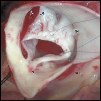
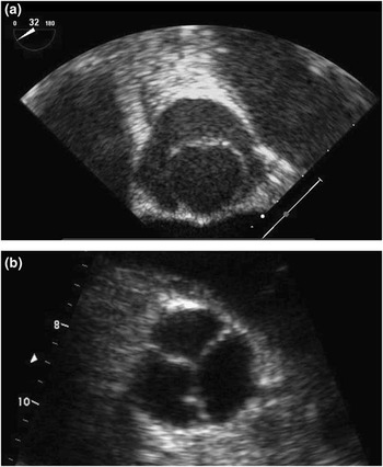
Unicusp aortic valves have a male predominance and are rare, occurring in 3–5% of individuals. As shown from the surgeon’s view at operation (Fig 1), such valves commonly have an asymmetric opening due to a single commissure between the non- (N) and left (L) coronary leaflets, with abnormally low fusion of the tubercles between the left–right (LR) and right–non-coronary (RN) leaflets (NCS = non-coronary sinus). This results in two short, rudimentary commissures/raphes, below the level of the coronary ostia. Preoperative, transesophageal imaging in a short-axis view (Fig 2a) from the 16-year-old male patient, who presented with aortic stenosis and insufficiency, shows the resulting echocardiographic “toilet seat” appearance. This may be difficult to diagnose preoperatively and is frequently misclassified as a bicuspid valve with rheumatic disease. The valve was repaired on cardiopulmonary bypass with cardioplegic arrest of the heart. After complete transection of the aorta with an incision extended into the non-coronary sinus, the rudimentary leaflets were separated, debrided, and augmented with autologous pericardium. Neo-commissures were then created to shape the sinuses of Valsalva.Reference Baird and Del Nido1 This resulted in a competent, three-leaflet valve, which had neither stenosis nor insufficiency on immediate post-operative (Fig 2b) and 2-year follow-up echocardiography.
Figure 1

Figure 2