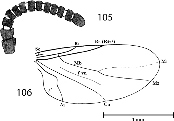
The Diptera from the late Eocene of the Isle of Wight are similar to the Recent fauna in generic composition, particularly in containing genera occurring today in subtropical regions. They are a very common and diversified group in the Insect Bed. Most important information about fossil Diptera from Isle of Wight were published by Cockerell (Reference Cockerell1915, Reference Cockerell1917a, Reference Cockerellb, Reference Cockerell1921); Cockerell & Haines (Reference Cockerell and Haines1921); Edwards (Reference Edwards1923); Jarzembowski (Reference Jarzembowski1976); Krzemiński (Reference Krzemiński1996); Vane-Wright (Reference Vane-Wright1967).
In years 2005–2007 thanks to financial support of INTAS (Project no. 03-51-4367) an international group of dipterologists, including Wiesław Krzemiński (Poland) – team leader, Dany Azar (Lebanon), Vladimir Blagoderow (Russia), Elena Lukashevich (Russia), Andre Nel (France), Ryszard Szadziewski (Poland), Sonja Wedmann (Germany), Vladimir Zlobin (Russia), had undertaken the task of determination and revision of dipteran materials from the Isle of Wight. Later Francis-Marie Collomb (France), David B. Nicholson (UK) and Alain Waller (France) also joined the project.
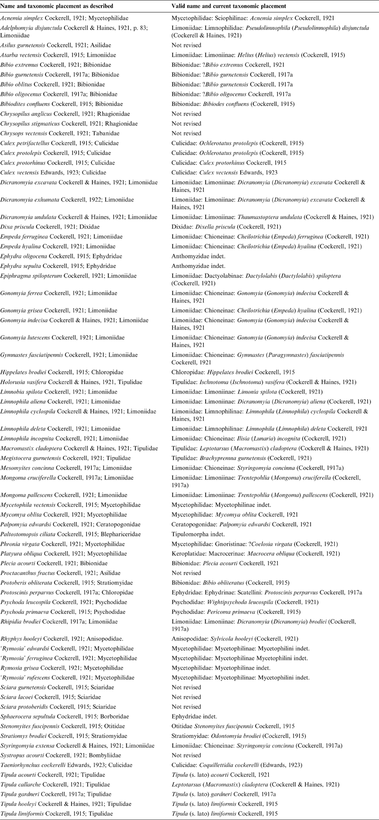
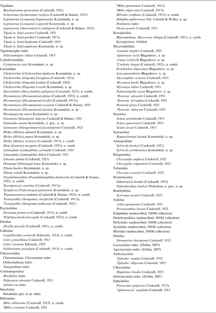
Our study on old and new collections housed in UK and USA museums and materials collected during field trips revealed over 1,700 fossil specimens, belonging to 32 families of the Diptera; however, only around 800 specimens were studied for this paper. Taxonomic results of study on higher flies from the Insect Bed are published elsewhere (Zlobin 2007). A summary of current taxonomic position of previously described species is presented in Table 1.
Current taxonomic position of the previously described species fossil Diptera form the Insect Limestone.

1. Material and methods
The fossil insects of the Isle of Wight originate from the late Eocene concretions or tabular bands of very fine-grained micrite, identified as Insect Limestone of the Isle of Wight. The unit where these concretions/bands occur is known as the Insect Bed, which lies towards the base of the Bembridge Marls Member (Solent Group: Bouldnor Formation) (Gale et al. Reference Gale, Huggett, Pälike, Laurie, Hailwood and Hardenbol2006; Ross & Self Reference Ross and Self2014).
The largest collection is housed in Natural History Museum (NHM), London. Most of the specimens at this museum belong to the A'Court Smith (purchased 1877, 1883), P.B. Brodie (purchased 1898) and R.W. Hooley (purchased 1924) collections. They are labelled ‘Gurnard Bay’ (which is an old name for Gurnet Bay); however, Smith collected specimens all the way from West Cowes to Newtown River on the NW side of the Isle of Wight (Jarzembowski Reference Jarzembowski1980). Most of the specimens probably came from Thorness Bay (Jarzembowski Reference Jarzembowski1976). Brodie and Hooley acquired parts of Smith's collection, so that parts and counterparts of individual insects have turned up in the three collections. The parts and counterparts often have different numbers because they were registered at different times. An additional collection was discovered at the Sedgwick Museum, Cambridge by A.J. Ross. This collection has also yielded counterparts of specimens at the NHM, which indicates that this is another part of the Smith collection. A label with ‘1883' on it suggests that the Sedgwick Museum acquired this collection in 1883, the same year that the NHM purchased specimens from Smith.
Prefixes I. and In. preceding the numerals of specimens indicate that they come from different series.
1.1. Number of specimens studied
Diptera Nematocera: Anisopodidae – five specimens; Bibionidae – 19 specimens; Ceratopogonidae – nine specimens; Chironomidae – 170 specimens; Culicidae – 140 specimens; Cylindrotomidae – one specimen; Dixidae – six specimens; Keroplatidae – 17 specimens; Limoniidae – 135 specimens; Mycetophilidae – 55 specimens; Psychodidae – 39 specimens; Scatopsidae – two specimens; Simuliidae – one specimen; Tipulidae – 32 specimens. Diptera Brachycera: Agromyzidae – two specimens; Anthomyzidae – 18 specimens; Chloropidae – three specimens; Dolichopodidae – 23 specimens; Empididae – two specimens; Ephydridae – four specimens; Heleomyzidae – 11 specimens; Lauxaniidae – 12 specimens; Otitidae – six specimens; Phoridae – two specimens; Stratiomyidae – 41 specimens; Syrphidae – four specimens. Cyclorrhaphous diptera were described in a separate publication (Zlobin Reference Zlobin2007). Also, Cockerell (Reference Cockerell1915, Reference Cockerell1921) described species of Asilidae, Blephariceridae (see below), Bombyliidae, Rhagionidae, Sciaridae and Tabanidae.
Fine-grained limestone often enables exceptional preservation of delicate structures, although many isolated wings and fragments of the body are preserved in the material. When taxonomically important structures like antennae, palps, legs, genitalia are missing or destroyed, not all families of the Diptera can be studied and determined on the same level.
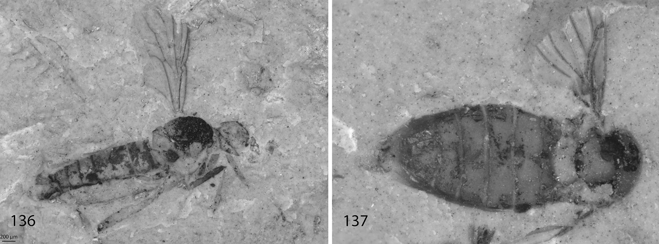
It has not been possible to re-describe all of Cockerell's types, which when originally described were only figured with a line drawing. However, all of his holotypes and other specimens that provide new family level records have been listed and imaged for completeness.
Classification of Diptera follows Pape et al. (Reference Pape, Blagoderov, Mostovski and Zhang2011). For an explanation of general morphological terminology of Culicidae and abbreviations see Dahl (Reference Dahl and Nilsson1997) and Szadziewski (Reference Szadziewski1998). For an explanation of general morphological terminology of Ceratopogonidae and abbreviations see Szadziewski (Reference Szadziewski1988, Reference Szadziewski1996). Nomenclature of radial and medial veins of Scatopsidae follows Cook (Reference Cook, McAlpine, Peterson, Shewell, Teskey, Vockeroth and Wood1981).
Fossil Diptera from the Isle of Wight are deposited in:
BMB – Booth Museum of Natural History, Brighton
CAMSM – Sedgwick Museum of Earth Sciences, University of Cambridge
IWCMS – Isle of Wight County Museum Service
NHMUK – Natural History Museum, London, Department of Palaeontology
MNEMG – Maidstone Museum & Bentlif Art Gallery
USNM – Department of Paleobiology, National Museum of Natural History, Smithsonian Institution, Washington DC
2. Systematic palaeontology
Order Diptera Linnaeus, Reference Linnaeus1758
Suborder Nematocera Dumeril, Reference Duméril1805
Superfamily Tipuloidea Latreille, Reference Latreille1802
by Wieslaw Krzemiński
Family Tipulidae Latreille, Reference Latreille1802
TIPULIDAE (Diptera Nematocera) are rather common and well preserved in Bembridge Marls of the Isle of Wight when compared to other contemporary faunas. These large Diptera are represented mostly by isolated wings or some wing fragments. Bodies are rare and mostly poorly preserved. In the collections from the Bembridge Marls eight species were described by Cockerell (Reference Cockerell1915, Reference Cockerell1917a, Reference Cockerell1921) and Cockerell & Haines (Reference Cockerell and Haines1921). Revision of some of these materials was made by Vane-Wright (Reference Vane-Wright1967) and Krzemiński (Reference Krzemiński1996); the latter demonstrated that Megistocera gurnetensis Cockerell, Reference Cockerell1921 belonged to the genus Brachypremna Osten Sacken, while Holorusia vasifera Cockerell & Haines Reference Cockerell and Haines1921 belongs to the genus Ischnotoma Skuse.
In the present paper further misinterpretations are dealt with: Tipula callarche Cockerell, Reference Cockerell1921 and Macromastix cladoptera Cockerell & Haines, Reference Cockerell and Haines1921 were described on the basis of part and counterpart of the same specimen. Also, no difference can be found between Tipula limiformis Cockerell, Reference Cockerell1915 and Tipula hooleyi Cockerell & Haines, Reference Cockerell and Haines1921. Therefore T. hooleyi is a younger synonym of T. limiformis. In conclusion, there are now six valid species described from the Bembridge Marls. Additionally, among undetermined material three additional species belonging to the genus Leptotarsus Guerin-Meneville and one to the genus Tipula Linnaeus were found. They are described below.
Today Tipulidae form a large family of Diptera Nematocera, with about 4000 species of worldwide distribution. Larvae live in freshwater, semi-aquatic and terrestrial habitats.
The oldest Tipulidae were described from the Lower Cretaceous (Krzemiński Reference Krzemiński1992). They are common in fossil materials from the Palaeogene and Neogene of Europa, Asia and North America.
Genus: Brachypremna Osten Sacken, Reference Osten Sacken1887
Type species. Tipula dispellens Walker, Reference Walker1861, p. 334, (designated by Coquillett Reference Coquillett1910)
Remarks. Recently the genus Brachypremna is represented by 37 species distributed only in tropical and subtropical regions of the world; most (34 sp.) known from the Neotropical Region, one from Nearctic and two from the Australian Region. Only three fossil species are known: from Eocene Baltic amber (Eocene), from the Dominican amber (Miocene) and from the early Oligocene Isle of Wight (Cockerell Reference Cockerell1921; Alexander Reference Alexander1931; Krzemiński Reference Krzemiński1996).
Brachypremna gurnetensis (Cockerell, Reference Cockerell1921)
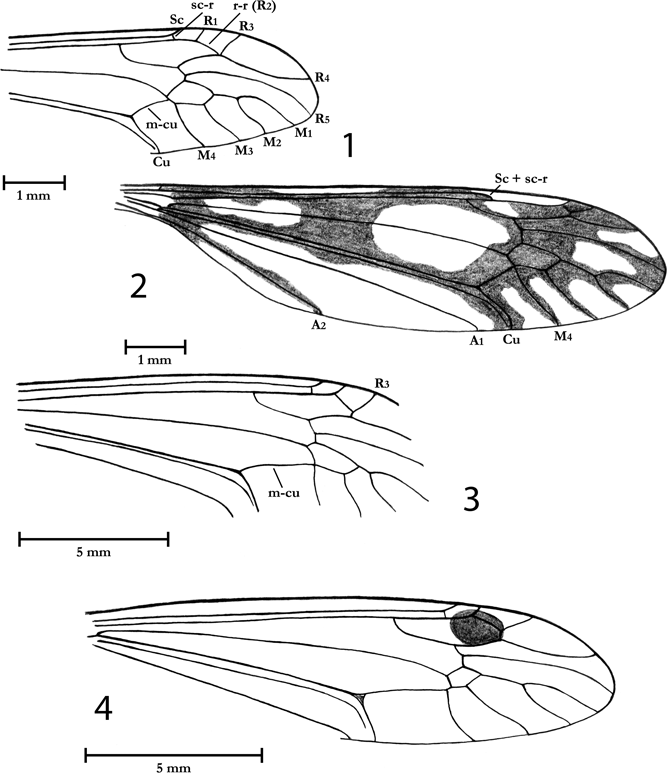
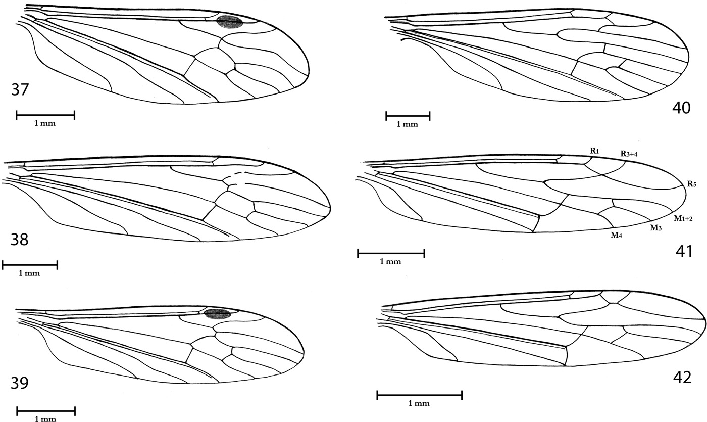
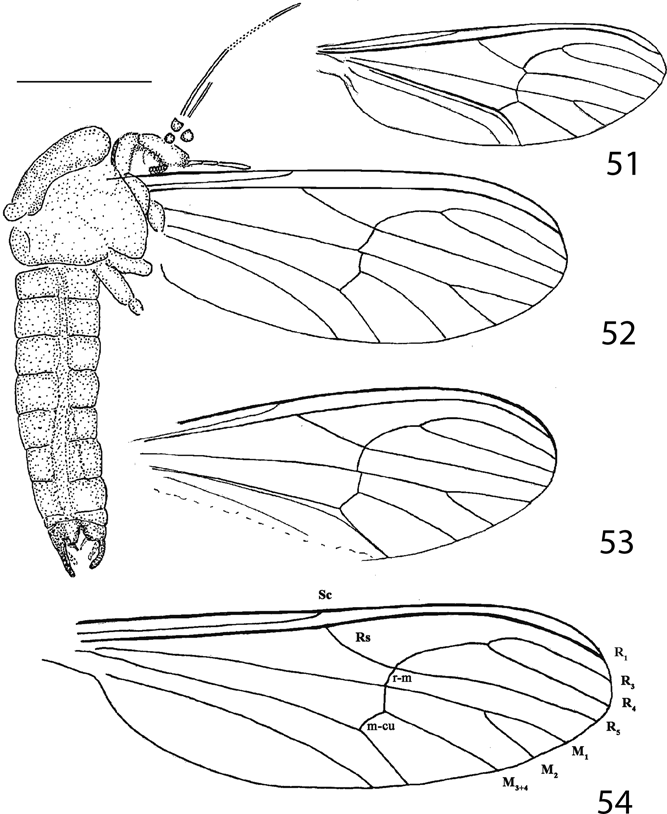
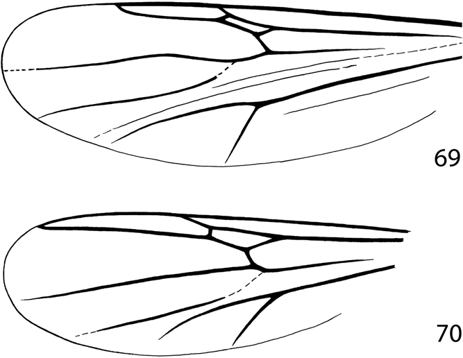
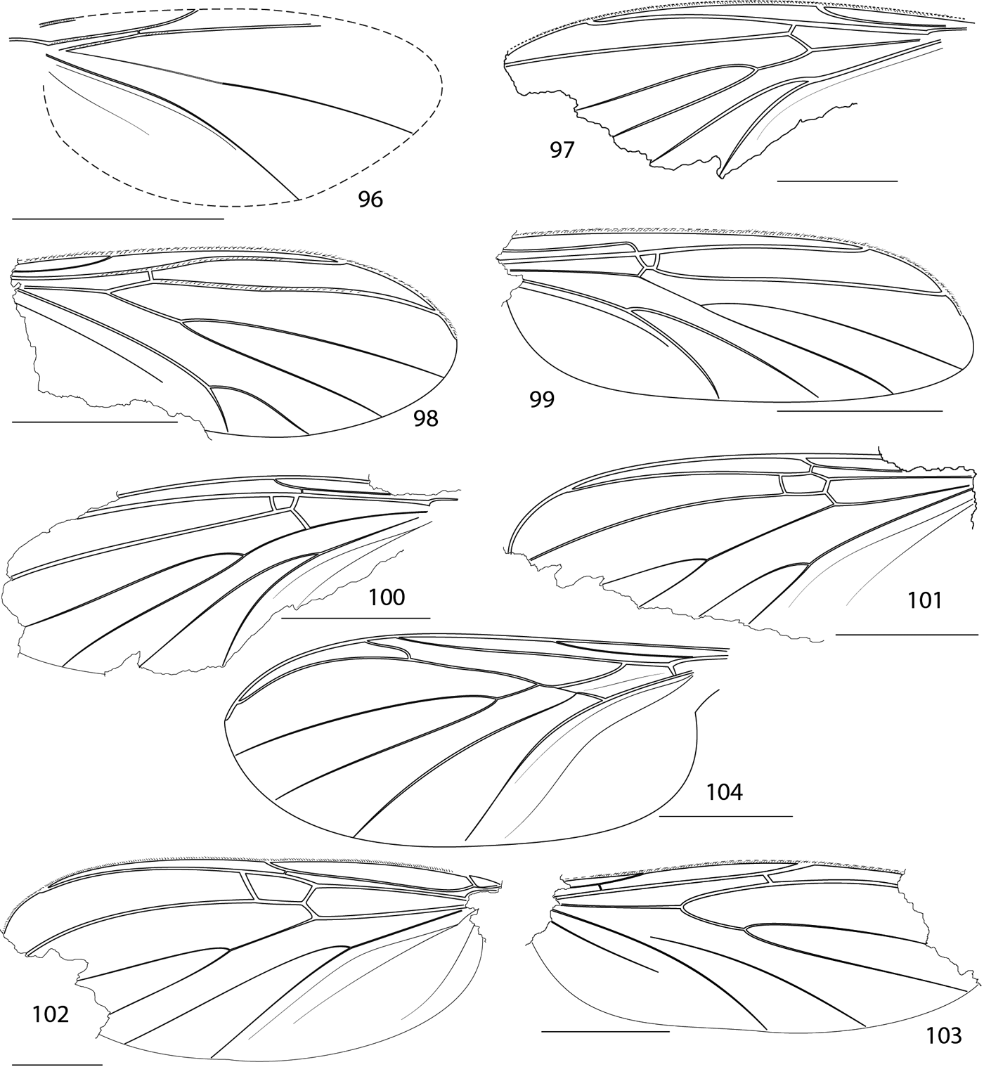
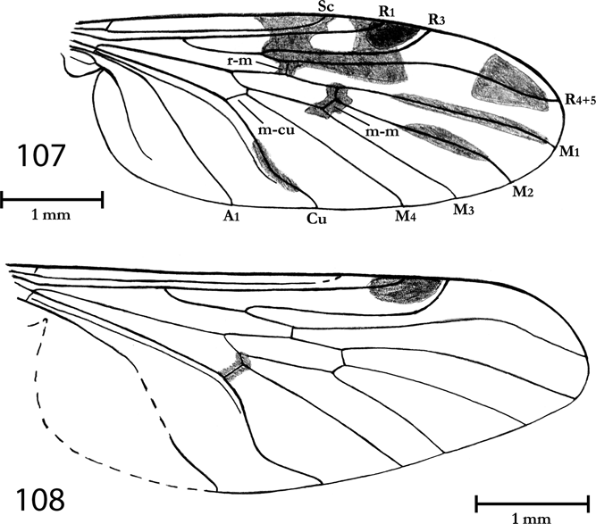
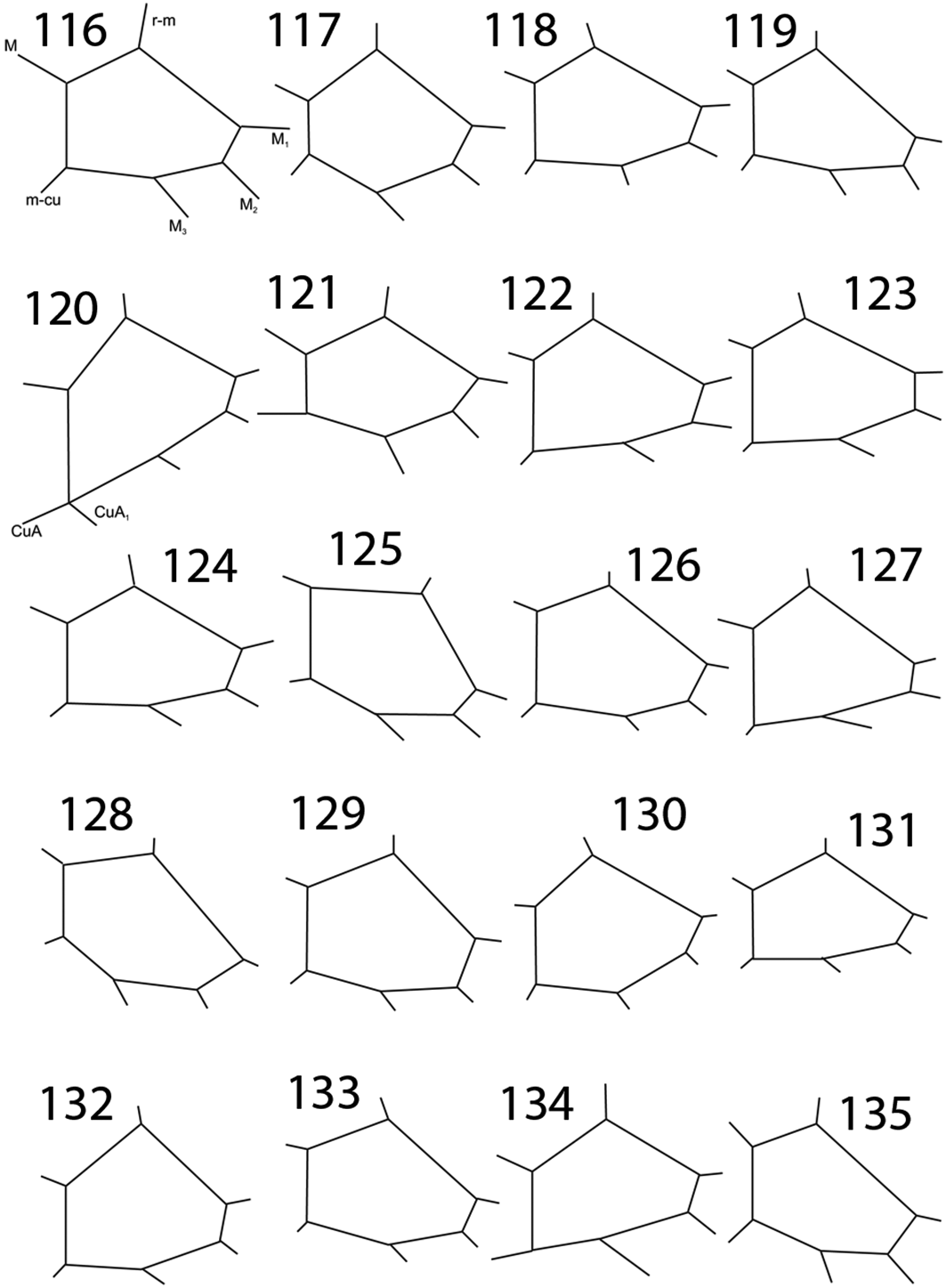
Tipulidae, wing venation. (1) Brachypremna gurnetensis (Cockerell, Reference Cockerell1921), holotype NHMUK In.17090. (2) Leptotarsus (Macromastix) cladoptera (Cockerell & Haines, Reference Cockerell and Haines1921), holotype NHMUK I.10379. (3) Leptotarsus (Longurio) fragmentatus Krzemiński, n. sp., holotype NHMUK In.25379. (4) Leptotarsus (Longurio) wegiereki Krzemiński, n. sp., holotype NHMUK In.17493.

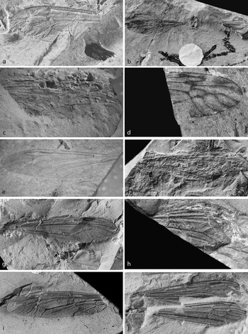
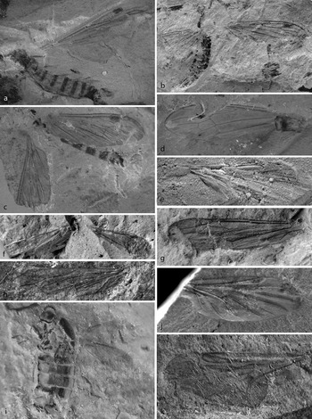
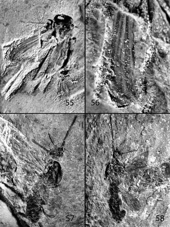
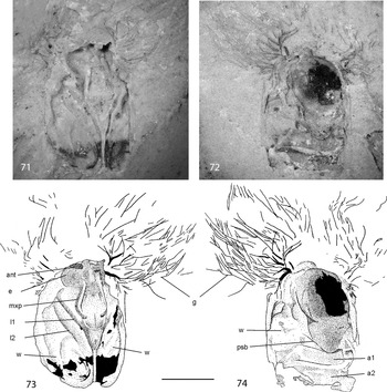
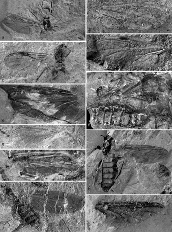
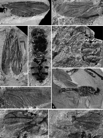
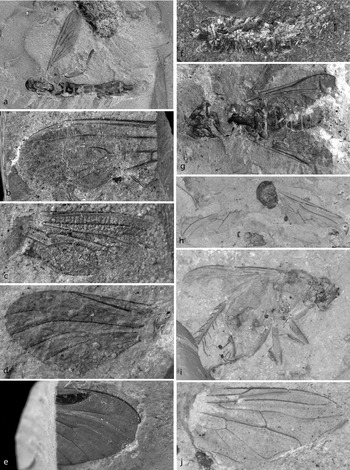
Tipulidae and Cylindrotomidae. (a) Brachypremna gurnetensis (Cockerell, Reference Cockerell1921) holotype part NHMUK In.17090. (b) Leptotarsus (Macromastix) cladoptera (Cockerell & Haines, Reference Cockerell and Haines1921), holotype part NHMUK I.10397. (c) Leptotarsus (Longurio) fragmentatus Krzemiński, n. sp., holotype NHMUK In.25379. (d) Tipula (s. lato) acourti Cockerell, Reference Cockerell1921, holotype part NHMUK I.8901. (e) Tipula (s. lato) anglicana Krzemiński, n. sp., holotype NHMUK In.64117. (f) Tipula (s. lato) gardneri Cockerell, Reference Cockerell1917a holotype NHMUK I.8663. (g, h) Tipula (s. lato) limiformis Cockerell, Reference Cockerell1915: (g) (type of Tipula hooleyi Cockerell & Haines, Reference Cockerell and Haines1921 syn. nov.), NHMUK In.24353; (h) holotype USNM 61438. (i) Ischnotoma (Ischnotoma) vasifera (Cockerell & Haines, Reference Cockerell and Haines1921), holotype NHMUK In.24352. (j) Cyttaromyia rossi Krzemiński, n. sp., holotype CAMSM X.50140.14.

1921 Megistocera gurnetensis Cockerell, p. 456, fig. 3
1931 Brachypremna gurnetensis Alexander, p. 8
1994 Brachypremna gurnetensis Evenhuis, p. 57
1996 Brachypremna gurnetensis Krzemiński, pp. 275–76, fig. 3
Holotype. NHMUK In.17090/In.17492 (part and counterpart), Smith Collection; Insect Bed; late Eocene; NW Isle of Wight, UK.
Remarks. Revision and redescription of this species was given by Krzemiński (Reference Krzemiński1996).
Genus: Leptotarsus Guerin-Meneville, Reference Guerin-Meneville and Duperrey1831
Type species. Leptotarsus macquartii Guerin-Meneville, Reference Guerin-Meneville and Duperrey1831: Atlas: pl. 20, fig. 1 (by monotypy)
Subgenus: Macromastix Osten Sacken, Reference Osten Sacken1887
Type species. Macrothorax ornatus Jaennicke, Reference Jaennicke1867, p. 320
Remarks. The subgenus Macromastix is represented in fossil state by two fossil species, from the Oligocene of France and from late Eocene Isle of Wight, England. Recently the subgenus comprises 94 species living in tropical and subtropical regions.
Leptotarsus (Macromastix) cladoptera (Cockerell & Haines, Reference Cockerell and Haines1921)
1921 Macromastix cladoptera Cockerell & Haines, p. 111–112, fig. I10397 (published May 1921)
1921 Tipula callarche Cockerell p. 455, fig. 1 (published June 1921), syn. nov.
1994 Leptotarsus (Macromastix) cladoptera Evenhuis, p. 56
1994 Tipula callarche Evenhuis, p. 58
Holotype. NHMUK I.10397(part), Brodie Collection, described as Macromastix cladoptera Cockerell & Haines, Reference Cockerell and Haines1921 and NHMUK In.17381(counterpart), Smith Collection (described as Tipula callarche Cockerell, Reference Cockerell1921); Insect Bed; late Eocene; NW Isle of Wight, UK.
Diagnosis. Wing wide, narrowing to petiolate basally, anal lobe very narrow; A2 very close and parallel to anal wing margin; colour pattern on wing membrane.
Redescription. Two wings (one incomplete) and small portion of thorax are preserved. Wing about 9.5mm long, with a well visible colour pattern. Venation: tip of Sc lost, and this vein together with sc-r terminates in Rl, opposite mid of Rs; R3 0.5 times length of R3+4; Rs little shorter than R3+4; petiole (most distal section) of Ml+2 as long as M2 and equal preceding section between cross-veins r-m and m-m; cross-vein m-cu in fork of M3+4; A2 close and parallel to anal wing margin; basal part of wing is narrow and almost without anal lobe.
Remarks. Macromastix cladoptera was described by Cockerell & Haines in May 1921 from one part of the specimen. One month later, in the same year, Cockerell described another species, Tipula callarche, from the counterpart of the same specimen (Cockerell Reference Cockerell1921, June). Therefore the latter species is a junior objective synonym of M. cladoptera.
Subgenus Longurio Loew, Reference Loew1869
Type species. Longurio testaceus Loew, Reference Loew1869 (by monotypy)
Remarks. Recently subgenus Longurio comprises 112 species living in tropical and subtropical regions and two fossil species from the Lower Cretaceous from Russia and China (Shih et al. Reference Shih, Kania, Liu and Krzeminski2015).
Leptotarsus (Longurio) fragmentatus Krzemiński, n. sp.
Holotype. NHMUK In.25379, Hooley Collection; Insect Bed; late Eocene; NW Isle of Wight, UK.
Diagnosis. Sc ending in Costa, opposite proximal third portion of R2+3+4; R3 strongly curved to costal wing margin; cross-vein m-cu about 1.25 times longer than d cell.
Description. Only a portion of the wing is preserved, but characteristic venation of radial sector allows to separate it from other Tipulidae from the Isle of Wight. Wing clear, 18–19mm long. Venation: Sc long, ending in the costal vein, opposite 1/3 of R2+3+4; cross-vein sc-r at its three lengths before tip of Sc; R3 short, and strongly curved to costal wing margin; Rs 1.5 times longer than R2+3+4; d cell long and narrow; petiole (distal section of M1+2) very short; upper margin of d cell four times longer than petiole; M1 about five times longer than petiole; cross-vein m-cu very long and about 1.25 times longer than d cell.
Leptotarsus (Longurio) wegiereki Krzemiński, n. sp.
Etymology. A new species is dedicated to Piotr Węgierek (Silesian University, Katowice, Poland) a paleoentomologist, specialist of Homoptera.
Holotype. NHMUK In.17493, Smith Collection; Insect Bed; late Eocene; NW Isle of Wight, UK.
Other material. CAMSM X.50140 (TN 76), CAMSM X.50140.89a (TN 141), Smith Collection; NHMUK In.25207, Hooley Collection.
Diagnosis. Sc ending in Costa, opposite Rs fork; d cell very small; cross-vein m-cu long, about three times longer than width of d cell.
Description. Wing length 18–19mm, without pattern, but anal part is destroyed in all specimens. Venation: Sc ending in Costal vein, opposite to fork of Rs; cross-vein sc-r at its three lengths before tip of Sc; R2+3+4 1.5 times shorter than Rs; R4 twice longer than R2+3+4; d cell very small; petiole of M1+2 twice longer than preceding section between cross-veins r-m and m-m; M1 about twice longer than M1+2; cross-vein m-cu 2.5 times longer than width of d cell.
Remarks. Venation pattern in this new species is unique by Sc being complete (tip not atrophied) and ending in the Costa, small d cell, and long cross-vein m-cu. All these characters are only exceptionally met in the Tipulidae, such as some Recent species of the genus Leptotarsus.
Genus: Tipula Linnaeus, Reference Linnaeus1758
Type species. Tipula oleracea Linnaeus, Reference Linnaeus1758, p. 585 (designated by Latreille Reference Latreille1810)
Remarks. The genus Tipula (Diptera Nematocera) is very diverse; currently about 1,200 Recent species in 34 subgenera are known, distributed worldwide. Most of the species within the subgenera are uniform in body structure and wing venation pattern. Some species and groups of species however are characteristic by having infuscated wings and by the proportions and shape of the veins and cells, which allows us to determine fossil species by wings only. One new species from the Isle of Wight is described below and three are redescribed.
Tipula (s. lato) acourti Cockerell, Reference Cockerell1921
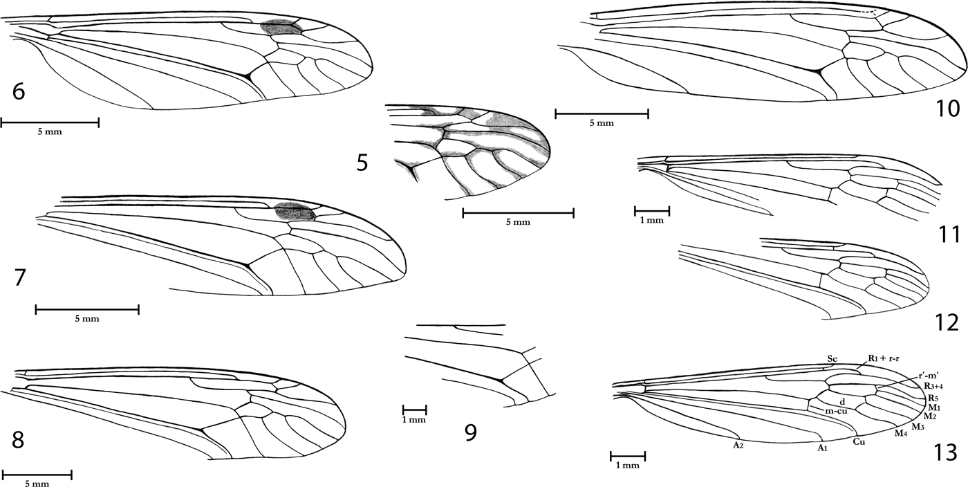
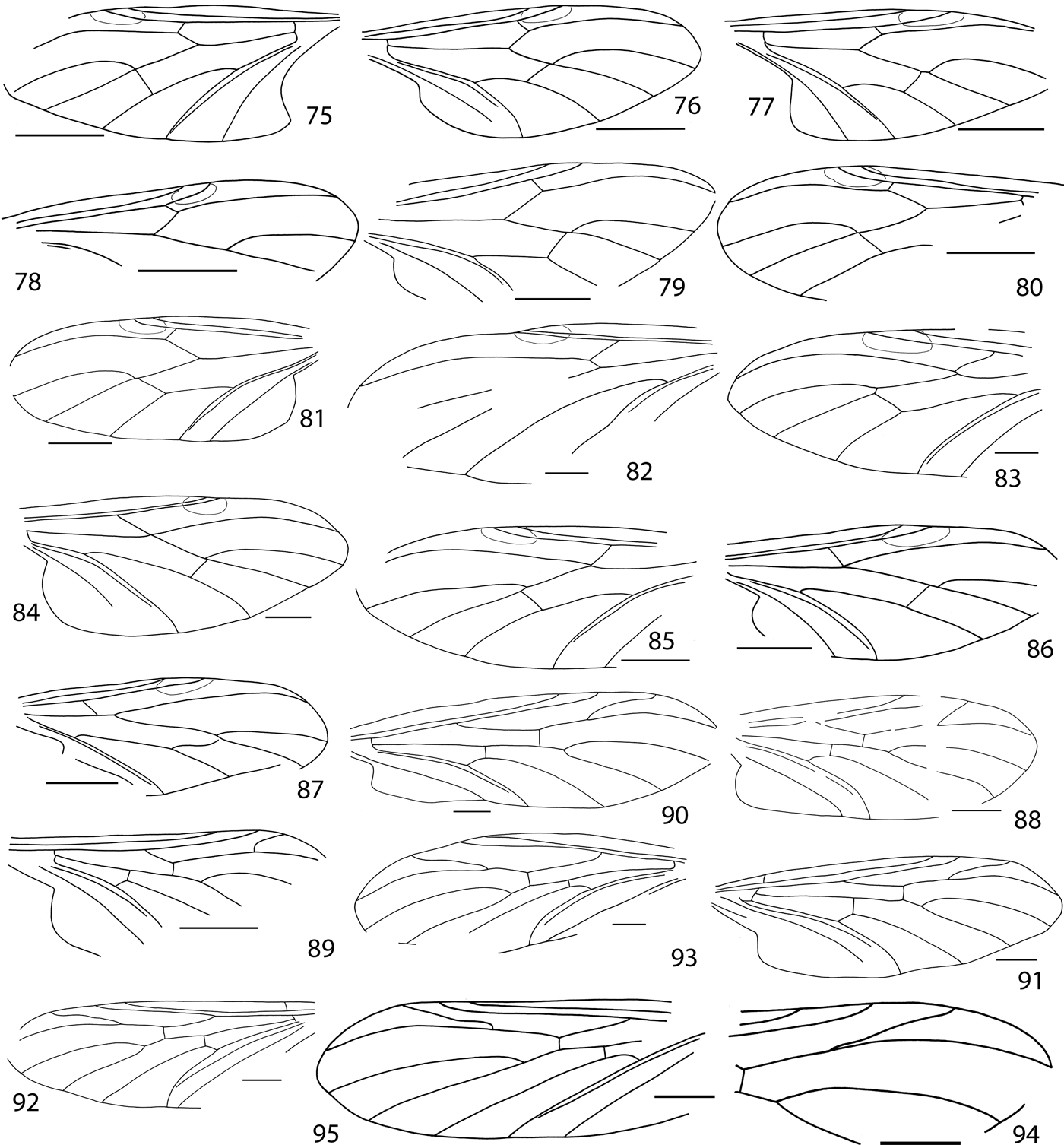
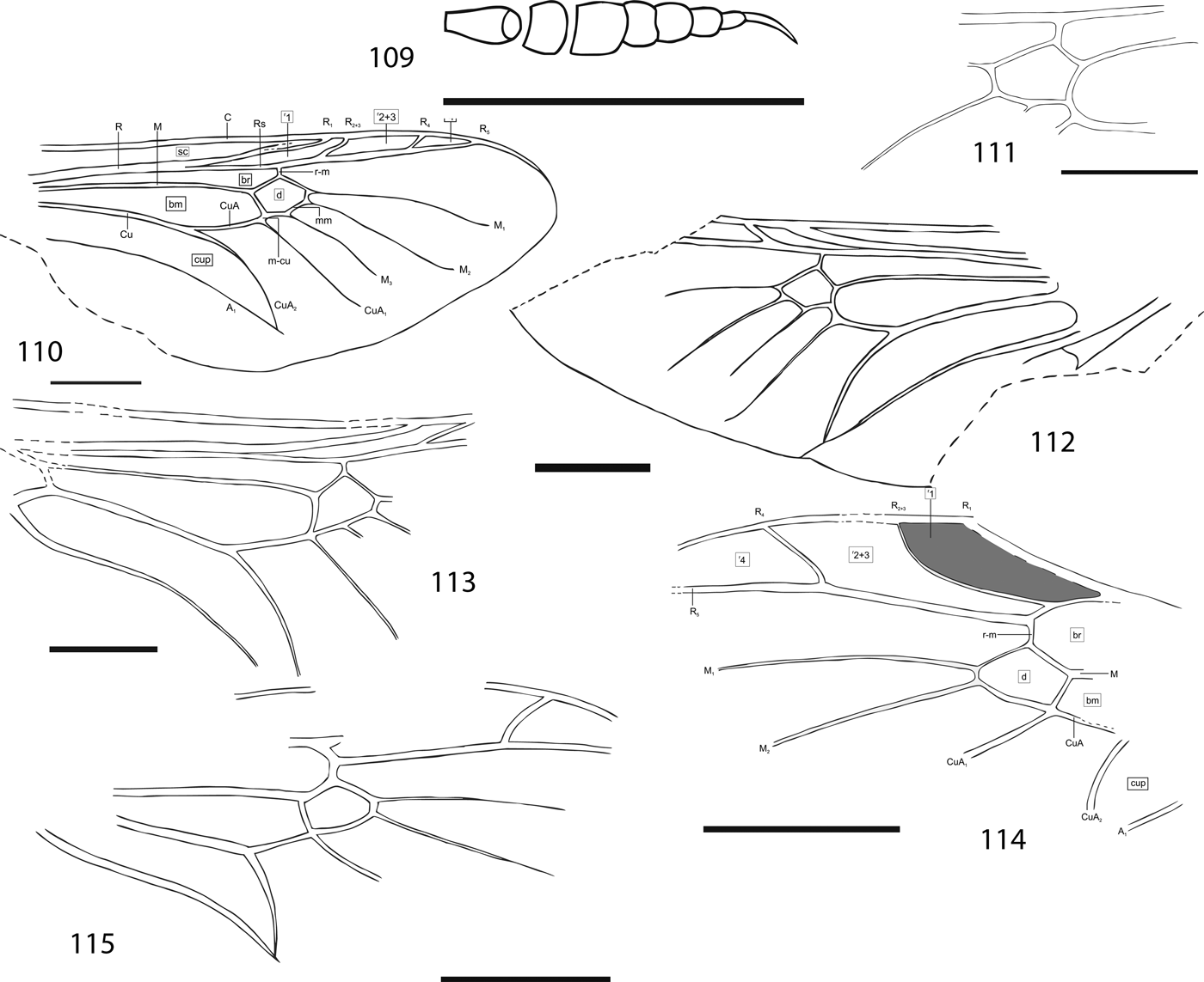
Tipulidae and Cylindrotomidae, wing venation. (5) Tipula (s. lato) acourti Cockerell, Reference Cockerell1921, holotype NHMUK I.8901. (6) Tipula (s. lato) anglicana Krzemiński, n. sp. holotype NHMUK In.64117. (7) Tipula (s. lato) gardneri Cockerell, Reference Cockerell1917a, holotype NHMUK I.8663. (8), (9) Tipula (s. lato) limiformis Cockerell, Reference Cockerell1915: (8) – (type of Tipula hooleyi Cockerell & Haines, Reference Cockerell and Haines1921 syn. nov.), NHMUK In.24353; (9) Holotype of Tipula limiformis USNM 61438. (10) Ischnotoma (Ischnotoma) vasifera (Cockerell & Haines, Reference Cockerell and Haines1921), holotype NHMUK In.24352. (11)–(13) Cyttaromyia rossi Krzemiński, n. sp. – wing venation; holotype CAMSM X.50140.14 (TN 63): (11) left wing; (12) right wing; (13) reconstruction based on both wings.

1921 Tipula acourti Cockerell, pp. 455–456, fig. 2
1994 Tipula acourti Evenhuis, p. 57
Holotype. NHMUK I.8901/In.25471 (part and counterpart), Brodie/Hooley Collections; Insect Bed; late Eocene; NW Isle of Wight, UK.
Other material. NHMUK I.8667, Brodie Collection; In.64102, In.64103, In.64109, In.64116, In.64118, In.25338, In.25471/In.64126 (part and counterpart), Hooley Collection.
Diagnosis. Wing venation very strong and additionally underlined by dark clouds accompanying veins. Sc ending in Rl opposite and near Rs fork; d cell large and broad; cu cell of same width in basal and distal part.
Redescription. Wing about 18–20mm, with strong veins and with dark clouds close to veins. Venation: tip of Sc lost, Sc terminally fused with sc-r and ending in Rl; Rs twice longer than R3+4 and as long as R4; d cell rather large and broad; petiole short, twice shorter than upper margin of d cell between cross-veins r-m and mm; Ml about three times longer than petiole; medial cells very broad; cross-vein m-cu in fork of M3+4; cell m-cu equally wide in basal and distal part.
Tipula (s. lato) anglicana Krzemiński, n. sp.
Holotype. NHMUK In.64117, Hooley Collection; Insect Bed; late Eocene; NW Isle of Wight, UK.
Other material. NHMUK In.25206/In.64100 (part and counterpart), Hooley Collection.
Diagnosis. Cell d very narrow; cu cell rectangular.
Description. Wing length 19mm, membrane little darkened but without colour pattern. All veins are very good visible. Venation: tip of Sc lost, Sc terminally fused with sc-r and ending in Rl, much before fork of Rs; R2+3+4 as long as R3; Rs about 1.5 times longer than distal M1+2; upper margin of d cell between cross-veins r-m and m-m about three times longer than petiole; d cell very narrow and rather small; cross-vein m-cu in fork of M3+4; mcu cell rectangular, equally wide over entire length.
Remarks. Venation T. (s. lato) anglicana n. sp differs from remaining species of Tipula from the Isle of Wight by the d cell and mcu cell shape.
Tipula (s. lato) gardneri Cockerell, Reference Cockerell1917a
1917a Tipula gardneri Cockerell, pp. 374, 384, pl. 31, fig. 1
1994 Tipula gardneri Evenhuis, Reference Evenhuis1994, p. 58
Holotype. NHMUK I.8663, Brodie Collection; Insect Bed; late Eocene; NW Isle of Wight, UK.
Other material. NHMUK In.17429 (part and counterpart), Smith Collection; In.25205, Hooley Collection.
Diagnosis. Wing membrane little darkened but without pattern; tip of Sc lost, Sc terminally fused with sc-r and ending in Rl, much before fork of Rs; distal M1+2 very short; preceding section between cross-veins r-m and mm about twice longer than distal M1+2; mcu cell distinctly more narrow in distal part.
Redescription. The species is represented by two separate wings of two specimens; wings are in good condition, only short basal part is missing. Wing length 20–22mm without dark pattern on wing membrane. Venation: tip of Sc lost, Sc terminally fused with sc-r and ending in Rl, well before fork of Rs; R2+3+4 twice shorter than Rs; d cell rather long and narrow; distal M1+2 very short, less than half as long as mid M1+2; Ml four times longer than distal M1+2; cross-vein m-cu in fork of M3+4; mcu cell in distal part narrowed.
Tipula (s. lato) limiformis Cockerell, Reference Cockerell1915
1915 Tipula limiformis Cockerell, p. 492, 499, pl. 61, fig. 11
1921 Tipula hooleyi Cockerell & Haines, p. 82, fig. H49, syn. nov.
1994 Tipula limiformis Evenhuis, p. 58
1994 Tipula hooleyi Evenhuis, p. 59
Holotype. USNM 61438 (Lacoe Collection No. 7523, Fig. 9, Pl. 1h); Insect Bed; late Eocene; NW Isle of Wight, UK.
Other material. In.24353 (Hooley Collection No. 49, Fig. 8, Pl. 1g) was described as Tipula hooleyi by Cockerell & Haines (Reference Cockerell1921), and In.25208.
Diagnosis. Tip of Sc lost, Sc terminally fused with sc-r and ending in Rl, well before fork of Rs; distal M1+2 very short, half as long as mid M1+2; mcu cell at anal margin narrowed (about 1/3).
Redescription. Wing 21–22mm long, without dark pattern. Venation: tip of Sc lost, Sc terminally fused with sc-r and ending in Rl, well before fork of Rs; R2+3+4 half as long as Rs; R4 rather long, little wavy and twice longer than R2+3+4; d cell rather short and broad; distal M1+2 very short, only half of mid M1+2; cross-vein m-cu in fork of M3+4; mcu cell about 1/3 narrower at anal margin than vein m-cu; anal part of wing in all specimens is missing.
Remarks. Tipula hooleyi described by Cockerell & Haines (Reference Cockerell and Haines1921) does not differ by a single character from the preserved wing fragment of Tipula limiformis, described some years earlier by Cockerell (Reference Cockerell1915); therefore this species is made a younger synonym of T. limiformis.
Genus Ischnotoma Skuse, Reference Skuse1890
Type species. Tipula serricornis Macquart, Reference Macquart1846 (designated by Alexander Reference Alexander1920)
Remarks. Recently the genus comprises about 50 species of Australian and Neotropical Regions.
It is noteworthy that most tipulid species from early Oligocene Isle of Wight belong to the genera and subgenera recently living in the tropical and subtropical regions.
Ischnotoma (Ischnotoma) vasifera (Cockerell & Haines, Reference Cockerell and Haines1921)
1921 Holorusia vasifera Cockerell and Haines, pp. 82–83, fig. H9
1967 Ischnotoma (Ischnotoma) vasifera Vane-Wright, pp. 529, 539–540
1994 Ischnotoma (Ischnotoma) vasifera Evenhuis, p. 56
Holotype. NHMUK In.24352/I.8852 (part and counterpart), Hooley/Brodie Collections; Insect Bed; late Eocene; NW Isle of Wight, UK.
Other material. Besides holotype, specimens NHMUK In.25210, In.25260/In.25493 (part and counterpart).
Diagnosis. Vein R4 strongly curved to upper wing margin; m-cu placed between fork of Mb and fork M3+4.
Redescription. Wing large, about 23mm long. Venation: Sc ending in C; sc-r at its two lengths before tip of Sc; R2+3+4 as long as R3; Rs 1.5 times longer than R2+3+4; R4 in distal part strongly curved to upper wing margin; d cell rather small; distal M1+2 very short; mid section of M1+2 is 2.5 times longer than distal; cross-vein m-cu placed between fork of Mb and fork M3+4.
Remarks. Holorusia vasifera described by Cockerell & Haines (Reference Cockerell and Haines1921) was transferred by Vane-Wright (Reference Vane-Wright1967) to the genus and subgenus Ischnotoma Skuse, Reference Skuse1890 on the basis of similarity in wing venation. The species is particularly very similar to the Australian Ischnotoma (Ischnotoma) par (Walker Reference Walker and Jekel1856).
Conclusion. Generally Tipulidae from the Isle of Wight are indicative of typical warm climate. Recent species from genera Brachypremna, Macromastix, Leptotarsus and Ischnotoma live in tropical and subtropical regions.
Family: Cylindrotomidae Schiner, Reference Schiner1863
Cylindrotomidae were until now unknown in the fossil material from the late Eocene of the Isle of Wight. There is one specimen belonging to the fossil genus Cyttaromyia Scudder. This genus has a characteristic wing venation and until now has been known only from nine fossil species from the Palaeogene of North America (Scudder Reference Scudder1877, Reference Scudder1894; Cockerell Reference Cockerell1924, Reference Cockerell1925), Denmark (Freiwald Reference Freiwald1991), France (Seguy Reference Seguy1934) and Russia (Freiwald & Krzemiński Reference Freiwald and Krzemiński1991) and one species from Baltic amber (Krzemiński Reference Krzemiński1998).
Recent Cylindrotomidae are a small family comprising 72 species inhabiting mainly Holartic and Oriental Regions. In the Afrotropical Region the family is not represented.
In the fossil state only 14 fossil species of the family have been described so far (Cockerell Reference Cockerell1920; Freiwald & Krzemiński Reference Freiwald and Krzemiński1991; Evenhuis Reference Evenhuis1994; Krzemiński Reference Krzemiński1998).
Genus Cyttaromyia Scudder, Reference Scudder1877
Type species. Cyttaromyia fenestrata Scudder, Reference Scudder1877, by monotypy
Cyttaromyia rossi Krzemiński, n. sp.
(Figs 11–13, Pl. 1j)
Derivation of name. The name of species is dedicated to Andrew Ross, with many thanks for his efficient help during all stages of our project.
Holotype. CAMSM X.50140.14 (TN 63) Smith Collection; Insect Bed; late Eocene; NW Isle of Wight, UK.
Diagnosis. R1 terminating in R3+4 (a generic character); R1 fused with r-r (R2) and terminating in R3+4 in 1/3 of its length; M3 as long as d cell; cross-vein m-cu distinctly before fork of Mb.
Description. Only two wings of the holotype are preserved; body absent. Wing length about 9mm, with distinct pterostigma.
Venation. Sc long, reaching beyond fork of Rs and terminating in the Costa; cross-vein sc-r at its four lengths before end of Sc; R1 terminating in R3+4 (diagnostic character of the genus); R1 fused with cross-vein r-r (R2) and terminating in proximal 1/3 of R3+4; addition cross-vein r′-m′ in mid of R5; d cell large , almost rectangular; four medial veins present; M3 as long as d cell length; cross-vein m-cu distinctly before fork of Mb; A2 rather long; anal lobe small.
Remarks. The new species Cyttaromyia rossi is most similar to Cyttaromyia frelloi Krzemiński, Reference Krzemiński1998 from the Eocene Baltic amber. The differences concern the length of R1 and M3; the former being shorter in C. rossi, while M3 is longer (of the length of d cell).
Conclusions. The presence of Cyttaromyia in the Isle of Wight suggests a rather warm climate of this taphocoenosis. All representatives of this fossil genus were known from subtropical climate (ex. Baltic amber), but generally Cyttaromyia are very rare in fossil materials (Freiwald & Krzemiński Reference Freiwald and Krzemiński1991; Krzemiński Reference Krzemiński1991, Reference Krzemiński1998).
Family Limoniidae Speiser, Reference Speiser and Sjöstedt1909
LIMONIIDAE (Diptera Nematocera) are common and very diverse among late Eocene insects from Bembridge Marls of the Isle of Wight. Most species are represented by isolated wings or some wing fragments, but the bodies are rather rare and poorly preserved. Fortunately, the patterns of wing venation are usually very stable within the genera and with numerous minor differences between species. In the Recent Limoniidae even small differences in the venation can accompany gross and important differences in the structure of genitalia, especially in the male.
Cockerell (Reference Cockerell1915, Reference Cockerell1917a, Reference Cockerellb, Reference Cockerell1921) and Cockerell & Haines (Reference Cockerell and Haines1921) described 23 fossil species, but some of them were incorrectly identified. The present revision includes all earlier described species. Between undetermined old collection and collections newly made during the present excavations there are representatives of one new genus and 12 species, described below. Together 30 species of Limoniidae are known from the Isle of Wight.
Recently Limoniidae are one of the largest family of the Diptera, with about 12,000 described species of worldwide distribution. Larvae usually live in running water, ponds and lakes; some are semiaquatic or terrestrial.
The oldest Limoniidae were described from the Middle and Upper Triassic of Asia and North America (Krzemiński Reference Krzemiński1992; Krzemiński et al. Reference Krzemiński, Krzemińska and Papier1994; Shcherbakov et al. Reference Shcherbakov, Lukashevich and Blagoderov1995; Krzemiński & Evenhuis Reference Krzemiński, Evenhuis, Papp and Darvas2000; Krzemiński & Krzemińska Reference Krzemiński and Krzemińska2003). They are common in most deposits with insects from the Lower Jurassic until the Neogene of Europe, Asia and North America. The oldest true limoniid is the Upper Triassic Architipula youngi Krzemiński (Reference Krzemiński1992) (about 210Ma) but from the Lower/Middle Triassic of Europe and from Middle Triassic of Asia flies have been described that are very similar to Limoniidae, which are probable ancestors of the Limoniidae (Shcherbakov et al. Reference Shcherbakov, Lukashevich and Blagoderov1995; Krzemiński & Evenhuis Reference Krzemiński, Evenhuis, Papp and Darvas2000; Krzemiński & Krzemińska Reference Krzemiński and Krzemińska2003).
Subfamily Limnophilinae Bigot, Reference Bigot1854
Genus Gurnardia Krzemiński, n. gen.
Diagnosis. Sc short, only little longer than half wing length, ending little before fork of Rs into R2+3+4 and R5; only three radial veins ending in C (R3 fused with R4); four medial vein present; cross-vein m-cu close to 1/3 of d cell.
Type species. Gurnardia corami n. sp. from the Insect Bed, NW Isle of Wight, late Eocene.
Derivation of name. The new genus is named after Gurnard Bay.
Description. Because only the type species is known, the description of the genus is the same as that of the species.
Gurnardia corami Krzemiński, n. sp.
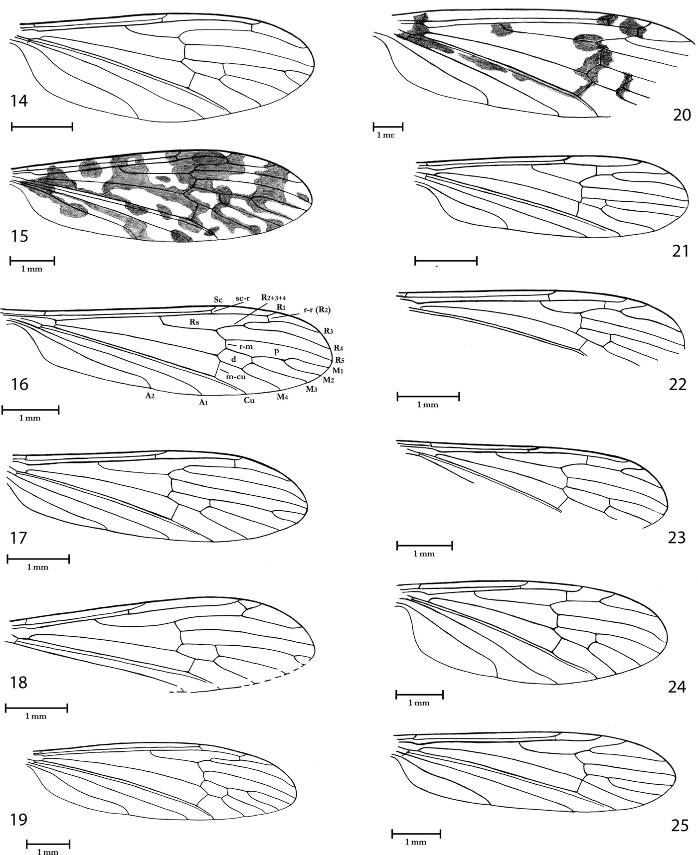
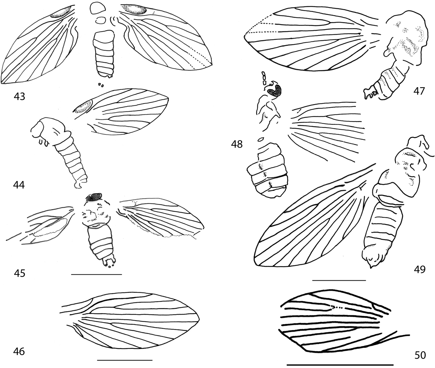
Limoniidae, wing venation. (14) Gurnardia corami Krzemiński, n. sp., holotype NHMUK In.64104. (15) Limnophila (Limnophila) cyclospila Cockerell & Haines, Reference Cockerell1921, holotype NHMUK I.9213. (16) Limnophila (Limnophila) deleta Cockerell, Reference Cockerell1921, holotype NHMUK I.9053. (17) Pilaria hooleyi Krzemiński, n. sp., holotype, NHMUK In.25254. (18) Pilaria volodii Krzemiński, n. sp., holotype NHMUK I.9167. (19) Pseudolimnophila (Pseudolimnophila) disjunctula (Cockerell & Haines, Reference Cockerell and Haines1921), holotype NHMUK. In.24355. (20) Dactylolabis (Dactylolabis) spiloptera (Cockerell, Reference Cockerell1921) n. comb., holotype NHMUK I.9015. (21) Cheilotrichia (Cheilotrichia) duplicata Krzemiński, n. sp., holotype NHMUK II 2763. (22) Cheilotrichia (Empeda) ferruginea (Cockerell, Reference Cockerell1921), holotype NHMUK I.8963. (23) Cheilotrichia (Empeda) hyalina (Cockerell, Reference Cockerell1921), holotype NHMUK I.24342. (24) Cheilotrichia (Empeda) szwedoi Krzemiński, n. sp., holotype NHMUK In.17082. (25) Gonomyia (Gonomyia) indecisa Cockerell & Haines, Reference Cockerell and Haines1921, holotype NHMUK. I.9087.

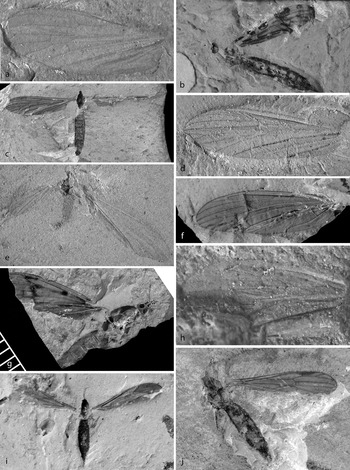
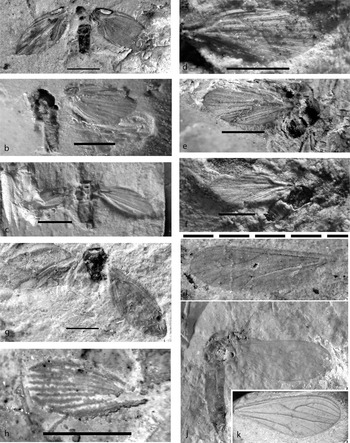
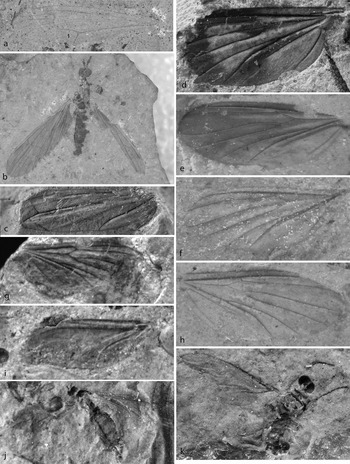
Limoniidae. (a) Gurnardia corami Krzemiński, n. sp., holotype NHMUK In.64104. (b) Limnophila (Limnophila) cyclospila Cockerell, Reference Cockerell1921, holotype NHMUK I.9213. (c) Limnophila (Limnophila) deleta Cockerell, Reference Cockerell1921, holotype NHMUK I.9053. (d) Pilaria hooleyi Krzemiński, n. sp., holotype NHMUK In.25254. (e) Pilaria volodii Krzemiński, n. sp., holotype NHMUK I.9167. (f) Pseudolimnophila (Pseudolimnophila) disjunctula (Cockerell & Haines, Reference Cockerell and Haines1921) n. comb., holotype NHMUK In.24355. (g) Dactylolabis (Dactylolabis) spiloptera (Cockerell, Reference Cockerell1921) n. comb., holotype NHMUK I.9015. (h) Cheilotrichia (Cheilotrichia) duplicata Krzemiński, n. sp., holotype NHMUK II 2763. (i) Cheilotrichia (Empeda) ferruginea (Cockerell, Reference Cockerell1921), holotype part NHMUK I.8963. (j) Cheilotrichia (Empeda) hyalina (Cockerell, Reference Cockerell1921), holotype NHMUK In.24342.

Holotype. NHMUK In.64104, Hooley Collection; Insect Bed; late Eocene; NW Isle of Wight, UK.
Additional material. NHMUK I.8982/I.9308 (female, part and counterpart), I.9491, I.9966, Brodie Collection; I.9188/In.25422 (part and counterpart), Brodie/Hooley Collections; In.25446, Hooley Collection.
Derivation of name. A new species is dedicated to Robert Coram (UK), collector and specialist of fossil insects.
Diagnosis. The same as that of the genus.
Description. Wing clear 5.0–5.8mm long.
Venation. Sc short, only little longer than half wing length, ending somewhat before fork of Rs into R2+3+4 and R5; cross-vein sc-r at its length before end of Sc; only three radial veins ending in C (R3 fused with R4); R1 long; cross-vein r-r (R2) at its length before end of R1; Rs rather short, about 1/4 shorter than R2+3+4 and as long as length as R3+4; four medial vein present; M2 short, about half as long as d cell; petiole of m1 cell three times longer than M2; d cell narrow; cross-vein m-cu about 1/3 of d cell; A2 rather long and wavy.
Remarks. The new genus is different from all other genera of the Limoniidae by the presence of three long radial and four medial veins. This composition of characters is unique among crane flies (Tipulomorpha). Unfortunately, in all specimens, legs are missing; hence, the presence of tibial spurs cannot be confirmed, which does not allow safe placement of the new genus Gunardia in the subfamily Limnophilinae. Although this character is very important to make the decision about the systematic position between Limoniidae on the subfamily level, the presence of four medial veins and position of cross-vein m-cu suggest that this new genus should be ascribed to the subfamily Limnophilinae.
Genus Limnophila Macquart, Reference Macquart1834
Type species. Limnobia pictipennis Meigen, Reference Meigen1818, p. 119 (designated by Westwood Reference Westwood1840)
Subgenus Limnophila Macquart, Reference Macquart1834
Type species. As for genus.
Remarks. One hundred and forty-six recent species of the subgenus Limnophila are known from Australian (87 sp.), Neotropic (39 sp.), Afrotropic (8 sp.) and Palaearctic (12 sp.) Regions. Fifteen fossil species are known from the Eocene and Oligocene of Europe (three from the Baltic amber) and four species from Palaeocene and Oligocene of North America.
Limnophila (Limnophila) cyclospila Cockerell & Haines, Reference Cockerell and Haines1921
1921 Limnophila cyclospila Cockerell & Haines, pp. 109–10, fig. I9213
1994 Limnophila cyclospila Evenhuis, p. 75
Holotype. NHMUK I.9213 (female), Hooley Collection), Insect Bed; late Eocene; NW Isle of Wight, UK.
Additional material. NHMUK I.9059, I.9868, I.9943, Brodie Collection; In.17491 (female), Smith Collection; In.24775, In.25209, In.25269, In.25270, Hooley Collection.
Diagnosis. Wing with conspicuous colour pattern; Sc ending a little before fork of Rs into R2+3+4 and R5; R2+3+4 very short; R4 16 times longer than R2+3+4; M1 long, 1.5 times longer than d cell; cross-vein m-cu in half of d cell.
Redescription. Wing length 7.0–7.8mm, with very distinct colour pattern (Fig. 15). Body is preserved partially only in two specimens (holotype I.9213 and In.17491). Antenna: only cylindrical scapus and broad, oval pedicel is preserved in one specimen, In.17491.
Venation. Sc ending before fork of Rs into R2+3+4 and R5; cross-vein sc-r at its length before end of Sc; R1 rather short; cross-vein r-r (R2) at its length before end of R1; R2+3+4 very short; R4 16 times longer than R2+3+4; Rs about 10 times longer than R2+3+4; d cell short and broad; petiole of m1 cell a little shorter than d cell; M1 about twice longer than petiole of m1 cell; cross-vein m-cu in half of d cell; A2 long and somewhat wavy.
Remarks. The original description of (Cockerell Reference Cockerell1921) of Limnophila cyclospila has many mistakes, the most important being the absence of vein Sc. Cross-vein m-cu is wrongly positioned in distal part of d cell position.
The colour pattern may indicate genus Austrolimnophila Alexander, but without a complete specimen preserved in good condition it is not possible to make this taxonomical decision.
Limnophila (Limnophila) deleta Cockerell, Reference Cockerell1921
1921 Limnophila deleta Cockerell, pp. 459, 462, fig. 9
1994 Limnophila deleta Evenhuis, 1994, p. 75
Holotype. NHMUK I.9053 (female), Brodie Collection, Insect Bed; late Eocene; NW Isle of Wight, UK.
Additional material. NHMUK, I.9019, I.9060, I.9067, I.9684, Brodie Collection; In.17115, In.17171, In.24414, Smith Collection; In.17484/In.64127 (part and counterpart), Smith/Hooley Collections; In.25217, In.25474, In.64127, Hooley Collection; PI. II. 2764, Palaeontological Institute, Moscow team, north end Thorness Bay, Isle of Wight.
Diagnosis. Sc rather long, ending opposite fork of Rs into R2+3+4 and R5; Rs twice longer than R2+3+4; d cell short; cross-vein m-cu little behind fork of Mb into M1+2 and M3+4 in 1/4 of d cell; A2 long and somewhat wavy.
Redescription. Wing length 5.4–8mm, without colour pattern. Few specimens are preserved with small portion of the body. Only holotype is preserved with body, but antennae and palps are missing.
Venation. Sc rather long, ending opposite fork of Rs on R2+3+4 and R5; cross-vein sc-r at its length before end of Sc; R1 rather long; cross-vein r-r (R2) at its length before end of R1; Rs about twice longer than R2+3+4; R3 about 1/4 longer than Rs; d cell small; petiole of m1 cell little longer than d cell; M1 about 1.4 times longer than petiole of m1 cell; cross-vein m-cu in quarter of d cell; A2 long and little wavy.
Remarks. Limnophila deleta is one of most common species of Limoniidae in the Isle of Wight. The specimens allotted herein are very variable and they may possibly represent more than one species, but based only on wing venation it is not possible separate them further.
Genus Pilaria Sintensis, Reference Sintensis1889
Type species. Limnobia pilicornis Zetterstedt, Reference Zetterstedt1851, p. 3885 (designated by Sharp Reference Sharp and Beddard1890).
Remarks. Forty-six recent species are known from Palaearctic (15 sp.), Nearctic (14 sp.), Afrotropic (8), Oriental (7 sp.), Australian (1 sp.) and Neotropic (1 sp.) Regions. Five fossil species are known from Eocene and Miocene of Europe (three from the Baltic amber).
Pilaria hooleyi Krzemiński, n. sp.
Holotype. NHMUK In.25254, Hooley Collection, Insect Bed; late Eocene; NW Isle of Wight, UK.
Additional material. NHMUK I.9032/In.64099 (part and counterpart), Brodie/Hooley Collections.
Derivation of name. A new species is dedicated to R.W. Hooley, the famous collector of fossil insects from the Isle of Wight.
Diagnosis. Sc ending opposite fork of Rs into R2+3+4 and R5; d cell long and narrow; petiole of m1 cell very long, about twice longer than M1 and 1.5 times longer than d cell.
Description. Wing length 2.9–5.2mm, without colour pattern.
Venation. Sc rather short, ending opposite fork of Rs; cross-vein sc-r twice its length before end of Sc; R1 rather long; cross-vein r-r (R2) at its length before end of R1; Rs five times longer than R2+3+4; R3 about 1.5 times longer than Rs; d cell long and narrow; petiole of m1 cell about twice longer than M1 and 1.5 times longer than d cell; cross-vein in third portion of d cell; A2 little curved.
Remarks. Basing only on wing venation it is very difficult to decide about generic position of this species, but in my opinion the general pattern of wing venation is more similar to the genus Pilaria than to Limnophila or Pseudolimnophila Alexander.
Pilaria volodii Krzemiński, n. sp.
Holotype. NHMUK I.9167/I.9201 (part and counterpart), Brodie Collection, Insect Bed; late Eocene; NW Isle of Wight, UK.
Derivation of name. A new species is dedicated to Vladimir (Volodia) Blagoderov (NHM, London), a paleoentomologist and specialist on Diptera.
Diagnosis. Sc very short, little shorter than half wing length, ending opposite 2/3 of Rs; R3 and M1 very short; petiole of m1 cell more than twice longer than d cell and about five times longer than M1.
Description. Wing length 4.7–5.0mm, without colour pattern; anal part of the wing destroyed.
Venation. Sc very short, little shorter than half wing length; cross-vein sc-r at three times its length before end of Sc; R1 long; cross-vein r-r (R2) at end of R1; R2+3+4 about 1/6 shorter than Rs and only little longer than R4; R3 very short, about 1/3 of R4; d cell long and narrow; M1 very short, petiole of m1 cell more than twice longer than d cell and about five times longer than M1; cross-vein m-cu in half length of d cell.
Remarks. Venation pattern of the wing venation with very short Sc, R3 and M1 is rather rare among Limoniidae. This new species is different from all Pilaria species from Baltic amber.
Genus Pseudolimnophila Alexander, Reference Alexander1919
Type species. Limnophila luteipennis Osten Sacken, Reference Osten Sacken1860, p. 236 (designated by Alexander Reference Alexander1920).
Subgenus Pseudolimnophila Alexander, Reference Alexander1919
Type species. As for genus.
Remarks. One hundred recent species of the subgenus Pseudolimnophila are known from Oriental (38 sp.), Nearctic (28 sp.), Neotropic (four sp.), Afrotropic (19 sp.), Palaearctic (seven sp.), Afrotropic (five sp.), Palaearctic (three sp.) Regions. Nine fossil species are known from Baltic amber (Eocene), one from Sicilian amber (Miocene) and one from Canada (Paleocene).
Pseudolimnophila (Pseudolimnophila) disjunctula (Cockerell & Haines, Reference Cockerell and Haines1921) n. comb.
1921 Adelphomyia disjunctula Cockerell and Haines, pp. 83–84, fig. H1398
1994 Adelphomyia disjunctula Evenhuis, Reference Evenhuis1994, p. 61
Holotype. NHMUK In.24355, Hooley Collection, Insect Bed; late Eocene; NW Isle of Wight, UK.
Additional material. NHMUK I.9050, Brodie Collection; In.17383, Smith Collection; In.64119, Hooley Collection.
Diagnosis. Sc long, ending opposite fork of Rs on R2+3+4 and R5; R1 long, ending opposite mid R3; M1 very long, twice longer than petiole of m1 cell and 2.5 times longer than petiole of m1 cell.
Redescription. Wing length 8.0–9.0mm, without colour pattern. Specimens I.9050 and In.64119 are preserved with portions of the body.
Venation. Sc long, ending opposite mid of R2+3+4; cross-vein sc-r little longer than end of Sc; R1 long, ending opposite mid of R3; cross-vein r-r (R2) about twice its length before end of R1; R3 little wavy, about 1.5 times longer than Rs and five times longer than R2+3+4; M1 very long, twice longer than petiole of m1 cell and 1.5 times longer than d cell; cross-vein m-cu close to mid of d cell; A2 little wavy.
Remarks. Cockerell & Haines (Reference Cockerell and Haines1921) placed this species in the genus Adelphomyia Bergroth, but absence of long Sc macrotrichia on wing membrane and presence of four medial veins indicate that this species cannot belong to this genus but rather to the genus Pseudolimnophila.
Subfamily Dactylolabinae Alexander, Reference Alexander1920
Genus Dactylolabis Osten Sacken, Reference Osten Sacken1860
Type species. Limnophila montana Osten Sacken, Reference Osten Sacken1860, p. 240 (by monotypy).
Subgenus Dactylolabis Osten Sacken, Reference Osten Sacken1860
Type species. As for genus.
Remarks. Fifty-seven recent species of the subgenus Dactylolabis are known only from Palaearctic (39 sp.) and Nearctic (18 sp.) Regions. Thirteen fossil species are described from the genus Dactylolabis but probably only four from the Oligocene and Miocene of Europe belong to the nominative subgenus. From Baltic amber no representative of this subgenus is known.
Dactylolabis (Dactylolabis) spiloptera (Cockerell, Reference Cockerell1921) n. comb.
1921 Epiphragma spilopterum Cockerell, pp. 458–59, fig. 8
1994 Epiphragma spilopterum Evenhuis, p. 70
Holotype. NHMUK I.9015, Brodie Collection, Insect Bed, late Eocene; NW Isle of Wight, UK.
Diagnosis. Wing with colour pattern; Sc very long, ending opposite mid of R2+3+4; Rs very long, longer than R3.
Redescription. Wing length 11.5mm, with colour pattern. Head, thorax, part of the abdomen and most parts of the wing are preserved.
Venation. Sc very long, ending opposite mid of R2+3+4; cross-vein sc-r at its length before end of Sc; R1 long; cross-vein r-r about twice its length before end of R1; Rs very long, little longer than R2+3+4 and R3 together; R2+3+4 2.5 times longer than R3; d cell very broad; cross-vein m-cu in about 1/3 of d cell; A2 rather long and straight.
Remarks. Cockerell (Reference Cockerell1921) placed this species in the genus Epiphragma Osten Sacken, but a careful study has shown that the cross-vein in the costal cell, the most important character of the genus Epiphragma, is missing here. General venation pattern indicates that this species most probably belongs to the genus Dactylolabis.
Subfamily Chioneinae Rondani, Reference Rondani1841
Genus Cheilotrichia Rossi, Reference Rossi1848
Type species. Erioptera imbuta Meigen, Reference Meigen1818, p. 114 (by monotypy).
Subgenus Cheilotrichia Rossi, Reference Rossi1848
Type species. As for genus.
Remarks. Thirty recent species of the subgenus Cheilotrichia are known from Palaearctic (18 sp.), Oriental (3 sp.), Afrotropic (3 sp.), Australian (2 sp.) and Nearctic (1 sp.) Regions. Till now no fossil species from the nominative subgenus was known.
Cheilotrichia (Cheilotrichia) duplicata Krzemiński, n. sp.
Holotype. NHMUK II 2763, collected by the Palaeontological Institute, Moscow team; Insect Bed; late Eocene; north end Thorness Bay, Isle of Wight, UK.
Diagnosis. Sc ending after 2/3 of Rs; Rs little shorter than R3; R4 twice longer than R2+3+4 and R3+4 together.
Description. Wing length 4.5mm. Middle part of wing broken and partially destroyed.
Venation. Sc long, ending behind 2/3 of Rs; cross-vein sc-r twice its length before end of Sc; R1 long, ending behind fork of R3+4 on R3 and R4; cross-vein r-r (R2) about five times before end of R1; R3 little longer than Rs and about twice longer than R2+3+4 and R3+4 together; R2+3+4 a little longer than R3+4; d cell short and narrow; M3 three times longer than d cell; cross-vein m-cu little before fork of Mb on M1+2 and M3+4; anal vein somewhat wavy.
Remarks. Cheilotrichia (Cheilotrichia) duplicata n. sp. is the first fossil representative of the subgenus Cheilotrichia. Recently the subgenus has been represented by 18 species distributed in Holarctic, Oriental, Australian and Ethiopian Regions, but most species are described from Palaearctic Region (nine sp.). Species of this subgenus prefer wet environments near streams and small ponds.
Subgenus Empeda Osten Sacken, Reference Osten Sacken1869
Type species. Empeda stigmatica Osten Sacken, Reference Osten Sacken1869, p. 184 (designated by Coquillett Reference Coquillett1910).
Remarks. One hundred and twelve recent species are described from Oriental (34 sp.), Neotropic (31 sp.), Palaearctic (22 sp.), Nearctic (14 sp.), Australian (nine species) and Afrotropic (two species) Regions.
Twelve fossil species are described, mostly (eight sp.) from Baltic amber (Eocene), two from the Isle of Wight (late Eocene), one from Sakhalin amber (Palaeocene) and one from New Jersey amber (Upper Cretaceous).
Cheilotrichia (Empeda) ferruginea (Cockerell, Reference Cockerell1921)
1921 Empeda ferruginea Cockerell, p. 457, fig. 6
1994 Cheilotrichia ferruginea Evenhuis, p. 65
Holotype. NHMUK I.8963/I.9084 (part and counterpart), Brodie Collection; Insect Bed; late Eocene; NW Isle of Wight, UK.
Additional material. I.9901/I.9139 (part and counterpart), Brodie Collection.
Diagnosis. Sc short, ending near after fork of Rs; Rs about 1.5 times longer than R3; R4 1.5 times longer than R2+3+4 and R3+4 together; d cell rather short.
Redescription. Wing length 4.4–4.7mm, clear. Anal part of the wing destroyed and terminal portion of Cu, A1 and A2 not preserved.
Venation. Sc rather short, ending a little behind fork of Rb into R1 and Rs; cross-vein sc-r at its length before end of Sc; R1 long, ending behind fork of R3+4 into R3 and R4; cross-vein r-r (R2) about three times before end of R1; Rs as long as R2+3+4 and R3+4 together, and about 1.5 times longer than R3; R4 about 1.5 times longer than Rs; d cell rather short and narrow; M1+2 about twice longer than d cell; cross-vein m-cu close to mid of d cell.
Remarks. Wing venation drawn by Cockerell (Reference Cockerell1921) is incomplete and with many mistakes. Recent revision of the holotype and two additional specimens found in the collection of NHMUK allowed a more complete reconstruction of the wing venation.
Cheilotrichia (Empeda) hyalina (Cockerell, Reference Cockerell1921)
1921 Empeda hyalina Cockerell, p. 457, fig. 5
1921 ‘Gonomyia' grisea Cockerell, p. 461, fig. 14, syn. nov.
1994 Cheilotrichia hyalina Evenhuis, p. 65
1994 Gonomyia? grisea Evenhuis, p. 89
Holotype. NHMUK I.24342, Hooley Collection; Insect Bed; late Eocene; NW Isle of Wight, UK.
Additional material. NHMUK In.24344, Hooley Collection, described as the holotype of the species Gonomyia grisea Cockerell, Reference Cockerell1921.
Diagnosis. Sc ending behind mid of Rs; Rs three times longer than R3; R2+3+4 and R3+4 together as long as Rs; and 1.5 times longer than R4; d cell long and narrow.
Redescription. Wing length 4.0–5.0mm. In both specimens anal part of the wings and last portion of Cu, A1 and A2 are not preserved.
Venation. Sc rather long, ending after mid of Rs; cross-vein sc-r three times his length before end of Sc; R1 long, ending behind fork of R3+4 into R3 and R4; cross-vein r-r (R2) about five times before end of R1; Rs as long as R2+3+4 and R3+4 together, and three times longer than R3; R2+3+4 as long as R3; d cell long and narrow, M1+2 only little longer than d cell; cross-vein m-cu in proximal 1/4 length of d cell.
Remarks. Cockerell (Reference Cockerell1921) described specimen In.24344 (Hooley number H. 1534) as Gonomyia grisea because he had overlooked the presence of cross-vein r-r (R2). This vein is the key character discerning genera Gonomyia and Cheilotrichia.
Cheilotrichia (Empeda) szwedoi Krzemiński, n. sp.
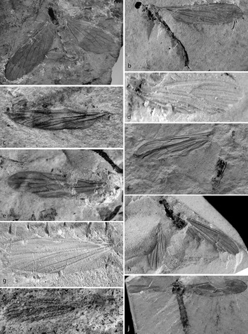
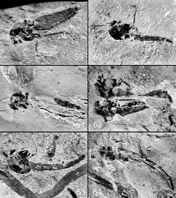
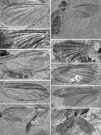
Limoniidae. (a) Cheilotrichia (Empeda) szwedoi Krzemiński, n. sp., holotype NHMUK In.17082. (b) Gonomyia (Gonomyia) indecisa Cockerell & Haines, Reference Cockerell and Haines1921, holotype NHMUK In.9087. (c) Gymnastes (Paragymnastes) fasciatipennis Cockerell, Reference Cockerell1921, holotype NHMUK I.10242. (d) Symplecta (Psiloconopa) gurnetensis Krzemiński, n. sp., holotype NHMUK In.25266. (e) Styringomyia concinna (Cockerell, Reference Cockerell1917a) n. comb., holotype NHMUK I.8666. (f) Thaumastoptera undulata (Cockerell & Haines, Reference Cockerell and Haines1921) n. comb., holotype NHMUK I.9146. (g) Orimarga (Orimarga) lenae Krzemiński, n. sp., holotype NHMUK In.25487. (h) Dicranomyia (Dicranomyia) aliena (Cockerell, Reference Cockerell1921) n. comb., holotype NHMUK I.9018. (i) Dicranomyia (Dicranomyia) brodiei (Cockerell, Reference Cockerell1917a) n. comb., holotype NHMUK I.8603. (j) Dicranomyia (Dicranomyia) excavata Cockerell & Haines, Reference Cockerell and Haines1921, holotype NHMUK I.9946.

Holotype. NHMUK In.17082, Smith Collection; Insect Bed; late Eocene; NW Isle of Wight, UK.
Derivation of name. A new species is dedicated to Jacek Szwedo (Poland), specialist of fossil Homoptera.
Diagnosis. Sc little shorter than half wing length; R1 short, ending before fork of R3+4 into R3 and R4; Rs long, 1.5 times longer than R2+3+4 and R3+4 together and four times longer than R3; M1+2 about 1.5 times longer than d cell.
Description. Specimen with most part of the body preserved, only last part of abdomen is lost. Wing length 6.1mm, with venation clearly visible.
Antenna: scapus and pedicel invisible; 12 flagellomeres preserved, basal three biggest and barrel like, next ones more slender; with short three to four bristles.
Venation. Sc short, less than half wing length; cross-vein sc-r 1.5 times its length before end of Sc; R1 very short, endingopposite mid of R3+4, far before R3+4 fork into R3 and R4; Rs long about 1.5 times longer than R2+3+4 and R3+4 together, and four times longer than R3; R2+3+4 as long as R3; d cell rather long; M1+2 about 1.5 longer than d cell; cross-vein m-cu in proximal 1/3 of d cell length; anal field broad; A2 little wavy, terminal section strongly curved.
Remarks. This new species is represented only by one specimen, but this very characteristic venation (especially length of Sc and R1) separates it from the remaining species of the subgenus Empeda from the Isle of Wight.
Genus Gonomyia Meigen, Reference Meigen1818
Type species. Limnobia tenella Meigen, Reference Meigen1818, p. 146 (by monotypy).
Subgenus Gonomyia Meigen, Reference Meigen1818
Type species. As for genus.
Remarks. Two hundred and sixteen recent species worldwide distributed from cold to tropical zone. Five fossil species of the genus Gonomyia are known, but probably only two belong to the nominative subgenus: one from Italy (Miocene) and one from Germany (Oligocene). Only one fossil species from the subgenus Electrogonomyia Alexander is described from Baltic amber.
From the Isle of Wight Cockerell & Haines (Reference Cockerell and Haines1921) described Gonomyia indecisa; in the same year Cockerell (Reference Cockerell1921) described G. ferrea, G. grisea and G. lutescens. Revision of the holotypes demonstrated that they all belong to one very common though somewhat variable species. Since G. indecisa was described as the first, G. ferrea and G. lutescens are junior synonyms of G. indecisa. G. grisea does not belong to the genus Gonomyia but to Cheilotrichia and is a junior synonym of C. (Empeda) hyalina.
Gonomyia (Gonomyia) indecisa Cockerell & Haines, Reference Cockerell and Haines1921
1921 Gonomyia indecisa Cockerell and Haines, p. 111, fig. I9087
1921 ‘Gonomyia' ferrea Cockerell, p. 460, fig. 12, syn. nov.
1921 ‘Gonomyia' lutescens Cockerell, p. 461, fig. 13, syn. nov.
1994 Gonomyia? ferrea Evenhuis, p. 89
1994 Gonomyia? lutescens Evenhuis, p. 89
Holotype. NHMUK I.9087, Brodie Collection; Insect Bed; late Eocene; NW Isle of Wight, UK.
Additional material. In.24343 (described as G. ferrea – old number H.835, Hooley Collection); I.9217/I.9409 (part described as G. lutescens), I.9044(1), I.9283, Brodie Collection; In.24632, In.24812, In.25420, In.25429, In.64120, In.64129, Hooley Collection; I.9728/In.17216 (part and counterpart), Brodie/Smith Collections; In.17248, Smith Collection.
Diagnosis. Sc short, ending opposite 2/3 of Rs; Rs about four times longer than R2+3+4 and twice longer than R3; d cell open.
Description. Wing in very bad condition, only distal part (about 3mm) is preserved, total length probably 5.5–6.0mm.
Venation. Sc short, ending opposite 2/3 of Rs; cross-vein sc-r at its length before end of Sc; R1 rather short; R2 absent; Rs about four times longer than R2+3+4 and twice longer than R3; d cell open by atrophy of cross-vein m3-m4; position of cross-vein m-cu variable: near before, in or little behind fork of Mb into M1+2 and M3+4; A2 somewhat wavy.
Remarks. Gonomyia indecisa is common in the materials from the Isle of Wight. Position of cross-vein m-cu and proportions between veins are so much variable that is not possible to find the border between species described by Cockerell (Reference Cockerell1921) and Cockerell & Haines (Reference Cockerell and Haines1921). In this situation wing venation cannot be used to establish a different species.
Genus Gymnastes Brunetti, Reference Brunetti1911
Type species. Gymnastes violaceus Brunetti, Reference Brunetti1911, p. 282 (by monotypy).
Remarks. The genus Gymnastes is represented in Recent fauna by three subgenera and 43 species in Oriental (25 sp.), Australian (11 sp.), Afrotropic (three sp.), Palaearctic (three sp.) one species is known from Neotropic Region. Only one fossil species is known.
Subgenus Paragymnastes Alexander, Reference Alexander1922
Type species. Limnobia fascipennis Thomson, Reference Thomson1869, p. 447 (original designation).
Remarks. Thirty-one recent species are known from Oriental, Australian and Eastpalaearctic Regions
Gymnastes (Paragymnastes) fasciatipennis Cockerell, Reference Cockerell1921
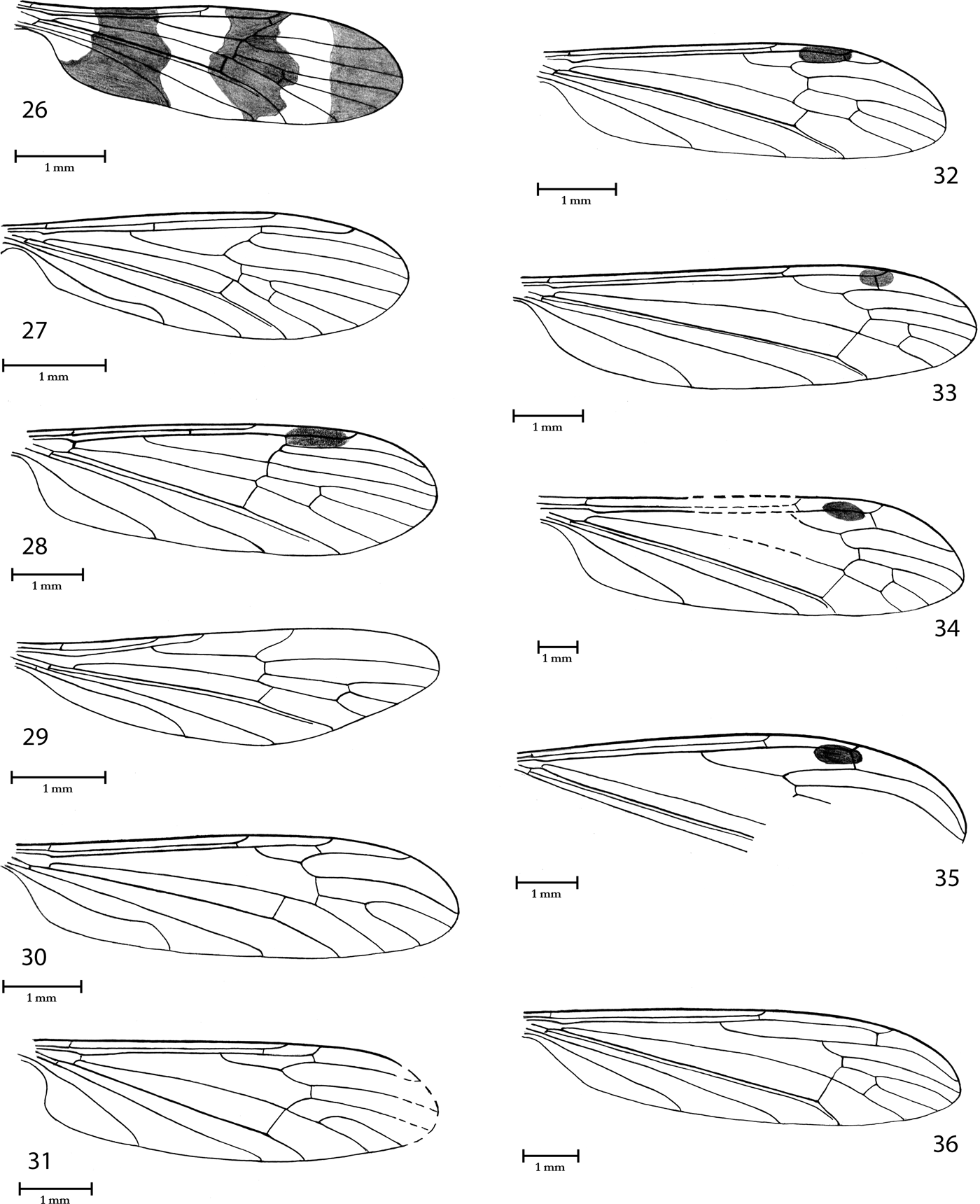
Limoniidae, wing venation. (26) Gymnastes (Paragymnastes) fasciatipennis Cockerell, Reference Cockerell1921, holotype, NHMUK I.10242. (27) Symplecta (Psiloconopa) gurnetensis Krzemiński, n. sp., holotype NHMUK In.25266. (28) Ilisia (Lunaria) incognita (Cockerell, Reference Cockerell1921), holotype NHMUK I.9186. (29) Styringomyia concinna (Cockerell, Reference Cockerell1917a), holotype NHMUK I.8666. (30) Thaumastoptera undulata (Cockerell & Haines, Reference Cockerell and Haines1921), holotype NHMUK I.9146. (31) Orimarga (Orimarga) lenae Krzemiński, n. sp., holotype NHMUK In.25487. (32) Dicranomyia (Dicranomyia) aliena (Cockerell, Reference Cockerell1921), holotype NHMUK I.9018. (33) Dicranomyia (Dicranomyia) brodiei (Cockerell, Reference Cockerell1917a), holotype NHMUK I.8603. (34) Dicranomyia (Dicranomyia) excavata Cockerell & Haines, Reference Cockerell and Haines1921, holotype NHMUK I.9946. (35) Dicranomyia (Dicranomyia) fasciata Krzemiński, n. sp., holotype NHMUK I.9058. (36) Limonia spilota (Cockerell, Reference Cockerell1921), holotype NHMUK In.17114.

1921 Gymnastes fasciatipennis Cockerell, pp. 456–457, fig. 4
1931 Gymnastes fasciatipennis Edwards in Marshall & Staley, p. 38
1994 Gymnastes fasciatipennis Evenhuis, p. 75
Holotype. NHMUK I.10242, Brodie Collection; Insect Bed; late Eocene; NW Isle of Wight, UK.
Additional material. NHMUK I, 9282, I.9297 (female), I.9959, Brodie Collection; I.17169 (male), In.17494, Smith Collection; In.25386, In.25408, I, 64105, In.64114, In.64115, I.64121, Hooley Collection; II 2848, Emma Ross Collection, north end Thorness Bay; MIWG 3685 (female).
Diagnosis. Wings with three broad dark bands; Sc shorter than half wing, ending opposite mid Rs; cross-vein r-r (R2) lost; R2+3 very short.
Redescription. Wing 3.2–4.8mm long, with three broad dark grey bands, clearly separated. Some specimens with body partially preserved. Head, thorax and abdomen dark brown or intensive black. Antennae and palps not preserved.
Venation. Sc very short, shorter than half wing, ending opposite mid Rs; cross-vein sc-r poorly visible, probably twice its length before end of Sc; R1 short; cross-vein r-r (R2) twice its length before end of R1; Rs twice longer than R2+R3; R4 1.5 times longer than Rs and three times longer than R2+3; cross-vein r-m little before or in fork of Rs on R2+3+4 and R5; d cell very narrow and rather long; M1+2 little more than twice longer than d cell; cross-vein m-cu little before fork of Mb into M1+2 and M3+4; A1 slightly wavy; A2 rather straight.
Remarks. Gymnastes fasipennis is one of the most common fossil Limoniidae from the Isle of Wight. Recently the subgenus Paragymnastes is known only from the Oriental (16 sp.), Australian (11 sp.) and Palaearctic (two sp.) Regions. All species prefer warm climate. Presence of the representative of this genus indicates tropical or subtropical climate in the late Eocene.
Genus Symplecta Meigen, Reference Meigen1830
Type species. Limnobia punctipennis Meigen, Reference Meigen1818, p. 147 (designated by Westwood Reference Westwood1840).
Subgenus Psiloconopa Zetterstedt, Reference Zetterstedt1838
Type species. Psiloconopa meigeni Zetterstedt, Reference Zetterstedt1838, p. 847 (by monotypy).
Remarks. Sixty-one recent species are known from Nearctic (29 sp.), Palaearctic (17 sp.), Oriental (11 sp.), Australian (two sp.), Afrotropic (one sp.) and Neotropic (one sp.) Regions.
Only two fossil species are known from the Oligocene and Miocene of Europe.
Symplecta (Psiloconopa) gurnetensis Krzemiński, n. sp.
Holotype. NHMUK In.25266, Hooley Collection); Insect Bed; late Eocene; NW Isle of Wight, UK.
Derivation of name. The name species origin from the old name of Gurnard Bay (Gurnet Bay).
Diagnosis. Sc rather long, ending behind cross-vein r-r (R2); cross-vein sc-r opposite proximal 1/4 Rs; cross-vein very close to fork of Mb into M1+2 and M3+4.
Description. Wing length 4.1mm without colour pattern. Wing folded.
Venation. Sc rather long, ending behind cross-vein r-r (R2); cross-vein sc-r opposite proximal 1/4 Rs, about 17 times its length before end of Sc; R1 long; cross-vein r-r (R2) 13 times its length before end of R1; Rs 3.5 times longer than R2+3+4; R4 about 1.5 times longer than Rs and about six times longer than R2+3+4; d cell short; M3 2.3 times longer than d cell; cross-vein m-cu somewhat before fork of Mb into M1+2 and M3+4; A2 long and strongly wavy in terminal section.
Remarks. Representatives of the genus Symplecta generally are very rare in fossil material. Until now, they were described only from the Oligocene of France (Theobald Reference Theobald1937), Miocene of Italy (Gentilini Reference Gentilini1984; Krzemiński & Gentilini Reference Krzemiński and Gentilini1992) and Miocene of Spain (Krzemiński and Arillo Reference Krzeminski and Arillo2008).
Genus Ilisia Rondani, Reference Rondani1856
Type species. Erioptera maculata Meigen, Reference Meigen1804, p. 51 (original designation).
Subgenus Lunaria Savchenko, Reference Savchenko1973
Type species. Ilisia (Lunaria) idiophallus Savchenko, Reference Savchenko1973, p. 38 (original designation).
Remarks. Only two recent species are known from the Palaearctic Region.
Ilisia (Lunaria) incognita (Cockerell, Reference Cockerell1921) n. comb.
1921 Limnophila incognita Cockerell, p. 462, Fig. 16
1994 Limnophila incognita Evenhuis, p. 75
Holotype. NHMUK I.9186, Brodie Collection; Insect Bed; late Eocene; NW Isle of Wight, UK.
Additional material. NHMUK In.64111, Hooley Collection.
Diagnosis. Sc short, ending before fork of Rs into R2+3+4 and R5; cross-vein opposite proximal 1/3 of Rs; M3 1.5 times longer than d cell; cross-vein m-cu behind fork of Mb into M1+2 and M3+4.
Redescription. Most parts of body preserved, without palps, legs and genitalia. Wing length 6.3mm.
Antenna. scapus rather long; pedicel short and rounded; only three flagellomeres are preserved: first two short, broad and next narrow and elongate, little widened basally; all with bristles shorter than flagellomeres.
Venation. Sc short, ending before fork of Rs into R2+3+4 and R5; cross-vein opposite proximal 1/3 of Rs, about nine times its length before end of Sc; R1 rather long; cross-vein r-r (R2) eight times its length before end of R1; Rs long, only about 1/3 shorter than R5; R2+3+4 and R2+3 very short, R2+3+4 only twice longer than R2+3; d cell narrow; M3 1.5 times longer than d cell; cross-vein little behind fork of Mb into M1+2 and M3+4; A2 slightly wavy.
Remarks. Ilisia (Lunaria) incognita is the first fossil representative of the genus Ilisia. Recently, Ilisia is a small genus whose species live mostly in Palaearctic and Oriental Regions, (some are also in Afrotropical and Australian Regions). Classification of the species from the Isle of Wight in the subgenus Lunaria is based only on wing venation.
Genus Styringomyia Loew, Reference Loew1845
Type species. Styringomyia venusta Loew, Reference Loew1845, p. 6 (by monotypy) (fossil).
Remarks. One hundred and sixty-five recent species are known mostly from tropical and subtropical regions of the world. Only four fossil species are known from the Eocene, Oligocene and Holocene of Europe. One species is described from Baltic amber.
Styringomyia concinna (Cockerell, Reference Cockerell1917a)
1917a Mesomyites concinnus Cockerell, pp. 377–78, 384, pl. 31, fig. 7
1917b Styringomyia concinna Cockerell, p. 141
1921 Styringomyia extensa Cockerell and Haines, p. 112, fig. H261, syn. nov.
1994 Styringomyia concinna Evenhuis, p. 84
1994 Styringomyia extensa Evenhuis, p. 84
Holotype. NHMUK I.8666, Brodie Collection; Insect Bed; late Eocene; NW Isle of Wight, UK.
Additional material. NHMUK In.24354, Hooley Collection (H 261), described as Styringomyia extensa; I.8998 (1), Brodie Collection; II 2,785 (2) collected by the Zoological Institute, St Petersburg team, Burnt Wood, Isle of Wight.
Diagnosis. Sc very short, about 1/3 wing length, ending just behind fork of Rb into R1 and Rs; R1 ending opposite proximal 1/3 of Rs; Rs twice times longer than R2+3+4.
Redescription. Wing length 3.5–4.8mm.
Venation. Sc very short, about 1/3 wing length, ending just behind fork of Rb into R1 and Rs; cross-vein sc-r absent; only three long radial veins present; R1 very short, ending opposite 1/3 of Rs; cross- vein r-r (R2) and vein R3 probably lost by atrophy; Rs twice longer than R2+3+4; R5 1.75 times longer than Rs; d cell long and narrow, about twice longer than M3; cross-vein m-cu in 1/5 of d cell; A2 long and little wavy.
Remarks. Cockerell (Reference Cockerell1917a) described Mesomyites concinnus and mistakenly included this species in the family Empididae (Diptera Brachycera) and together with Haines (Cockerell & Haines Reference Cockerell and Haines1921) described new species Styringomyia extensa. Study of the holotypes of these species shoved that it is the same species from the genus Styringomyia and S. extensa is the junior synonym.
Subfamily Limoniinae Speiser, Reference Speiser and Sjöstedt1909
Genus Thaumastoptera Mik, Reference Mik1866
Type species. Thaumastoptera calceata Mik, Reference Mik1866, p. 303 (original designation).
Subgenus: Thaumastoptera Mik, Reference Mik1866
Type species. As for genus.
Remarks. Eight recent species are known from Palaearctic (three sp.), Oriental (two sp.), Afrotropic (two sp.) and Nearctic (one sp.) Regions. Only two fossil species are known from the Baltic amber (Eocene).
Thaumastoptera undulata (Cockerell & Haines, Reference Cockerell and Haines1921)
n. comb.
1921 Dicranomyia undulata Cockerell and Haines, p. 110, fig. I9146
1931 Dicranomyia undulata Alexander, pp. 8, 41
1994 Dicranomyia undulata Evenhuis, p. 68
Holotype. NHMUK I.9146, Brodie Collection; Insect Bed; late Eocene, NW Isle of Wight, UK.
Diagnosis. Sc short, ending opposite fork of Rb into R1 and Rs: Rb forked behind mid wing; cross-vein m-cu at its length before fork of Mb into M1+2 and M2+3.
Redescription. Wing length 6.0mm.
Venation. Sc very short, only a little longer than half wing length, ending opposite fork of Rb into R1 and Rs; cross-vein sc-r twice its length before end of Sc; Rb forked behind mid wing; R1 rather short; cross-vein r-r(R2) twice its length before end of R1; Rs very short , only twice longer than R2+3+4; R3+4 three times longer than R2+3+4 and twice longer than Rs; R5 three times longer than Rs; d cell open by atrophy of cross-vein between M3 and M4; M3 1.5 times longer than M1+2; cross-vein m-cu at its length before fork of Mb into M1+2 and M3+4; A2 strongly wavy terminally.
Remarks. Cockerell & Haines (Reference Cockerell and Haines1921) erroneously included T. undulata in the genus Dicranomyia.
Recently genus Thaumastoptera is not represented in Australian and Neotropical Regions; most species live in the Holarctic Region. Until now only two species from Baltic amber have been described (Krzemiński, Reference Krzemiński1985).
Genus Orimarga Osten Sacken, Reference Osten Sacken1869
Type species. Limnobia alpina Zetterstedt, Reference Zetterstedt1851, p. 3894 (designated by Coquillett Reference Coquillett1910).
Subgenus Orimarga Osten Sacken, Reference Osten Sacken1869
Type species. As for genus.
Remarks. One hundred and thirty-seven recent species are known from Neotropic (42 sp.), Oriental (41 sp.), Palaearctic (21 sp.), Australian (15 sp.), Afrotropic (14 sp.) and Nearctic (four sp.) Regions.
Orimarga (Orimarga) lenae Krzemiński, n. sp.
Holotype. In.25487 Hooley Collection; Insect Bed; late Eocene; NW Isle of Wight, UK.
Derivation of name. The new species is dedicated to Lena Lukashevich (Russia, Moscow), specialist of fossil Diptera.
Diagnosis. Sc ending opposite fork of Rs into R2+3+4 and R5; cross-vein m-cu little before fork of Mb into M1+2 and M3+4.
Description. Sc rather short, ending opposite fork of Rs into R2+3+4 and R5; cross-vein sc-r three time its length before end of Sc; R1 short, ending opposite cross-vein of m-r; cross-vein r-r (R2) at its length before end of R1; R3+4 three times longer than R2+3+4 and little more than twice Rs; d cell open by atrophy of cross-vein m-m between M1+2 and M3; cross-vein m-cu somewhat before fork of Mb into M1+2 and M3+4; A2 short and rather straight.
Remarks. Orimarga(Orimarga) lenae is the first fossil representative of the genus Orimarga.
Genus Dicranomyia Stephens, Reference Stephens1829
Type species. Limnobia modesta Meigen, Reference Meigen1818, p. 134 (designated by Coquillett Reference Coquillett1910).
Subgenus Dicranomyia Stephens, Reference Stephens1829
Type species. As for genus.
Remarks. Eight hundred and fifty-seven recent species worldwide distributed from cold to tropical zones. About 30 fossil species are known from the Upper Cretaceous to Miocene. Unfortunately, representatives of the nominal subgenus cannot be identified without knowledge of morphology of male genitalia, and only wings are presented. Four species are known from Baltic amber.
Dicranomyia (Dicranomyia) aliena (Cockerell, Reference Cockerell1921) n. comb.
1921 Limnophila aliena Cockerell, pp. 461–62, fig. 15
1931 Limnophila aliena Alexander, p. 8
1994 Limnophila aliena Evenhuis, p. 75
Holotype. NHMUK I.9018/I.9420 (part and counterpart), Brodie Collection; Insect Bed; late Eocene; NW Isle of Wight, UK.
Additional material. NHMUK, I.9758, Brodie Collection.
Diagnosis. Sc rather long, ending opposite mid Rs; d cell long and broad; cross-vein m-cu in proximal 1/3 of d cell.
Description. Wing length 5.4–7.0mm, basal part and small parts of medial and cubital field destroyed.
Venation. Sc rather long, ending opposite mid of Rs; cross-vein sc-r at its length before end of Sc; R1 rather long, ending opposite fork; of R2+3+4 into R3 and R3+4; Rs as long as R2+3+4 and 1.5 times longer than R3+4; R5 three times longer than R5; d cell long and broad; M3 1.3 longer than d cell; cross-vein m-cu in proximal 1/3 of d cell; A1 straight; A2 rather short and straight.
Remarks. Cockerell (Reference Cockerell1921) erroneously included this species in the genus Limnophila.
Dicranomyia (Dicranomyia) brodiei (Cockerell, Reference Cockerell1917a) n. comb.
1917a Riphidia brodiei Cockerell, pp. 373, 384, pl. 31, fig. 7
1917b Rhipidia brodiei Cockerell, p. 141
1994 Rhipidia brodiei Evenhuis, p. 84
Holotype. NHMUK I.8603, Brodie Collection; Insect Bed; late Eocene; NW Isle of Wight, UK.
Additional material. In.17486 (part and counterpart), Smith Collection; In.25460, In.25477/In.26051 (part and counterpart), Hooley Collection.
Diagnosis. Sc short, ending opposite fork of Rb on R1 and Rs; d cell short and broad; cross-vein m-cu in or little before fork of Mb on M1+2 and M3+4.
Redescription. Wing 6.1–7.0mm, with big, dark stigma.
Venation. Sc short ending opposite fork of Rb into R1 and Rs; cross-vein sc-r twice its length before end of Sc; R1 rather long; R3+4 2.3 times longer than Rs and 2.5 times longer than R2+3+4; R5 2.5 times longer than Rs; d cell short and broad; M3 as long as d cell; cross-vein m-cu in fork of Mb into M1+2 and M3+4, but in some specimens this cross vein is positioned little before this fork; A1 straight or little wavy; A2 little wavy.
Remarks. Cockerell (Reference Cockerell1917a, Reference Cockerellb) erroneously included this species in the genus Rhipidia. More detailed study of the holotype and additional materials has shown that species described as Rhipidia brodei belongs to the genus Dicranomyia.
Dicranomyia (Dicranomyia) excavata Cockerell & Haines, Reference Cockerell and Haines1921
1921 Dicranomyia excavata Cockerell & Haines, pp. 110–11, fig. I9946
1921 Dicranomyia excavata Cockerell, p. 458, fig. 7 (junior homonym of Dicranomyia excavata Cockerell & Haines Reference Cockerell and Haines1921)
1922 Dicranomyia excavata Cockerell, p. 17
1922 Dicranomyia exhumata Cockerell, p. 17 (new replacement name for Dicranomyia excavata Cockerell Reference Cockerell1921), syn. nov.
1994 Dicranomyia excavata Evenhuis, p. 67
1994 Dicranomyia exhumata Evenhuis, p. 67
Holotype. NHMUK I.9946 (male), Brodie Collection; Insect Bed; late Eocene; NW Isle of Wight, UK.
Additional material. NHMUK I.8884 (described as the holotype of Dicranomyia exhumata Cockerell, Reference Cockerell1922), I.9766, I.8862, Brodie Collection; In.64096/In.64108 (part and counterpart), Hooley Collection.
Diagnosis. Sc ending little behind fork of Rb on R1 and Rs; d cell short and broad; cross-vein m-cu behind fork of Mb into M1+2 and M3+4.
Redescription. Wing length 6.0–6.5mm, with big dark stigma. Holotype with wing partially destroyed in middle part but most parts of the thorax and abdomen preserved.
Venation. Sc ending little behind fork of Rb into R1 and Rs; cross-vein sc-r at its length before end of Sc; R1 long; R3+4 1.3 times longer than Rs and 2.3 times longer than R2+3+4; R5 2.3 times longer than Rs; d cell short and broad; M3 little shorter than d cell; A1 straight; A2 rather short and little wavy.
Remarks. Cockerell & Haines (Reference Cockerell1921) described Dicranomyia excavata and in this same year Cockerell (June 1921) described another species under the same name. When he realised his mistake, he changed the name of the latter species to Dicranomyia exhumata (Cockerell Reference Cockerell1922). Present detailed study has shown that differences between both species are small and can be ascribed to intraspecific variability. Thus D. exhumata becomes a junior synonym.
Dicranomyia (Dicranomyia) fasciata Krzemiński, n. sp.
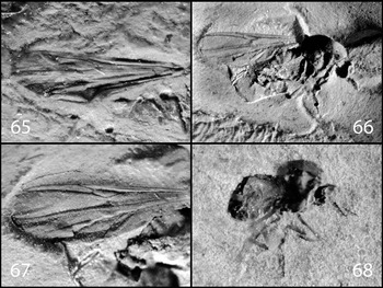
Limoniidae, Scatopsidae and Anisopodidae. (a) Dicranomyia (Dicranomyia) fasciata Krzemiński, n. sp., holotype NHMUK I.9058. (b) Limonia spilota (Cockerell, Reference Cockerell1921), holotype NHMUK In.17114. (c) Helius (Helius) edmundi Krzemiński, n. sp., holotype NHMUK I.9810. (d) Helius (Helius) popovi Krzemiński, n. sp., holotype NHMUK I.9392. (e) Dicranoptycha staryi Krzemiński, n. sp., holotype NHMUK In.24787. (f) Helius (Helius) vectensis (Cockerell, Reference Cockerell1915) n. comb., holotype USNM 61439. (g) Trentepohlia (Mongoma) pallescens (Cockerell, Reference Cockerell1921), NHMUK I.8898. (h) Trentepohlia (Mongoma) cruciferella (Cockerell, Reference Cockerell1917a), holotype NHMUK I.8635. (i) Regmoclemina haennii Krzemiński, n. sp., holotype NHMUK In.9512. (j) Sylvicola hooleyi (Cockerell, Reference Cockerell1921), holotype NHMUK In.24346. (k) Sylvicola problematica Krzemiński, n. sp., holotype NHMUK In.25675.

Holotype. NHMUK I.9058, Brodie Collection; Insect Bed; late Eocene; NW Isle of Wright, UK.
Diagnosis. Sc long, ending opposite 3/4 of Rs; R3+4 very long and strongly curved.
Description. Wing length 8.0mm, with big, dark stigma. Wing in medial, cubital and annal part destroyed; most parts of the body are preserved.
Head. antenna with big tubular scapus and small flat pedicel; flagellomeres short and rounded, with short bristles, not longer than flagellomere.
Thorax partially preserved, with only one strongly destroyed wing.
Venation. Sc long, ending opposite 3/4 of Rs; cross-vein sc-r at its length before end of Sc; R3+4 very long and strongly curved, 2.3 times longer than Rs and about twice longer than R2+3+4; R5 very long, strongly curved, and more than twice longer than Rs. Remaining veins in most parts are destroyed.
Abdomen. Male genitalia with outer dististyle large, balloon like; inner dististyle invisible; penis short and broad basally.
Remarks. Despite poor state of wing preservation, the venation retained (especially shape of radial veins) and male genitalia show distinct differences between this species and all other fossil species of the genus Dicranomyia.
Genus Limonia Meigen, Reference Meigen1803
Type species. Limonia tripunctata Fabricius, Reference Fabricius1781, p. 405 (autom.).
Remarks. Two hundred and twenty eight species are known from Palaearctic (72 sp.), Oriental (88 sp.), Australian (39 sp.), Afrotropic (16 sp.) and Nearctic (13 sp.) Region. Evenhuis (Reference Evenhuis1994) included 17 fossil species in his catalogue, but many of them need to be revised, some may belong to Dicranomyia.
Limonia spilota (Cockerell, Reference Cockerell1921)
1921 Limnobia spilota Cockerell, pp. 459–60, fig. 10
1994 Limonia spilota Evenhuis, p. 77
Holotype. NHMUK In.17114/In.20538 (part and counterpart), Smith Collection; Insect Bed; late Eocene; NW Isle of Wight, UK.
Additional material. In.25498, Hooley Collection.
Diagnosis. Sc ending opposite 1/3 of Rs; d cell short and broad; M3 1.5 times longer than d cell; cross-vein in fork of Mb into M1+2 and M3+4.
Redescription. Wing length 8.0mm, without colour pattern. Two wings, parts of head, thorax and abdomen are preserved; antennae, palps and hypopygium are missing.
Venation. Sc short, ending opposite 1/3 of Rs; cross-vein sc-r twice its length before end of Sc; R1 long; cross-vein r-r (R2) at its length before end of R1; R3+4 about twice longer than R2+3+4; R5 about 1.3 times longer than Rs; d cell short and broad; M3 1.5 times longer than d cell; cross-vein m-cu in fork of Mb into M1+2 and M3+4; A2 straight.
Remarks. Recently genus Limonia is represented in most regions of the Earth, except the Neotropical Region. In the fossil state the oldest species was described from Baltic amber (late Eocene). Some species of Dicranomyia are sometimes included in Limonia.
Genus Helius Lepeletier & Seville, Reference Lepeletier, Serville, Latreille, Lepeletier, Serville and Guerin Meneville1828
Type species. Limnobia longirostris Meigen, Reference Meigen1818, p. 146 (autom.).
Subgenus Helius Lepeletier & Seville, Reference Lepeletier, Serville, Latreille, Lepeletier, Serville and Guerin Meneville1828
Type species. As for genus.
Remarks. One hundred and eighty recent species are known from Oriental (70 sp.), Neotropic (51 sp.), Afrotropic (23 sp.), Palaearctic (20 sp.), Australian (14 sp.) and Nearctic (two sp.) Regions. Seven fossil species are described from Upper Cretaceous through Oligocene.
Helius (Helius) edmundi Krzemiński, n. sp.
Limoniidae, wing venation. (37) Helius (Helius) edmundi Krzemiński, n. sp., holotype NHMUK I.9810. (38) Helius (Helius) popovi Krzemiński, n. sp., holotype NHMUK I.9392. (39) Helius (Helius) vectensis (Cockerell, Reference Cockerell1915), holotype USNM 61439. (40) Dicranoptycha staryi Krzemiński, n. sp., holotype NHMUK In.24787. (41) Trentepohlia (Mongoma) cruciferella (Cockerell, Reference Cockerell1917a), holotype NHMUK I.8635. (42) Trentepohlia (Mongoma) pallescens (Cockerell, Reference Cockerell1921), holotype NHMUK I.8898.

Holotype. NHMUK I.9810, Brodie Collection; Insect Bed; late Eocene; NW Isle of Wight, UK.
Derivation of name. A new species is dedicate to Edmund Jarzembowski (UK) specialist of fossil insects.
Diagnosis. Sc ending opposite fork of Rs into R2+3+4 and R5; R5 twice longer than Rs, in mid part strongly wavy and d cell widened in proximal broaden in 1/3.
Description. Wing length 5.0mm, with small black stigma. Most parts of thorax, abdomen and two wings are preserved; head is destroyed.
Venation. Sc rather long, ending opposite fork of Sc into R2+3+4 and R5; cross-vein four times its length before end of Sc; R1 long; R2+3+4 1.25 times of Rs; R5 twice longer than Rs, strongly wavy in mid part; d cell roughly rhomboid, with rounded upper margin and widened in 1/3 length; M1+2 strongly wavy; M3 1.25 times longer than d cell; A1 little wavy in distal part; A2 only slightly curved to anal margin of the wing.
Remarks. Strong and wavy veins R5 and M1+2 separate this single species from all other representatives of Helius.
Helius (Helius) popovi Krzemiński, n. sp.
Holotype. NHMUK I.9392, Brodie Collection; Insect Bed; late Eocene; NW Isle of Wight, UK.
Derivation of name. The new species is dedicated to the late Yuri Popov (Russia), specialist of fossil Heteroptera.
Diagnosis. Sc ending opposite fork of Rs into R2+3+4 and R5; R5 twice longer than Rs; d cell rectangular.
Description. Wing length 6.0mm, with dark stigma. Only small parts of thorax and abdomen are preserved.
Venation. Sc rather long, ending opposite fork of Rs into R2+3+4 and R5; R1 long; R2+3+4 1.3 times longer than Rs; R5 twice longer than Rs; d cell rectangular, broad; M3 1.3 times longer than d cell; A1 and A2 little wavy in distal part.
Remarks. H. popovi differs from two other Helius species from the Isle of Wight by the regular rectangular shape of d cell.
Helius (Helius) vectensis (Cockerell, Reference Cockerell1915) n. comb.
1915 Atarba vectensis Cockerell, pp. 492–93, 499, pl. 61, fig. 10
1994 Atarba vectensis Evenhuis, p. 64
Holotype. USNM 61439 (Lacoe Collection 7513); Insect Bed; late Eocene; NW Isle of Wight, UK.
Additional material. NHMUK I.10169 (female), Brodie Collection; In.25252/In.25512 (female, part and counterpart), In.25464/In.25383 (part and counterpart), In.64097, Hooley Collection.
Diagnosis. Sc ending opposite 2/3 of Rs; R5 2,5 times longer than Rs; d cell broaden in 1/3 of its length.
Redescription. Wing length 4.8–7.8mm, with small, dark stigma. Some specimens preserved with body.
Head with long rostrum, about twice longer than head; of the antenna only scapus, pedicel and three flagellomeres are preserved. Scapus long and tubular; pedicle half as long as scapus and more broad; flagellomeres short and oval.
Venation. Sc ending opposite 2/3 of Rs; cross-vein sc-r twice its length before end of Sc; R1 long; R2+3+4 1.5 times longer than Rs; R5 2,5 times longer than Rs; d cell rhomboid, widened in 1/3 length; M3 1.3 times longer than d cell; cross-vein m-cu just behind fork of Mb into M1+2 and M3+4; terminal section of A1 wavy; A2 rather straight.
Abdomen. Some specimens preserved with ovipositor.
Remarks. Helius vectensis is the most common species of the genus. Cockerell (Reference Cockerell1915) used old genus name Atarba, a synonym of Helius. Recently the genus is rich in species and distributed in all regions on the Earth.
Genus Dicranoptycha Osten Sacken, Reference Osten Sacken1860
Type species. Dicranoptycha germana Osten Sacken, Reference Osten Sacken1860, p. 217 (designated by Coquillett Reference Coquillett1910).
Remarks. Eighty-nine recent species are known from Afrotrocic (31 sp.), Palaearctic (22 sp.), Nearctic (22 sp.), Oriental (six sp.) and Neotropic (three sp.) Regions. Five fossil species are known from Upper Cretaceous through Miocene.
Dicranoptycha staryi Krzemiński, n. sp.
Holotype. NHMUK In.24787, Hooley Collection; Insect Bed; late Eocene; NW Isle of Wight, UK.
Derivation of name. A new species is dedicated to Jaroslav Stary (Czech Republic), specialist of recent Limoniidae (Diptera).
Diagnosis. R5 2.3 length of Rs; cross-vein m-cu in fork of Mb into M1+2 and M3+4.
Description. Wing length 7.0mm.
Venation. Sc ending opposite 1/4 of R2+3+4; cross-vein sc-r at its length before end of Sc; R1 long; cross-vein r-r (R2) long, twice longer than last section of Sc; R3+4 1.25 times longer than R2+3+4 and only a little shorter than Rs; R5 2.5 times longer than Rs; d cell big and broad with rounded upper margin; M3 as long as d cell; cross-vein m-cu in fork of Mb into M1+2 and M3+4; A1 a little curved.
Genus Trentepohlia Bigot, Reference Bigot1854
Type species. Limnobia limnobioides Bigot, Reference Bigot1854, p. 456 (original designation).
Subgenus Mongoma Westwood, Reference Westwood1881
Type species. Mongoma fragillima Westwood, Reference Westwood1881, p. 364 (by monotypy).
Remarks. One hundred and forty-one recent species are known from tropical and subtropical regions. Two fossil species are known.
Trentepohlia (Mongoma) cruciferella (Cockerell, Reference Cockerell1917a)
1917a Mongoma cruciferella Cockerell, p. 460, fig. 11
1994 Trentepohlia (Mongoma) cruciferella Evenhuis, p. 87
Holotype. NHMUK I.8635, Brodie Collection; Insect Bed; late Eocene; NW Isle of Wight, UK.
Additional material. In.17116, In.17130, In.17259, Smith Collection; In.17264/I.9200 (female, part and counterpart) Smith/Brodie Collections; In.25505, In.64123, Hooley Collection.
Diagnosis. Cross-vein r-r (R2) twice longer than last section of R1 and escaping in to fork of R2+3+4 into R3+4 and R5; d cell long and narrow, about 1.3 times longer than M3.
Redescription. Wing length 5.0–6.0mm. Female In.17264/I.9200 preserved with body.
Head rather small, preserved without antennas and palps. Thorax in good condition, but without legs and only one wing is visible.
Venation. Sc rather short, ending opposite 1/3 of R2+3+4+5 on R2+3+4 and R5; R1 rather short; r-r (R2) long, twice longer than last section of R1 and escaping in fork of R2+3+4+5; Rs 1.25 times longer than R2+3+4+5, a little more than twice R3+4 and a little shorter than R5; d cell long and narrow, and about 1.3 longer than M3; cross-vein m-cu in fork of Mb into M1+2 and M3+4 and strongly curved basally; Cu strongly curved in last section, connected with tip A1; A2 very short and wavy.
Abdomen of specimen In.17264/I.9200 with well preserved ovipositor.
Trentepohlia (Mongoma) pallescens (Cockerell, Reference Cockerell1921)
1921 Mongoma pallescens Cockerell, p. 460, fig. 11
1994 Trentepohlia (Mongoma) pallescens Evenhuis, p. 87
Holotype. NHMUK I.8898, Brodie Collection; Insect Bed; late Eocene; NW Isle of Wight, UK.
Additional material. In.17117/I.9051 (part and counterpart) Smith/Brodie Collections; In.17123, Smith Collection; In.25431, In.25439, In.64113, Hooley Collection.
Diagnosis. Cross-vein r-r (R2) about three times longer than last section of R1 escaping in R2+3+4+5, before fork of this vein; d cell short and narrow, about half of M3.
Redescription. Sc rather short, ending opposite 1/4 of R2+3+4+5; cross-vein sc-r at its length before end of Sc; R1 rather short; cross-vein r-r (R2) about three times longer than last section of R1 and escaping directly in R2+3+4+5; Rs as long as R2+3+4+5 and R3+4+5 together, three times longer than R3+4 and about half as long as R5; d cell short and narrow, about half as long as M3; cross-vein in fork of Mb into M1+2 and M3+4, strong curved basally; Cu strongly curved in distal section, connected with tip of A1; A2 very short and wavy.
Remarks. These two species of the subgenus Mongoma are common at the site and indicate tropical or subtropical climate. All recent representatives live in warm climate zones.
Superfamily Psychodoidea Newman, Reference Newman1834
by Dany Azar
Family Psychodidae Newman, Reference Newman1834
Psychodidae are a cosmopolitan family habitually associated with moist or sylvan habits. Most are nocturnal and can be found resting in shaded places in the daylight hours.
Fossil Psychodidae fossilised in the amber are sufficiently well preserved in pristine detail for accurate comparisons with the extant representatives of the family. Unfortunately, the study of insects' fossil impressions in marl or limestone is mainly based on the wing venation.
Cockerell (Reference Cockerell1915, Reference Cockerell1921) described very succinctly two psychodid flies (Psychoda primaeva Cockerell, Reference Cockerell1915, and Psychoda leucospila Cockerell, Reference Cockerell1921).
The recent revision of the Isle of Wight fossil insect material kept in the NHMUK and the Maidstone Museum and Art Gallery (Maidstone) revealed a new species of fossil psychodid flies and led to improvement of our knowledge of P. primaeva Cockerell, Reference Cockerell1915, and P. leucospila Cockerell, Reference Cockerell1921.
Herein I redescribe P. leucospila Cockerell, Reference Cockerell1921, and P. primaeva Cockerell, Reference Cockerell1915, as Wightipsychoda leucospila (Cockerell Reference Cockerell1921) n. comb.; Pericoma primaeva (Cockerell Reference Cockerell1915) n. comb. One more species is recorded but unnamed. A key for fossil psychodid flies from the Isle of Wight is given.
Below I follow the wing venational nomenclature of McAlpine (Reference McAlpine, McAlpine, Peterson, Shewell, Teskey, Vockeroth and Wood1981)
Subfamily Psychodinae Newman, Reference Newman1834
Genus Wightipsychoda Azar, n. gen
Derivation of name. After the Isle of Wight and Psychoda.
Type species. Psychoda leucospila Cockerell, Reference Cockerell1921.
Diagnosis. Presence of incomplete eye bridge. Wings shape and venation very similar to those of the genus Psychoda with pointed wings; radial fork beyond medial, but with the difference of R4 ending at tip instead of R5. Males with white oval prominent tuft of scales shortly beyond costal node and between C and Sc. CuA2 well developed, strongly curved basally and reaching costal margin.
Wightipsychoda leucospila (Cockerell, Reference Cockerell1921) n. comb.
Psychodidae. (43)–(45) Wightipsychoda leucospila (Cockerell, Reference Cockerell1921) n. comb.: (43) holotype, NHMUK I.9463, male; (44) NHMUK I.10010; (45) NHMUK I.9384. (46)–(49) Pericoma primaeva (Cockerell, Reference Cockerell1915) n. comb.: (46) holotype, USNM 61441; (47) NHMUK In.25345, male; (48) NHMUK I.8987; (49) NHMUK I.9165, female? (50) Psychodinae? NHMUK In.26069(2). Scale bars = 1mm.

Psychodidae and Dixidae. (a–c) Wightipsychoda leucospila (Cockerell, Reference Cockerell1921) n. comb.: (a) holotype NHMUK I.9463, male; (b) NHMUK I.10010; (c) NHMUK I.9384. (d–g) Pericoma primaeva (Cockerell, Reference Cockerell1915) n. comb.: (d) NHMUK I.9177, male; (e) NHMUK I.9149; (f) NHMUK I.9290, male; (g) holotype, USNM 61441. (h) Unnamed new ?Psychodinae, NHMUK In.26069 (2). Scale bars = 1mm. (i–k) Dixella priscula (Cockerell, Reference Cockerell1921) n. comb.: (i) NHMUK In 24345, holotype; (j) NHMUK In 17402, male; (k) NHMUK In 26055.

1921 Psychoda leucospila Cockerell, p. 466, fig. 25
1994 Psychoda leucospila Evenhuis, p. 194
Holotype. NHMUK I.9463, Brodie Collection, male (Fig. 43, Pl. 5a); Insect Bed; late Eocene; NW Isle of Wight, UK.
Additional material. NHMUK I.10010, male (Fig. 44, Pl. 5b), I.9384, male (Fig. 45, Pl. 5c), I.9265 (female), I.9974, Brodie Collection; In.17204 (probably male), Smith Collection; In.25340, Hooley Collection.
Diagnosis. As for the genus.
Description. Eyes with a developed eye bridge. Eyes separated by a distance of 0.017mm. Wing 2.2mm long, 1.02mm wide. Chalky-white large oval prominent tuft of scales shortly beyond costal node and between C and Sc. Subcostal vein Sc distally reaching the costal margin. R1 reaching the costal margin 1.4mm from wing base. Branching of R2+3 0.65mm distal of wing base. R2 and R3 separated 1.1mm distally. R2 and R3 reaching costal margin. R4 reaching wing apex. Fork of M into M1+2 and M3 0.8mm distal of wing base. M3 reaching wing margin at 1.45mm from wing base. CuA1 reaching wing margin 1.1mm distal of wing base. CuA2 rather developed, strongly curved basally. Thorax (after specimen I.10010) 0.65mm long, 0.5mm high. Abdomen 1mm long, and 0.45mm wide.
Remarks. Wightipsychoda leucospila (Cockerell Reference Cockerell1921), shares with the Psychoda species the general known features of the genus such as (1) the presence of eye bridge; (2) pointed wing; (3) radial fork beyond medial; and (4) males with prominent tuft of scales shortly beyond costal node and between C and Sc. It differs from Psychoda with R4 ending at wing apex instead of R5. The latest character allows the attribution of this species to a new genus Wightipsychoda. Cockerell (Reference Cockerell1921, p. 466) stated that no species could be found among living forms of psychodid flies with the presence of ‘chalky-white costal patch'. A minute examination of this ovoid patch indicates that it is formed of prominent tuft of scales. This character is known in males of several taxa belonging to Psychoda genus as could be seen in Duckhouse (Reference Duckhouse1966, p. 194), like Psychoda pennicillata Satchell, Reference Satchell1950; Psychoda mundula Duckhouse, Reference Duckhouse1966; Psychoda cristata Duckhouse, Reference Duckhouse1966; Psychoda gracilipenis Duckhouse, Reference Duckhouse1966; Psychoda disaccus Duckhouse, Reference Duckhouse1966; Psychoda salicorna Quate, Reference Quate1954, etc. In females this patch is present but smaller and without scales. Excluding the general features of the family, Wightipsychoda leucospila does not share special similarities with any of the known Tertiary fossils moth flies.
Genus Pericoma Haliday, Reference Haliday and Walker1856
Type species. Pericoma trifasciata (Meigen Reference Meigen1804) = Trichoptera trifasciata Meigen, Reference Meigen1804, by subsequent designation of Coquillett (Reference Coquillett1910).
Pericoma primaeva (Cockerell, Reference Cockerell1915) n. comb.
1915 Psychoda primaeva Cockerell, pp. 493, 494, plate 62, fig. 5
1994 Psychoda leucospila Evenhuis, p. 194
Holotype. USNM 61441 (Fig. 46, Pl. 5g), Lacoe Collection; Insect Bed; late Eocene; NW Isle of Wight, UK.
Additional material. NHMUK I.8987 (Fig. 48), I.9011/I.9272 (part and counterpart), I.9011, I.9149 (Pl. 5e), I.9165 (female?, Fig. 49), I.9177 (male, Pl. 5d), I.9290 (male, Pl. 5f), I.9319, I.9388, I.9600, I.9746, I.10403, Brodie Collection; In.25341, In.25342, In.25344, In.25345 (male, Fig. 47), In.25346, In.26057, Hooley Collection; In.64535, Jarzembowski Collection (Thorness Bay); In.25456, Hooley collection; I.10069, Brodie collection; In.25343, Hooley collection; I.9254, Brodie collection; II.2719, Azar Collection (Thorness Bay, May 2005); MNEMG 2018.6.0517; 2018.6.1522; 2018.6.0545/2018.6.0549 (part and counterpart); 2018.6.0159; 2018.6.0411; 2018.6.0684 and 2018.6.0761.
Diagnosis. Presence of incomplete eye bridge. Antenna with barrel-shaped flagellomeres. Wings obtusely pointed; radial fork level very slightly distal to radial one. R5 ending in wing margin beyond the apex.
Description. Eyes presenting a well-developed but incomplete eye bridge. Eyes separated with a distance of 0.025mm (after specimens I.8987 and I.9319). Antenna with barrel-shaped flagellomeres. The first one nearly twice longer than the other flagellomeres (after specimen I.9319). Wings obtusely pointed 2.3–3mm long (the holotype is 2.8mm long), 0.9–1.2mm wide (the holotype is 1.08mm wide). Subcostal vein Sc distally reaching the costal margin. R1 reaching the costal margin 1.5–1.62mm from wing base. Branching of R2+3 0.5–0.56mm distal of wing base. R2 and R3 separated 0.95 to 1mm distally. R2 and R3 reaching costal margin. Fork of M into M1+2 and M3 0.94mm distal of wing base. M3 reaching wing margin at 1.65–1.68mm from wing base. CuA1 reaching wing margin 1.42–1.45mm distal of the wing base. CuA2 reaching wing margin at 0.83–1mm from wing base. Anal vein well developed reaching wing margin at 0.375–0.43mm apically. Thorax 0.6–0.85mm long, 0.55–0.75mm wide. Abdomen 0.9–1.4mm long, and 0.5mm wide.
Remarks. Cockerell (Reference Cockerell1915, pp. 493–94) described Pericoma primaeva (Cockerell Reference Cockerell1915) n. comb. and attributed it to the genus Psychoda Latreille, 1796 because the ‘rather pointed wingtip and general appearance indicate the reference to Psychoda'. But for all the species attributed to Psychoda, R5 ends at wingtip, and the antennal flagelomeres are not barrel-shaped but rather nodiform, thus Pericoma primaeva cannot belong to the genus Psychoda. According to the keys to genera given by Quate & Vockeroth (Reference Quate, Vockeroth, McAlpine, Peterson, Shewell, Teskey, Vockeroth and Wood1981), Pericoma primaeva (Cockerell Reference Cockerell1915) n. comb. falls into the genera Pericoma Haliday, Reference Haliday and Walker1856 for the following reasons: (1) eyes with eye bridge; (2) flagellomeres barrel-shaped; (3) R5 ending beyond wing apex; (4) radial fork on the same level as medial fork or slightly distal to it.
The relatively abundance of the fossils belonging to Pericoma primaeva allows a better understanding of this species; it indicates that there is a large range of morphological polymorphism (not due to sexual dimorphism) in wing sizes and measurements. At a first examination I decided to split this species into two different ones on the basis of the difference in wing length based on a 30 % of difference in size: a group with large wings and another one with small wings. But the minute study of all the specimens shows clearly a gradualism in size from small (wings of 2.3mm) to large individuals (wings of 3mm) with a clear dominance of specimens of middle size (six specimens with wings of 2.6mm). Unfortunately, except the wing venation measurements, no preserved features could be utilised to verify if the observed difference is only due to polymorphism. Except the common features of the genus, Pericoma primaeva doesn't share particular similarities with any of the known Tertiary fossils moth flies, but is much larger.
Subfamily ?Psychodinae Newman, Reference Newman1834
The examination of all the Isle of Wight psychodid material allows the finding of one more fossil moth fly species represented by the unique specimen NHMUK In.26069(2), Hooley Collection (Fig. 50, Pl. 5h). This species is very small (wing not more than 1.35mm long) and quite different from all the known fossil psychodid flies from the Isle of Wight. The radial fork apically well beyond the medial. Unfortunately the badly preserved state of the specimen does not allow any further consideration beyond the subfamily level.
Key to the fossil Eocene moth fly species from the Isle of Wight
(1) Wings smaller in length than 1.5mm…the herein unnamed species
Wings greater than 1.5mm…2
(2) Wing with R4 ending in wing apex, and radial fork distal to medial fork…Wightipsychoda leucospila
R4 and R5 beyond wing apex, and level radial fork slightly distal to medial one….Pericoma primaeva
Superfamily Culicoidea Latreille, 1817
Family Dixidae Schiner, Reference Schiner and von Wuellerstorf-Urbain1868
by Elena Lukashevich
Members of the extant cosmopolitan family Dixidae are delicate nematocerous flies, commonly called meniscus midges. Larval Dixidae usually inhabit running water with shady water edges and the non-feeding adults are found resting on vegetation nearby. Worldwide, seven extant genera and about 175 species are recognised (Peters Reference Peters and Evenhuis1989).
The fossil representatives of the family have been recorded from the Jurassic and Lower Cretaceous of Asia and Europe (Lukashevich Reference Lukashevich, Coram and Jarzembowski1996; Lukashevich et al. Reference Lukashevich, Coram and Jarzembowski2001) and Cenozoic of Europe and America (Evenhuis Reference Evenhuis1994). Mesozoic dixids (known from isolated wings only) have been assigned to extinct genera, but Cenozoic ones (known from compression fossils and amber inclusions) are placed in extant genera and belong to Dixa Meigen, Reference Meigen1818 or Dixella Dyar & Shannon, Reference Dyar and Shannon1924 (like the majority of recent species).
Seven species of Cenozoic Dixidae have been described, and one specimen from Dominican amber (Oligocene/Miocene) was mentioned (Poinar Reference Poinar1992). Four species are known from Baltic amber (Eocene/Oligocene). Three of them are representatives of Dixella: D. succinea (Meunier Reference Meunier1906), D. distans (Hennig Reference Hennig1966) and D. filiforceps (Hennig Reference Hennig1966). The members of Dixa are known from inclusions – D. minuta Meunier, Reference Meunier1906 (Baltic amber) and from impressions – D. tertiaria (Meunier Reference Meunier1915) (=D. hyalipennis Theobald Reference Theobald1937) (Aix en Provence, France; Oligocene), D. priscula Cockerell, Reference Cockerell1921 (Bembridge Marls Insect Bed of the Isle of Wight, England, late Eocene) and D. cimbrica Ansorge, Reference Ansorge1992 (Fur-Formation (Moler) of Jylland, Denmark; Upper Paleocene/Lower Eocene). See below for discussion about generic position of these species.
The holotype of D. priscula was described by T. D. A. Cockerell from the R. W. Hooley Collection In.24345 (old number H 708). Later, three other specimens of Dixidae were discovered there: In.25478 (H 535), In.25470 (H 567) and In.26055 (H 1518). However, meniscus midges are also known in other collections from Gurnard Bay, P. B. Brodie collection (I.8896) and E. J. A'Court Smith collection (In.17402); so one can conclude that Dixidae in Bembridge Marls are rare, but regularly found.
Genus Dixella Dyar & Shannon, Reference Dyar and Shannon1924
Type species. Dixella lirio Dyar & Shannon, Reference Dyar and Shannon1924, by monotypy.
Dixella priscula (Cockerell, Reference Cockerell1921) n. comb.
Dixidae. (51) Dixidae incertae sedis NHMUK In.25478, wing. (52)–(54) Dixella priscula (Cockerell, Reference Cockerell1921) n. comb.: (52) NHMUK In.17402, male; (53) NHMUK In.26055, wing; (54) NHMUK In.24345, holotype, wing. Scale bar=1 mm.

1921 Dixa priscula Cockerell, p. 466, fig. 26
1923 Dixa priscula Cockerell; Edwards, p. 142
1929 Dixa priscula Cockerell; Martini, p. 16
1932 Dixa priscula Cockerell; Edwards, p. 6
1992 Dixa priscula Cockerell; Ansorge, p. 516
1994 Dixa priscula Cockerell; Evenhuis, p. 222
Holotype. NHMUK In.24345 (Fig. 54, Pl. 5i), Hooley Collection; poorly preserved wing; Insect Bed; late Eocene; NW Isle of Wight, UK.
Material. NHMUK In.17402 (well preserved male without legs, Fig. 52, Pl. 5j), In.25470 (partly preserved wing), In.26055 (well preserved wing without anal lobe, Fig. 53, Pl. 5k), Hooley Collection.
Description. First antennal segments indistinctly separated (probably the basal flagellomere elongate, cylindrical and no segments of flagellum fusiform). Scutum glabrous. Wing transparent, without spots. Sc not reaching midwing, ending just before Rs origin. R3+4 fork about 1.5–2 times the length of its stem. Crossvein r-m just below Rs furcation, M forking just below r-m or slightly distal. M1+2 aligned with M stem. M1+2 fork somewhat shorter than its stem. Abdomen glabrous. Gonocoxite with elongate apical lobe, at least half as long as gonostylus. Approximate lengths (mm): partly preserved antenna 1.4; 3 distal palpomere 0.5; thorax 1.1; abdomen 2.3; wing 3.3–4.0.
Remarks. Unfortunately, the extant genera are indistinguishable by wing characters, so the isolated wing described by Cockerell could in fact belong to any of them. However, the well preserved male (In.17402) may be a member of Dixella based on indistinctly separated first antennal segments (the basal flagellomere is probably elongated and cylindrical) and elongated apical lobe of gonocoxite. We suppose that the male is conspecific with D. priscula in spite of differences in size and several details of venation. Smaller size of this male compared with the holotype (wing length 3.3mm versus 4.0mm) can be explained by sexual dimorphism (dixid females are usually larger). Position of the base of M fork in poorly preserved holotype, mentioned as m-cu proximal to r-m by Cockerell and considered to be a diagnostic feature by him, is very obscure and seems to be just below r-m. In the male and two additional wings M is forked slightly distally of r-m, but similar variation of venation is known in the recent species of Dixidae (sometimes the left wing is different from right one, e.g., in D. autumnalis Meigen, Reference Meigen1838 – Disney, Reference Disney1975, pl. 2M, N).
The wing colour pattern of the Cockerell holotype remains unclear due to poor preservation, but in the male In.17402 and wing In.26055 spots on the wing are clearly absent.
Among described fossils of Dixidae only Dixa minuta is undoubtedly not a member of Dixella (Hennig Reference Hennig1966); Dixa tertiaria (=D. hyalipennis) probably also belongs to Dixa sensu stricto because of first flagellomere slightly fusiform and clearly separated (Theobald Reference Theobald1937, pl. XXIII, fig. 15a), so we cannot compare these species with D. priscula. However, Dixa cimbrica is known from an isolated wing so, in fact, can belong to any of extant genera, including Dixella. Dixa cimbrica and Dixella priscula are similar in size (wing length about 4mm), with wing membrane without spots; the only difference of the latter compared with the former, the position of r-m beyond Rs furcation, cannot be a diagnostic character of the species taking into account the variability of venation in the recent species (Disney Reference Disney1975).
Among amber species described or redescribed by Hennig, D. priscula resembles Dixella filiforceps and Dixella distans in the structure of genitalia (Hennig Reference Hennig1966, Abb. 12, 13; more detailed comparison with inclusions is impossible due to different type of preservation of the male from Bembridge). However, at least in D. distans the scutum and abdomen are covered with short macrotrichia (pers. obs.), instead of glabrous in D. priscula (in Bembridge fossils pubescence is usually preserved). So at the present stage of knowledge it is impossible to decide if Dixella priscula is not conspecific with D. filiforceps and D. cimbrica.
Family Dixidae? Schiner, Reference Schiner and von Wuellerstorf-Urbain1868
Specimens NHMUK In.25478 (well preserved impression of isolated wing 2.6mm long – Fig. 51) and I.8896 (partly preserved body and wing) are not conspecific with D. priscula due to much smaller size and can be identified only as Dixidae inc. sedis.
Family Culicidae Meigen, Reference Meigen1818
by Ryszard Szadziewski
The Culicidae is a well known family of nematocerous flies. In the extant world fauna over 3200 species are known. They are frequent parasites of mammals and birds as females feed on their blood. Mosquito larvae and pupae inhabit exclusively shallow stagnant waters.
Despite of the fact that the phylogenetic history of mosquitoes goes back to the Mesozoic era, they are rarely preserved as fossils. The oldest true mosquitoes are reported from the Upper Cretaceous in Canadian amber (Poinar et al. Reference Poinar, Zavortink, Pike and Johnston2000) and Burmese amber (Borkent & Grimaldi Reference Borkent and Grimaldi2004).
Fossil mosquitoes from the Tertiary are reviewed by Edwards (Reference Edwards1923), Statz (Reference Statz1944a), Evenhuis (Reference Evenhuis1994) and Szadziewski (Reference Szadziewski1998). Some named fossils are barely preserved and the visible characters show nothing more than that they are mosquitoes. Mosquitoes are very rare in Eocene Baltic amber (Szadziewski Reference Szadziewski1998; Podenas Reference Podenas1999), while they are more common in younger Miocene Dominican amber (Szadziewski & Grogan Reference Szadziewski and Grogan1994; Poinar Reference Poinar2005), Miocene deposits of Rott (Statz Reference Statz1944a) or the Oligocene Insect Bed of the Isle of Wight described here.
Dipterans of the family Culicidae are common fossils among insects from the Bembridge Marls (Cockerell Reference Cockerell1915; Edwards Reference Edwards1923). Over 140 specimens of adult mosquitoes and one pupa have been found in the collection of the NHM (Edwards Reference Edwards1923; A. Ross, pers. comm.). This paper presents results of an examination of the collection of mosquitoes housed at the NHM, London.
Subfamily Culicinae Latreille, 1817
Genus Ochlerotatus Lynch Arribálzaga, Reference Lynch Arribálzaga1891
Type species. Ochlerotatus confirmatus Lynch Arribálzaga, Reference Lynch Arribálzaga1891 (=Aedes scapularis Rondani, Reference Rondani1848).
Ochlerotatus protolepis Cockerell, Reference Cockerell1915, n. comb.
1915 Culex protolepis Cockerell, p. 488, pl. 62, fig. 1 (Gurnet Bay, wing, sex not indicated)
1915 Culex petrifactellus Cockerell, 489, pl. 61, fig. 12 (Gurnet Bay, wing; sex not indicated)
1923 Aedes protolepis (Cockerell); Edwards, p. 142, figs 1, 2; pl. 7: figs 2–4 (Gurnet Bay, combination, syn. C. petrifactellus, male, female)
1944b Aedes protolepis (Cockerell); Statz: 114 (comments)
1994 Aedes protolepis (Cockerell); Evenhuis, p. 232 (in catalogue)
1998 Aedes protolepis (Cockerell); Szadziewski, p. 234 (comments)
Holotype. USNM. 61429, Lacoe Collection; Insect Bed; late Eocene; NW Isle of Wight, UK.
Description (from Edwards Reference Edwards1923). Wing length 3.2–4.0mm. Wing scales slender; subcostal vein short, ending at level of base of vein R2+3; vein R2+3 relatively long, only 1.4 times shorter then R2 (10:7). Male palpi are longer than proboscis with fourth and fifth segments of almost equal length. Male genitalia barely preserved; gonostyli rather long, slender, curved, with single apical tooth.
Discussion. Cockerell (1915) briefly described Culex protolepis and Culex petrifactellus from single incomplete wings housed in the Smithsonian Institution, Washington. Sex was not indicated.
Edwards (Reference Edwards1923) suggested that C. protolepis is a female while C. petrifactellus a male of the same species. In the collection of the NHM he determined a further 24 males and 14 females and completed a description of the species which was assigned to the genus Aedes. Edwards (l.c.) suggested that another 40 incomplete specimens probably belong to the same species.
Reinert (Reference Reinert2000) proposed to divide the composite genus Aedes Meigen into two genera Aedes and Ochlerotatus and this proposition is widely accepted. The great majority of the extant Holarctic species belongs to the genus Ochlerotatus. The oldest fossil species in the genus is Ochlerotatus serafini (Szadziewski Reference Szadziewski1998) from Eocene Baltic amber (Szadziewski & Giłka Reference Szadziewski and Giłka2011).
In Ochlerotatus the gonostyli are long, narrow, somewhat curved, with a single gonostylar tooth while in Aedes the gonostyli have the distal portion expanded, bifurcated and usually with more than one apical claw. Drawings of male genitalia of Aedes protolepis determined by Edwards (Reference Edwards1923, fig. 1c–e) show that gonostyli are rather long, slender, curved, with single apical tooth as in the genus Ochlerotatus. This indicates that the species should be placed in this genus.
Genus Coquillettidia Dyar, Reference Dyar1905
Type species. Culex perturbans Walker, Reference Walker and Jekel1856.
Coquillettidia cockerelli (Edwards, Reference Edwards1923) n. comb.
1923 Taeniorhynchus (?) cockerelli Edwards, p. 151, fig. 4b (Gurnet Bay, female, fig. wing)
1944b Mansonia cockerelli Edwards; Statz, p. 114 (combination)
1994 Ochlerotatus cockerelli Edwards; Evenhuis, p. 234 (combination, in catalogue)
1998 Mansonia cockerelli Edwards; Szadziewski, pp. 234, 243 (comments, in key)
Holotype. NHMUK I.10106/I.17190 (part and counterpart, female), Brodie/Smith Collection; Insect Bed; late Eocene; NW Isle of Wight, UK.
Description. Female. Body length 6.3mm. Proboscis length 2.0mm. Wing length 2.57mm, veins with broad scales, vein R2+3 short, 2.9 times shorter than R2. Thorax and abdomen barely preserved. Male unknown.
Discussion. Edwards (Reference Edwards1923) provisionally placed this fossil in the Ochlerotatus Lynch Arribálzaga, Reference Lynch Arribálzaga1891 (=Taeniorhynchus Lynch Arribálzaga Reference Lynch Arribálzaga1891) treating as a subgenus of Aedes and now is as a distinct genus. This fossil mosquito with broad wing scales was included in genus Mansonia Blanchard, Reference Blanchard1901 by Statz (Reference Statz1944b) and Szadziewski (Reference Szadziewski1998). Recently the subgenus Coquillettidia was removed from the genus Mansonia and elevated to a distinct genus (Dahl Reference Dahl and Nilsson1997). Mansonia is absent in the extant fauna of Europe while Coquillettidia is represented by one species, C. richiardii (Ficalbi Reference Ficalbi1889). Larvae and pupae of species from both genera pierce water plants and take air from plant tissues. They are able to inhabit lakes and ponds with fish, hiding against predators among plants in the littoral zone. Two other species described by Statz (Reference Statz1944b) in the genus Mansonia from the early Miocene deposits of Rott in Germany. Their generic position was determined by an expert in that family, Prof. E. Martini of Hamburg. In the present paper these species are also included in the genus Coquillettidia as new combinations: C. martinii (Statz Reference Statz1944b), n. comb. and C. varivestita (Statz Reference Statz1944b), n. comb. The oldest report of the genus is from Eocene Baltic amber (Szadziewski & Giłka Reference Szadziewski and Giłka2011).
Genus Culex Linnaeus, Reference Linnaeus1758
Type species. Culex pipiens Linnaeus, Reference Linnaeus1758.
Culex protorhinus Cockerell, Reference Cockerell1915
1915 Culex protorhinus Cockerell, p. 488, pl. 62, fig. 2 (Gurnet Bay, male, no wings, head, thorax and abdomen in lateral view, apex absent)
1923 Culex protorhinus Cockerell; Edwards, p. 148, fig. 3 (male, female)
1944b Culex protorhinus Cockerell; Statz, p. 114 (comments)
1994 Culex protorhinus Cockerell; Evenhuis, p. 233 (in catalogue)
1998 Culex protorhinus Cockerell; Szadziewski, p. 243 (in key)
Holotype. USNM 61430, Lacoe Collection; Insect Bed; late Eocene; NW Isle of Wight, UK.
Additional material. NHMUK I.8998(2), (male), Brodie Collection.
Description. Female and male. Body length about 6mm. Wing length 4.3–5.2mm; scales slender; subcostal vein short, reaching level of base of R2+3; vein R2+3 about 1.7 times shorter than R2 in male, and 1.8 times in female. Length of male proboscis 3.7mm, palpi 3.3mm. Palpi probably shorter than proboscis (Edwards Reference Edwards1923).
Discussion. Cockerell (1915) described the species from the collection of the Smithsonian Institution, Washington, from a barely preserved male devoid of wings and genitalia. Subsequently, Edwards (Reference Edwards1923) determined 14 further specimens in the collection of the NHM, London, and redescribed both sexes. The species is similar to O. protolepis in having the same wing venation (see key).
Culex vectensis Edwards, Reference Edwards1923
Culicidae. (55), (56) Culex vectensis Edwards, Reference Edwards1923, holotype female, NHMUK I.9324: (55) habitus; (56) wing. (57), (58) Coquillettidia cockerelli (Edwards, Reference Edwards1923), holotype female, part and counterpart: (57) NHMUK I.10106; (58) NHMUK I.17190.

1923 Culex vectensis Edwards, p. 151, fig. 4a (Gurnet Bay, female, wing, apex of abdomen missing)
1944b Culex vectensis Edwards; Statz, p. 114 (comments)
1994 Culex vectensis Edwards; Evenhuis: 233 (in catalogue)
1998 Culex vectensis Edwards; Szadziewski, Reference Szadziewski1998, p. 244 (in key)
Holotype. NHMUK I.9324, (female) Brodie Collection; Insect Bed; late Eocene; NW Isle of Wight, UK.
Description. Female. Body length about 2.9mm. Proboscis and antennae incomplete (Fig. 55). Second segment of palpus 1.6 times longer than the first [probably 1+2]. Distal part of wing preserved 1.66mm long. Subcostal vein long, reaching almost level of base of vein R2. Vein R2 2.8 times longer than vein R2+3. Wing scales slender (Fig. 56). Tip of abdomen missing. Male unknown.
Discussion. Mosquitoes are common fossils in the Bembridge Marls. However, they are barely preserved and their determinations should be treated with a certain caution. These materials are not comparable with extant forms or with fossils from ambers. Among over 140 preserved mosquitoes, four species are determined in three extant genera Coquillettidia, Ochlerotatus and Culex of worldwide distribution. These genera are common inhabitants of shallow reservoirs of stagnant water in present-day Europe. Larvae and pupae of Coquillettidia live among water plants, pierce them by siphon or thoracic trumpet and take air from plant tissues. In this way they are able to inhabit the littoral of lakes and ponds with fish.
Key to Mosquitoes from Bembridge Marls
(1) Wing veins with broad scales…Coquillettidia cockerelli (Edwards Reference Edwards1923)
Wing veins with slender scales…2
(2) Subcostal vein long, reaching level of vein R2 base…Culex vectensis Edwards Reference Edwards1923 (female)
Subcostal vein short, ending at level of vein R2+3 base…3
(3) Female cerci slender, apex of abdomen pointed. Wing length 3.2–4.0mm….Ochlerotatus protolepis (Cockerell Reference Cockerell1915)
Female cerci not visible. Wing length 4.3–5.2mm…Culex protorhinus Cockerell Reference Cockerell1915
Superfamily Chironomoidea Newman, Reference Newman1834
Family Chironomidae Newman, Reference Newman1834
by Ryszard Szadziewski
Dipterans of the family Chironomidae are rare and barely preserved fossils among insects from Bembridge Marls and were not examined previously.
Chironomidae (Diptera) form a large family of nematocerous flies common in all types of freshwater habitats. Some species of the subfamily Orthocladiinae are semiaquatic and live in moist soil close to permanent water bodies and a few are truly terrestrial. The family includes over 5000 extant species (Giłka, pers. comm.) and over 170 fossil species (Evenhuis Reference Evenhuis1994). Chironomids are common in the fossil record, and sometimes well preserved.
This paper presents results of an examination of a small collection of 35 adult chironomids housed at the NHMUK. Better preserved specimens are determined to the subfamilies Tanypodinae (four specimens), Orthocladiinae (one) and Chironominae (14). The other 16 barely preserved specimens remain undetermined.
Family Chironomidae Newman, Reference Newman1834
Chironomidae. (59) Tanypodinae indet., female, NHMUK I.9921. (60) Orthocladiinae indet., male, NHMUK I.17390. (61) Chironominae indet., male, NHMUK I.9665. (62) Chironomini indet., male and female, NHMUK I.8912. (63) Chironomini indet., male, NHMUK I.9611. (64) Chironomini indet., male, NHMUK In.17388.

Indeterminate material. NHMUK I.8475, one female, lateral position; I.8530, one female, lateral position, no wings, length of preserved body 1.4mm; I.8542, one female, lateral position, no wings; I.8991, one male, lateral position, no wings; I.9660, barely preserved remnants of three specimens; I.9782, one male, lateral position, no wings; I.9793, one female, dorsal view, no wings; I.9880, one male, partly preserved; I.9964, one male, lateral position, no wings, body length 3.4mm; I.10060, one female, lateral view, no wings, body 2.2mm long; I.10217, one female, lateral position, thorax and abdomen 4.0mm, all Brodie Collection; In.17470, one female, barely visible, dorsal position, Smith Collection; In.25394, one male, lateral position, wings not preserved; In.25759, one male, barely preserved, body length 5.0mm, Hooley Collection; all from the Insect Bed; late Eocene; NW Isle of Wight, UK.
Subfamily Tanypodinae Frey, 1927
Material. NHMUK I.9308, one female, wing 1.9mm long; I.9589, one female, lateral position, flagellum short, with numerous flagellomeres, body length 2.7mm; I.9750, one male, dorsal position, wings partly preserved, body 4.3mm long; I.9921 (Fig. 59), one female, lateral position, flagellum composed of numerous flagellomeres, wings absent, all Brodie Collection.
Subfamily Orthocladiinae Kiefer, 1911b
Material. I.17390(1), one male, body length without flagellum 1.9mm (Fig. 60), together with Stilobezzia indet. (Ceratopogonidae), Smith Collection.
Subfamily Chironominae Newman, Reference Newman1834
Material. NHMUK I.9665, one male with preserved genitalia, body 4.5mm long, in lateral position (Fig. 61); I.10071, two females, lateral position, wing in one specimen visible, Brodie Collection.
Tribe Chironomini Newman, Reference Newman1834
Material. NHMUK I.8912, one male, body 3.6mm long, one female, body 2.7mm long, without wings (Fig. 62); I.9611, one male, lateral position, wings not preserved, body length 4.0mm (Fig. 63); I.9623, one female, lateral position, wings absent, body 4.2mm long, Brodie Collection; In.17109, one male, body 7.1mm long, wings and genitalia not preserved; In.17388, three males, two females, one male very large, abdomen plus thorax 9.4mm long (Fig. 64), Smith Collection; In.26040, part and counterpart, one female, lateral position, no wings, body 2.8mm long, Hooley Collection.
Comments. Chironomids are rare fossils preserved in the Insect Bed of the Bembridge Marls. They are poorly preserved and have little taxonomic value. Among better preserved compression fossils from the early Miocene deposits of Rott in Germany the chironomids were represented by adults and pupae assigned to the same subfamilies (Statz Reference Statz1944b).
Family Ceratopogonidae Newman, Reference Newman1834
by Ryszard Szadziewski
Biting midges (Diptera: Ceratopogonidae) form a small family of nematocerous flies which is relatively well known in the recent fauna and has well studied fossil record from the Lower Cretaceous to the Miocene. The family includes 5,471 extant species and 253 fossil species (Borkent & Wirth Reference Borkent and Wirth1997; Borkent, personal comm.). Fossil species from the Lower and Upper Cretaceous as well as from many Tertiary deposits were described within 19 fossil and 25 extant genera. The best known is the fossil fauna of biting midges in Eocene Baltic amber (Szadziewski Reference Szadziewski1988).
Hong (Reference Hong1981) described within the family Chironomidae a new fossil genus Aspinus Hong, Reference Hong1981, p. 52, with type-species Aspinus orientalus Hong, Reference Hong1981, p. 53 from Eocene Fushun amber. Evenhuis (Reference Evenhuis1994) discovered that the Aspinus was preoccupied and replaced it with the name Amberaspinus Evenhuis, Reference Evenhuis1994, p. 260. The single known male of Amberaspinus orientalus (Hong), based on the description and illustration by Hong (Reference Hong1981), is clearly a member of the genus Forcipomyia Meigen, Reference Meigen1818 of the family Ceratopogonidae. The overall habitus, presence of 13 flagellomeres (10–13 elongated), elongated proboscis with relatively short palpi and the shape of gonocoxites and gonostyli indicate this generic placement. The wing venation and the shape of tergite IX in genitalia are probably incorrect in the drawings. The type species of Aspinus is here therefore named Forcipomyia orientalus, n. comb. and genera Amberaspinus Evenhuis, Reference Evenhuis1994 and Aspinus Hong, Reference Hong1981 are recognised as new junior synonyms of Forcipomyia. However, two other species described by Hong (Reference Hong1981) from females in the genus Aspinus (A. amblopteres Hong, Reference Hong1981 and A. stenopteres Hong, Reference Hong1981) actually are members of the Chironomidae. I propose to leave these species in the original combination within the Chironomidae as species incertae sedis.
Dipterans of the family Ceratopogonidae are rare fossils among insects from the Bembridge Marls. Until now only one biting midge Palpomyia edwardsi has been described from the collection of the NHM by Cockerell (Reference Cockerell1921).
Nine fossils of Ceratopogonidae from the NHMUK, were examined. Nine of them were determined to genera, one barely preserved specimen (NHMUK In.25394) remains undetermined.
Tribe Ceratopogonini Newman, Reference Newman1834
Genus Stilobezzia Kieffer, Reference Kieffer1911a
Type species. Stilobezzia festiva Kieffer, Reference Kieffer1911a.
Stilobezzia sp. indet.
Ceratopogonidae. (65) Stilobezzia sp. indet., NHMUK I.9809. (66), (67) Palpomyia edwardsi Cockerell, Reference Cockerell1921, NHMUK I.9757. (68) Dasyhelea sp. indet., NHMUK I.9453.

Material. NHMUK I.9210, female, preserved one wing, part of abdomen; I.9798, female, preserved one wing, abdomen in dorsal view, scutum; I.9809, probably female, one wing and part of thorax preserved, Brodie Collection; In.17390(2), female?, wing incomplete, part of thorax preserved; In.17471, female, one wing, thorax plus abdomen in lateral view preserved, Smith Collection; all from the Insect Bed; late Eocene; NW Isle of Wight, UK.
Description. Female. Wing length 2.40–2.51mm, costal ratio CR 0.73–0.81. Venation typical of the genus Stilobezzia Kieffe with median veins M1 and M2 petiolate and both first radial cells well developed (Figs 65, 69). Macrotrichia on wing membrane not visible.
Male unknown.
Discussion. Preserved wings of the fossils allow us to determine them as members of the extant genus Stilobezzia. However, they are poorly preserved females devoid of subgeneric and specific features which indicate the subgeneric position or allow distinguishing them from other fossil species known from Tertiary ambers and rocks. It seems that all specimens examined from the Bembridge Marls belong to one species.
Fossil Stilobezzia were reported from Paleocene amber of Sakhalin (Szadziewski Reference Szadziewski1990, one species indet.), Eocene Baltic amber (Szadziewski Reference Szadziewski1988, Reference Szadziewski1993, four sp.), Miocene Dominican amber (Szadziewski & Grogan Reference Szadziewski and Grogan1998a, two sp.), and Miocene compression fossils from Rott (Statz Reference Statz1944c; Szadziewski Reference Szadziewski1993, one sp.).
Stilobezzia is a worldwide distributed genus with 310 extant species (Borkent & Wirth Reference Borkent and Wirth1997). Females are predators on other small insects. Predatory larvae are good swimmers which live in small water bodies and semiaquatic habitats.
Tribe Palpomyiini Enderlein, Reference Enderlein, Brohmer, Ehrmann and Ulmer1936
Genus Palpomyia Meigen, Reference Meigen1818
1818 Palpomyia Meigen, p. 82. Type species Ceratopogon flavipes Meigen, Reference Meigen1804
2000 Eopalpomyitis Hong, Guo & Ren, p. 226. Type species Palpomyia unca Hong, Reference Hong1981, by original designation. Eopalpomyitis: Hong, Reference Hong2002, p. 165. Syn. nov.
2002 Palpoimyitis Hong, p. 165. Nomen nudum
2002 Palpomyitis Hong, p. 165. Nomen nudum
Discussion. Hong (Reference Hong1981) described and illustrated a female of Palpomyia unca from Eocene amber of Fushun. Subsequently Hong et al. (Reference Hong, Guo and Ren2000) proposed a new genus Eopalpomyitis for this species. Actually the female of the type species has all femora armed with ventral spines, enlarged fore femora, two first radial cells well developed, sessile median veins, tarsal claws moderately long, equal to subequal, with basal inner tooth and other characters typical of the genus Palpomyia (Szadziewski Reference Szadziewski1988; Grogan, pers. comm.). Other characters used by Hong et al. (Reference Hong, Guo and Ren2000) for diagnosing a new genus like 14-segmented flagellum, first flagellomere with sensillum coeloconicum, costal vein prolonged to M1, sensory pit on third palpal segment are most probably a result of misinterpretations of structures not clearly visible in amber. In the biting midges antenna in both sexes never includes more than 13 flagellomeres, and within the tribe Palpomyiini the costal vein is never prolonged beyond R2+3, sensilla coeloconica are absent on first flagellomere as well as the sensory pit is not developed on the third palpal segment. I suggest placing the generic name Eopalpomyitis proposed by Hong et al. (Reference Hong, Guo and Ren2000) among synonyms of the large, worldwide distributed extant genus Palpomyia.
Palpomyia edwardsi Cockerell, Reference Cockerell1921
(Figs 66, 67, 7016)
Ceratopogonidae. (69) Stilobezzia sp. indet., NHMUK I.9809. (70) Palpomyia edwardsi Cockerell, Reference Cockerell1921, NHMUK I.9757.

1921 Palpomyia edwardsi Cockerell, p. 469, fig. 31 (Gurnet Bay, fig. wing, thorax)
1988 Palpomyia edwardsi Cockerell; Szadziewski, p. 240
1994 Palpomyia edwardsi Cockerell; Evenhuis, p. 251
1997 Palpomyia edwardsi Cockerell; Borket and Wirth, p. 131
Holotype. NHMUK I.9757. One wing, thorax and partly abdomen in lateral view preserved, Brodie Collection; Insect Bed; late Eocene; NW Isle of Wight, UK.
Description. Female. Body length about 4.4mm. Wing length 2.74mm, CR 0.92. Second radial cell 2.47 times longer than first one. Median vein M2 forked proximal of cross-vein r-m (Figs 66, 67, 70). Male unknown.
Remarks. The generic position of this fossil determined solely by wing characters is not certain. Similar wing venation can be found also in some other genera of predatory biting midges within the tribes Sphaeromiini or Heteromyiini. However, legs which are necessary for final determination of the genus are not preserved. The relatively broad wing and stout preserved part of abdomen suggest that it is a female.
Palpomyia is an extant genus of biting midges which evolved during Tertiary (Szadziewski Reference Szadziewski1996). It includes 11 fossil species reported from Eocene Baltic amber (Szadziewski Reference Szadziewski1988, Reference Szadziewski1993, four sp.), Eocene Chinese amber (Hong Reference Hong1981, one sp.), Oligocene Bembridge Marls (one sp., see above), Miocene Dominican amber (Szadziewski & Grogan Reference Szadziewski and Grogan1997, one sp.) and Miocene nodules from California (Pierce Reference Pierce1966, four sp.).
Palpomyia is a genus distributed worldwide that includes 234 recent species (Borkent & Wirth Reference Borkent and Wirth1997). Females are predators on small insects. In most cases the prey consists of males of Chironomidae, or small Ephemeroptera. Swimming larvae of Palpomyia are common inhabitants of different types of aquatic and semiaquatic habitats where they feed on other small animals.
Subfamily Dasyheleinae Lenz, Reference Lenz and Lindner1934
Genus Dasyhelea Kieffer, Reference Kieffer1911c
Type species. Dasyhelea halophila Kieffer, Reference Kieffer1911c.
Dasyhelea sp. indet.
(Fig. 68)
Material. NHMUK I.9453, female, wings absent, thorax and abdomen in lateral view and flagellum preserved, doubtful determination, Brodie Collection; In.25374/In.25405 (part and counterpart), female, one wing, thorax and abdomen partly preserved; Insect Bed; late Eocene; NW Isle of Wight, UK.
Description. Female. Body length 1.1mm, wing length 0.8mm, costal ratio CR about 0.4. Radial cells barely visible.
Remarks. The shape of partly preserved flagellum, wing venation and total habitus show that most probably it is a species of the genus Dasyhelea. The oldest records of Dasyhelea are from Eocene Baltic amber (Szadziewski Reference Szadziewski1988, Reference Szadziewski1993, four sp.). The genus was also reported from Miocene Dominican amber (Szadziewski & Grogan Reference Szadziewski and Grogan1998b, four sp.), Mexican amber (Szadziewski & Grogan Reference Szadziewski and Grogan1996, one species indet.) and Miocene nodules from California (Palmer Reference Palmer1957; Pierce Reference Pierce1966, six sp.).
The phyto- and/or saprophagous larvae of Dasyhelea are aquatic and usually inhabit small and shallow bodies of water. The genus is distributed worldwide and includes 466 recent species (Borkent & Wirth Reference Borkent and Wirth1997). Adults of both sexes feed on honeydew or visit flowers for nectar.
Discussion. Biting midges are rare fossils preserved in the Insect Bed of Bembridge Marls. They are poorly preserved and have a little taxonomic value. The examined Ceratopogonidae represent large extant genera of worldwide distribution: Palpomyia, Stilobezzia and Dasyhelea. Their larvae live in aquatic and semiaquatic habitats. Larvae and female adults of Palpomyia and Stilobezzia are predaceous while those of Dasyhelea phyto- and/or saprophagous. Females of the latter genus feed on nectar of flowers and honeydew like males of biting midges.
Among better preserved compression fossils from the early Miocene deposits of Rott in Germany, the biting midges were represented by five genera: Atrichopogon Kieffer, Bezzia Kieffer, Culicoides Latreille, Stilobezzia Kieffer and Serromyia Meigen (Statz Reference Statz1944c). As in Bembridge Marls they are exclusively extant genera of worldwide distribution which are common in recent semiaquatic and aquatic habitats.
Family Simuliidae Newman, Reference Newman1834
by Sonja Wedmann
Fossil black flies are very rare and fossils of immature simuliids are rarer still. Compared to the more than 2100 described extant species (Adler & Crosskey Reference Adler and Crosskey2011), there are only very few fossil specimens recorded. Evenhuis (Reference Evenhuis1994) gave an overview of the fossil record of Simuliidae. Updated information on fossil simuliids (excluding subfossil specimens) is discussed by Currie & Grimaldi (Reference Currie, Grimaldi and Grimaldi2000). Up to now, there are only 12 fossil simuliid species described, which are listed in Adler & Crosskey (Reference Adler and Crosskey2011). But in the private collection of Christel and Hans Werner Hoffeins there are several yet undescribed inclusions of adult black flies in Baltic and Bitterfeld amber (C. & H. W. Hoffeins 2007, pers. comm.). The oldest undisputed simuliid fossil records are from the Jurassic/Cretaceous of Siberia (Currie & Grimaldi Reference Currie, Grimaldi and Grimaldi2000).
Additionally, there are a few undetermined immature simuliid fossils, e.g., from the Lower Cretaceous of Australia (Jell & Duncan Reference Jell, Duncan, Jell and Roberts1986; Currie & Grimaldi Reference Currie, Grimaldi and Grimaldi2000). Because of the scarcity of records of immature simuliids, the fossil record of the pupa from the late Eocene Insect Bed of the Isle of Wight is interesting even if its exact systematic placement cannot be determined.
Material: The single simuliid pupa was collected from late Eocene deposits of the Isle of Wight, southern England. It is deposited in the palaeontological collection of the NHMUK under collection no. In.36778. Both part and counterpart are present. On the same pieces of limestone is a specimen of a female Culex protorhinus Cockerell Reference Cockerell1915 (Culicidae) is located which was mentioned by Edwards (Reference Edwards1923) under its old collection no. H. 540 (former Hooley Collection, now included in the collection of the NHMUK).
Methods: The fossil was examined with a Leica MZ 9.5 Stereomicroscope. Drawings were produced directly from the specimen with a camera lucida attachment on a Leica MZ 9.5 Stereomicroscope. Photographs were made with a Leica MZ 9.5 Stereomicroscope, and a Nikon Coolpix 4500 digital camera.
Terminology and systematics: Morphological terminology follows Adler et al. (Reference Adler, Currie and Wood2004). Current systematic placement of Simuliidae is in Culicomorpha (Yeates & Wiegmann Reference Yeates, Wiegmann, Yeates and Wiegmann2005). A recent molecular analysis by Beckenbach & Borkent (Reference Beckenbach and Borkent2003) suggests that Simuliidae is the sister group of (Ceratogogonidae+Chironomidae). For Simuliidae the phylogeny given in Adler et al. (Reference Adler, Currie and Wood2004) is followed.
Family Simuliidae Newman, Reference Newman1834
Subfamily Simuliinae Newman, Reference Newman1834
Gen. et sp. indet.
Simuliid pupa gen. et sp. indet. (71), (72) Photographs. (73), (74) Drawings. In the drawings, black colour indicates areas where dark pigment is present in the fossil, dotting illustrates three-dimensional preservation. (71), (73) NHMUK In.36778a, showing the ventral side of the fossil. (72), (74) NHMUK In.36778b, showing the dorsal side of the fossil. Scale bar = 1mm. Abbreviations: a1 = abdominal segment 1; a2 = abdominal segment 2; ant = antennal sheath; e = eye; g = gill; l1 = sheath of leg 1; l2 = sheath of leg 2; mxp = sheath of maxillary palp; psb = postscutellar bridge; w = wing sheath.

Description. The fossil pupa is preserved in the special three-dimensional mode of preservation typically found in insects from the Isle of Wight. The counterparts of the fossil show completely different aspects of the fossil. The former body left behind a cavity which reveals morphological features rich in detail. In this fossil probably also remains of the former cuticle are preserved which have a dark colour.
Length of the visible part of the body (without gills) is about 2.5mm, both from the ventral side (In.36778a, H. 540) and from the dorsal side (In.36778b, H. 229). Present are head, thorax and two basal segments of the abdomen. The apical abdominal segments are not visible, probably they are still embedded in the sediment. The estimated length of the complete pupa probably would have been 5mm. Maximum width of the preserved thorax is 1.75mm.
In.36778a shows the pupa from its ventrolateral view (Figs 71, 73). The pupa is of the obtect type, its appendages are not free but soldered to the body.
The head lies anteroventral to the thorax. An imprint of the basal part of one antennal sheath is visible at the uppermost part of the head. Both eyes, parts of the mouthparts and other structures are discernable in the head and in the ventral thoracic region. Imprints of the sheaths of the rather long maxillary palps can be seen clearly. No special surface sculpture on head and thorax is discernible, and no cephalic or thoracic trichomes can be identified.
As in In.36778b the prothoracic gills are partly visible. They are arborescent, with many filaments preserved only fractionally. The imprints of the leg sheaths of the first and second pair of legs are conspicuous. The imprints of the wing pads are preserved completely and show an uneven surface. In the tips of the wing sheaths there seem to be some dark patches of pigmented cuticle preserved. The rest of the body is missing. No trace of a cocoon is preserved.
In.36778b shows the pupa in dorsal view with most of the abdomen not visible (Figs 72, 74). The thorax is enlarged and most probably kind of hump-backed or arched, but no trichomes are discernible. The dark former integument preserved in the thoracic region laterally shows a structure consisting of small fissures, but this could also be secondary due to diagenetic processes during fossilisation.
At each anterolateral corner of the prothorax one gill is present which consists of many long, slender branches. In the fossil there is no basal stalk visible. The number of the main trunks of the gill cannot be seen, only two or three thickened basal parts are preserved. The number of secondary filaments is high, approaching 40 to 50 fine secondary filaments per gill. The surface sculpture of the gill is not discernible in detail, but with 60-fold magnification its surface seems to be quite smooth. Anterior to the first abdominal segment the postscutellar bridge is visible. Only the first and second basal abdominal segments are preserved. Most of the abdomen is not visible.
Owing to the very well preserved three-dimensional structure of this fossil imprint, it seems most probable that the fossil was a pupa from which the adult fly had not emerged yet. A moulted pupal skin could not generate such a good three-dimensional imprint. With the preserved structures it is not possible to determine the sex of the pupa.
Discussion: Systematic placement. The fossil pupa can be placed systematically in Simuliidae: Simuliinae because simuliid pupae are characterised by a pair of large prothoracic spiracular gills which are multibranched. Branched pupal gills are considered as an autapomorphy of the pupal groundplan of Simuliidae (Adler et al. Reference Adler, Currie and Wood2004, p. 131). It must be noted that branched pupal gills also occur convergently in some other groups of nematocerous Diptera (e.g., Hinton Reference Hinton1965), but the overall morphology with the big and arched thorax and the presence of the postscutellar bridge strongly suggest a systematic placement of this pupa in Simuliidae.
A systematic placement of the fossil pupa in Parasimuliinae can be ruled out because in this taxon the pupal gills consist of only three filaments (diagnostic feature, Adler et al. Reference Adler, Currie and Wood2004, p. 235). Another feature which might exclude the belonging of the fossil pupa to Parasimuliinae is the possible presence of pigmentation in the pupal integument. The extent of the pigmentation is indicated by black markings in Figs 72, 74. But it is also possible that these remains of pigmentation belonged to the adult fly developing in the pupa.
The pupal gills are characteristic for simuliid pupae, but within Simuliidae the number of filaments per gill is very variable. Intraspecific variation in the filament number is especially common in species that have more than 16 filaments (Adler et al. Reference Adler, Currie and Wood2004, p. 49). Multibranched gills as found in the fossil specimen occur in many different genera among Simuliinae, both in Prosimuliini and in Simuliini.
A more precise systematic placement seems not possible because insufficient morphological details are preserved in this fossil pupa, and because most of its abdominal segments are not visible.
Ecology. The immature stages of black flies are bound to flowing freshwater (Adler et al. Reference Adler, Currie and Wood2004, p. 71); that is, they usually need lotic conditions. The most typical larval habitat of blackflies is streams and rivers, but blackflies can develop in almost every kind of natural running water (Crosskey Reference Crosskey1990, p. 182ff.). The larvae mostly are attached to the substrate. They are filter-feeding organisms, but they can also scrape organic material from the substrate (Crosskey Reference Crosskey1990, p. 138). For pupation, simuliid larvae usually construct a cocoon which is made from silk. In extant basal taxa such as Parasimulium, the prosimuliines, and basal simuliines the cocoon is quite shapeless and saclike (Adler et al. Reference Adler, Currie and Wood2004, p. 50). A possible reason for the lack of a cocoon in this fossil might be that the pupa was ripped out of its cocoon and got carried away by strong water currents. There are also several simuliid taxa whose pupae are fixed to the substrate by only a few threads of cocoon material (Crosskey Reference Crosskey1990, p. 296), and this might be another explanation for the lack of a cocoon in the fossil specimen.
The length of pupal life varies with the water temperature of the habitat. Under tropical conditions the adult fly can emerge within two days of pupation, in cold climates this period rarely exceeds 14 days (Crosskey Reference Crosskey1990, p. 320).
The adult flies feed on the nectar of a wide variety of plants (Crosskey Reference Crosskey1990, p. 388). Additionally, the females of many species feed on the blood of warm-blooded vertebrates (Crosskey Reference Crosskey1990, p. 411).
Superfamily Bibionoidea Fleming, 1821
by André Nel, François-Marie Collomb and Alain Waller
Family Bibionidae Fleming, 1821
The past diversity of Bibionidae is difficult to estimate. Their systematic is far from being easy as these insects have an important intraspecific variability, in body size and wing venation (see above). Also, several characters that can be used for recent species (male anal appendages, body and wing coloration) cannot be applied for many compression fossils from nearly all lacustrine outcrops because of problems of preservation. Species described from one outcrop can hardly be recognised in other places. This has led to an unsupported multiplication of the fossil species. Thus it is necessary to revise the very old and poorly described taxa before any new attempt of description of new species, especially for the genera Bibio and Plecia. Therefore we thought it unnecessary to add new species descriptions, except in very particular unambiguous cases. Herein we revise and describe bibionid species from the late Eocene of the Isle of Wight, together with a few other species of special interest from other outcrops.
Subfamily Bibioninae Fleming, 1821
Tribe Dilophini Pinto & Amorim, Reference Pinto and Amorim2000
Genus Dilophus Meigen, Reference Meigen1803
Type species. Tipula febrilis Linnaeus, Reference Linnaeus1758, by subsequent designation of Latreille (Reference Latreille1810, p. 442).
Dilophus andrewrossi Nel, Colomb & Waller, n. sp.
(Figs 75–78, Pl. 6a, Table 2)
Bibionidae. (75–78) Dilophus andrewrossi Nel, Colomb & Waller, sp. n.: (75) holotype counterpart NHMUK I.9235; (76) paratype NHMUK In.26048; (77) holotype part NHMUK I.9073; (78) paratype NHMUK I.9148. (79–81) ?Bibio gurnetensis Cockerell, Reference Cockerell1917a: (79) holotype part NHMUK I.8641; (80) NHMUK I.9604 (type of Bibio oblitus Cockerell, Reference Cockerell1921 syn. nov.); (81) holotype counterpart NHMUK I.8900. (82–84) ?Bibio oligocenus Cockerell, Reference Cockerell1917a: (82) holotype NHMUK I.8650; (83) NHMUK In.32283; (84) NHMUK In.25243. (85) ?Bibio extremus Cockerell, Reference Cockerell1921, holotype NHMUK I.8860. (86) ?Bibio sp., NHMUK I.9363. (87) Bibiodes confluens (Cockerell, Reference Cockerell1915) n. comb., NHMUK In.43454 (scale bar = 4mm). (88) Plecia acourti Cockerell, Reference Cockerell1921, holotype NHMUK In.17098/In.17500. (89–92) Plecia sp.: (89) NHMUK In.26049; (90) NHMUK In.17230/In.17474; (91) NHMUK In.25249; (92) NHMUK In.20558/In.20557. (93) Plecia undans Zeuner, Reference Zeuner1941, holotype NHMUK In.37274. (94, 95) Penthetria sp.: (94) NHMUK In.24800; (95) NHMUK In.25246. (Scale bars = 1mm if not stated otherwise.)

Bibionidae. (a) Dilophus andrewrossi Nel, Colomb & Waller, n. sp. holotype counterpart NHMUK I.9235. (b–d) ?Bibio gurnetensis Cockerell, Reference Cockerell1917a: (b) holotype part NHMUK I.8641. (c) NHMUK I.9604 (type of Bibio oblitus Cockerell, Reference Cockerell1921 syn nov.). (d) holotype counterpart NHMUK I.8900. (e) ?Bibio oligocenus Cockerell, Reference Cockerell1917a, holotype NHMUK I.8650. (f) Bibio obliteratus (Cockerell, Reference Cockerell1915) n. comb., holotype USNM 61442. (g) ?Bibio extremus Cockerell, Reference Cockerell1921, holotype NHMUK I.8860. (h) ?Bibio sp., NHMUK I.9363. (i) Bibiodes confluens (Cockerell, Reference Cockerell1915) n. comb., NHMUK In.43454. (j) Plecia acourti Cockerell, Reference Cockerell1921, holotype counterpart NHMUK In.17500. (k) ?Plecia sp., NHMUK In.26049.

Dilophus andrewrossi sp. n. Main wing dimensions of the type material. Abbreviations: L wing=wing length; W wing=wing width; L r-m=length of r-m; L Rs base=length of basal part of Rs; L bm-cu=length of bm-cu.

Holotype. NHMUK I.I.9073/9235 (part and counterpart, Figs 75, 77, Pl. 6a), Brodie Collection; Insect Bed; late Eocene; NW Isle of Wight, UK.
Paratypes. In.26048 (Fig. 76), I.9148 (Fig. 78).
Derivation of name. After Dr Andrew Ross.
Diagnosis. Two small apical spines and no strong spur on fore tibia; wing hyaline, 3.85mm long; r-m cross-vein two times as long as basal part of Rs.
Description (see Table 2). The body of these small flies are either missing or poorly preserved, with nearly no available character. Nevertheless the fore tibia of the holotype has two small apical spines and no strong apical spur. The wing is hyaline, 3.85mm long in mean, 1.45mm wide; pterostigma elliptic, clearly sclerified, 0.60mm long, 0.13mm wide; C ends 0.15mm distal of apex of R4+5; Sc ends just basal of pterostigma; R1 reaches the costal margin in the pterostigma; basal part of R4+5 is short, 0.15mm long in mean, distinctly shorter than r-m; r-m is 0.305mm long in mean; distance between base of r-m and fork of M into M1 and M2 0.72mm in mean; cross-vein bm-cu slightly basal of fork of M or ending on it; bm-cu 0.30mm long; A1 well defined, reaching posterior wing margin.
Remarks. These fossils are attributable to the same species because of their very similar wing venation and proportions, and to Dilophus rather than to Bibio or Bibiodes because of the presence of only two small apical spines on fore tibia (Hardy Reference Hardy, McAlpine, Perterson, Shewell, Teskey, Vockeroth and Wood1981). The very long r-m cross-vein two times as long as basal part of Rs is a very secondary character that could be present in Dilophus, but that also occurs in the genus Bibio. This structure can be very variable in the same species of a recent Bibio, viz. B. hortulanus. Among the fossil bibionids from the Isle of Wight, Bibio gurnetensis Cockerell, Reference Cockerell1917a has also a very long r-m, about three times as long as basal part of Rs, but this species is distinctly larger than D. andrewrossi, with a wing 4.8mm long. Comparison of D. andrewrossi with the recent species is impossible to achieve because of the lack of the genital structures in our material. The fossil record of Dilophus is extremely scarce compared to that of Bibio, with eight species, Dilophus campbelli Harris, Reference Harris1983 (larva, New Zealand, Eocene), D. krantzii Heyden, Reference von Heyden1870 (Rott am Siebengebirge, Germany, Oligocene), D. magnus Dürrenfeldt, Reference von Dürrenfeldt1968 (Germany, Pliocene), D. priscus Loew, Reference Loew1850 (Baltic amber, Eocene/Oligocene), D. matilei Waller et al., Reference Waller, Nel and Menier2000 (Dominican Republic amber, Miocene), D. palaeofebrilis Skartveit, Reference Skartveit2009, D. crassicornis Skartveit, Reference Skartveit2009 and D. succineus Skartveit Reference Skartveit2009 (Evenhuis Reference Evenhuis1994; Waller et al. Reference Waller, Nel and Menier2000; Skartveit Reference Skartveit2009). Statz (Reference Statz1943, p. 56) added an undescribed record from the Late Oligocene of Aix-en-Provence, France. This record is very unlikely: in 26 years we have never collected any representatives of this genus among the thousands of bibionids from this outcrop.
The original description and figures of D. krantzii are not very convincing and this species should be redescribed. Nevertheless, its wing is distinctly longer than that of D. andrewrossi, i.e., about 7.9mm long, instead of 3.85mm in D. andrewrossi (Heyden Reference von Heyden1870, p. 255, pl. 45, fig. 24). The wings of D. matilei are shorter: 2mm long (Waller et al. Reference Waller, Nel and Menier2000). The wings of D. magnus are distinctly longer: 8.0mm long (Dürrenfeldt Reference von Dürrenfeldt1968, p. 50). D. palaeofebrilis, D. crassicornis and D. succineus have wing length similar to that of D. andrewrossi, between 3.15 and 3.80mm. The r-m cross-vein is distinctly more than two times as long as basal part of Rs in both D. palaeofebrilis and D. crassicornis. D. andrewrossi has only two small apical spines on fore tibia, instead of four or five in D. palaeofebrilis, D. crassicornis, and D. succineus. D. priscus has apparently never been revised and is very poorly known, by the following information concerning the strong thoracic setae: ‘Von der Gattung Dilophus ist mir nur ein einziges Weibchen vorgekommen, welches in der Stellung der dicken Dornen auf dem Thorax von allen bei uns einheimischen Arten abweicht. Ich nenne die Art Dilophus priscus.' (Loew Reference Loew1850, p. 38). Thus it is impossible to compare to our fossils.
Tribe Bibionini Frey, 1927
Subtribe Bibionina Frey, 1927
Genus Bibio Geoffroy, Reference Geoffroy1762
Type species. Tipula hortulana Linnaeus, Reference Linnaeus1758, by subsequent designation of Latreille (Reference Latreille1810, p. 442) (as ‘Hirtea hortulana Fabr.'). (Validated by I.C.Z.N. (1957a, p. 86).)
?Bibio gurnetensis Cockerell, Reference Cockerell1917a
1917a ?Bibio gurnetensis Cockerell, p. 375, 384, pl. 31, fig. 4
1921 Bibio oblitus Cockerell, p. 468, fig. 29 syn. nov.
1994 Bibio gurnetensis Evenhuis, p. 124
1994 Bibio oblitus Evenhuis, p. 125
Holotype. NHMUK I.8641/I.8900 (part and counterpart Figs 79, 81, Pl. 6b, d), Brodie Collection; Insect Bed; late Eocene; NW Isle of Wight, UK.
Additional material. I.9604 (type of Bibio oblitus, Fig. 80, Pl. 6c).
Description. Wing hyaline, 4.8–4.9mm long, 1.8–2.0mm wide; pterostigma elliptic, clearly sclerified, 0.7–0.8mm long, 0.2–0.3mm wide; C ends 0.1–0.2mm distal of apex of R4+5; Sc ends just basal of pterostigma; R1 reaches the costal margin in the pterostigma; basal part of R4+5 is short, 0.20–0.26mm long, three times shorter than r-m; r-m 0.60–0.64mm long; distance between base of r-m and fork of M into M1 and M2 0.9–1.0mm; cross-vein bm-cu slightly basal of fork of M or ending on it; bm-cu 0.44–0.49mm long; A1 well defined, reaching posterior wing margin.
Remarks. Cockerell (Reference Cockerell1917a, pp. 375, 384, pl. 31, fig. 4; 1921, p. 468, fig. 29) described B. gurnetensis and B. oblitus on the basis of rather poorly preserved wings. There is no correct character to separate to the two species. All these fossils are attributable to the same species because of their very similar wing venation and proportions, especially in the large wing and the very long r-m, about three times as long as basal part of Rs. Nevertheless, its attribution to the genus Bibio rather than Dilophus is impossible to establish as there is no wing character that would be sufficient to separate the two genera.
?Bibio oligocenus Cockerell, Reference Cockerell1917a
(Figs 82–84, Pl. 6e)
1917a Bibio oligocenus Cockerell, pp. 375, 384, pl. 62, fig. 3a–d
1994 Bibio oligocenus Evenhuis, p. 125
Holotype. NHMUK I.8650 (Fig. 82); Insect Bed; late Eocene; NW Isle of Wight, UK.
Description. Wing hyaline, about 9.1mm long, 3.7mm wide; pterostigma elliptic, clearly sclerified, 1.3mm long, 1.42mm wide; C ends just distal of apex of R4+5; Sc and R1 reach the costal margin in the pterostigma; basal part of R4+5 is long, 0.82mm long, three times longer than r-m; r-m 0.27mm long; distance between base of r-m and fork of M into M1 and M2 unknown; position of cross-vein bm-cu unknown; A1 well defined.
Remarks. Cockerell (Reference Cockerell1917a, pp. 375, 384, pl. 31, fig. 5) described and figured this species on the basis of an incomplete and rather poorly preserved wing. Its main character is its very large wing, with a long basal part of Rs. It is probably a Bibio but the possible attribution to Dilophus cannot be excluded. Two other specimens In.32283 (Fig. 83, Colenutt Collection) and In.25243 (Fig. 84, Hooley Collection) with very large wings could belong to the same species, but their wings are slightly shorter and narrower than that of the type of B. oligocenus and their r-m are very short or completely reduced.
Bibio obliteratus (Cockerell, Reference Cockerell1915) n. comb.
1915 Protoberis obliterata Cockerell, pp. 494, 499, pl. 63, figs A,B
1992 Protoberis obliterata Carpenter, p. 422
1994 Protoberis obliterata Evenhuis, p. 309
Remarks. Cockerell (Reference Cockerell1915, pp. 494, 499, pl. 63, fig. 1a, b) described and figured a very poor specimen (holotype USNM 61442) he attributed to the Stratiomyidae under the name Protoberis obliterata. A simple examination of the photo of the type specimen shows that it is a Bibioninae, and probably a Bibio. Its large size (wing length about 8mm) suggests that it could correspond to the same species as B. oligocenus, but we have no definite proof of this hypothesis. We propose to name it Bibio obliteratus n. comb.
?Bibio extremus Cockerell, Reference Cockerell1921
1921 Bibio extremus Cockerell, p. 486, fig. 30
1994 Bibio extremus Evenhuis, p. 124
Holotype. NHMUK I.8860, Brodie Collection; Insect Bed; late Eocene; NW Isle of Wight, UK..
Description. Wing hyaline, about 5.5mm long, 2.0mm wide; pterostigma elliptic, clearly sclerified, 0.82mm long, 0.27mm wide; apex of C not preserved; Sc and R1 reach the costal margin in the pterostigma; basal part of R4+5 is long, 0.42mm long, r-m completely reduced and Rs and M touching in one point; distance between this point and fork of M into M1 and M2 1.0mm; cross-vein bm-cu 0.42mm long; A1 well defined.
Remarks. The figure by Cockerell (Reference Cockerell1921, p. 468, fig. 30) is partly erroneous as he figured a short r-m cross-vein. It is smaller than B. oligocenus and longer than Bibiodes confluens (see below), but the complete reduction of r-m suggests possible affinities with Bibiodes, which cannot be definitely demonstrated. Another specimen (I.9363, see Fig. 86, Pl. 6h) has also its r-m completely reduced and Rs and M touching in one point, but its wing is shorter than that of B. extremus (4.5mm long instead of 5.5mm in the latter). I.9363 could correspond to a different species but this difference in size is not sufficient to clearly separate it from B. extremus. We prefer to consider it as a Bibio? species.
Subtribe Bibiodina Frey, 1927
Genus Bibiodes Coquillett, Reference Coquillett1904
1904 Bibiodes Coquillett, p. 171
1915 Bibiodites Cockerell, p. 493; syn. nov.
Type species. Bibiodes halteralis Coquillett, Reference Coquillett1904, by original designation.
The fossil record of this recent Nearctic genus comprises Bibiodes intermedia James, Reference James1937, B. nanus Skartveit, Reference Skartveit2009 and B. balticus Skartveit, Reference Skartveit2009. Cockerell (Reference Cockerell1915, p. 493) separated his fossil genus Bibiodites from Bibiodes on the basis of its shorter confluence between Rs and M. The ratio (length of confluent Rs-M/wing length) is 0.09 in the recent Bibiodes aestivus Melander, Reference Melander1912, 0.05 in the type specimen of Bibiodites confluens, 0.06 in specimen In.43454, 0.0325 in the type specimen of Bibiodes intermedia (Cockerell Reference Cockerell1915; James Reference James1937) and 0.03 in Bibiodes massiliensis sp. n. The differences are too weak to justify a generic separation. Thus we support the synonymisation of the two genera and the transfer of Bibiodites confluens Cockerell, Reference Cockerell1915 into the genus Bibiodes, as proposed by Skartveit (Reference Skartveit2009). The presence of the Nearctic genus Bibiodes in the Eocene and Oligocene of Europe is of great interest for its age and past distribution. Its relatively recent discovery in China (Bibiodes sinensis Yang & Luo Reference Yang and Luo1987) supports its ancient presence in the Palaearctic region (Yang & Luo Reference Yang and Luo1987).
Bibiodes confluens (Cockerell, Reference Cockerell1915) n. comb.
1915 Bibiodites confluens Cockerell, pp. 493, 499, pl. 62, fig. 3a–d
1992 Bibiodites confluens Carpenter, p. 414
1994 Bibiodites confluens Evenhuis, p. 126
Holotype. USNM 61440, Lacoe Collection; Insect Bed; late Eocene; NW Isle of Wight, UK.
Material. In addition to holotype specimen NHMUK In.43454 (Fig. 87, Pl. 6i), Hooley Collection.
Description. (Based on specimen In.43454) An isolated wing, hyaline, about 4.32mm long (about 5mm in the holotype), 1.66mm wide; pterostigma elliptic, clearly sclerified, 0.82mm long, 0.25mm wide; C ends just distal of apex of R4+5; Sc reaches the costal margin just basal of the pterostigma and R1 in the pterostigma; basal part of R4+5 is long, 0.27mm long; Rs fused with M for 0.25mm; cross-vein bm-cu deformed; A1 present.
Remarks. The attribution of In.43454 to Bibiodes confluens is based on its very close wing dimensions. Its fusion Rs-M slightly longer than in the type of this species supports its attribution to Bibiodes.
Subfamily Plecinae Duda, 1930
Genus Plecia Wiedmann, Reference Wiedmann1828
Type species. Hirtea fulvicollis Fabricius, Reference Fabricius1805, by subsequent designation of Blanchard (Reference Blanchard and de Laporte1840, p. 576).
Plecia acourti Cockerell, Reference Cockerell1921
1921 Plecia acourti Cockerell, pp. 467–68, fig. 28
1994 Plecia acourti Evenhuis, p. 129
Holotype. NHMUK In.17098/In.17500 (part and counterpart), Smith Collection; Insect Bed; late Eocene; NW Isle of Wight, UK.
Description. This species is based on a rather poorly preserved female specimen (abdomen, part of thorax and one wing) (Cockerell Reference Cockerell1921, pp. 467–68). Wing infuscate, 6.4mm long, 2.5mm wide; pterostigma not visible; basal part of Rs long, 1.0mm long, r-m 0.26mm long; R2+3 separates from R4+5 1.5mm distal of r-m; R2+3 nearly straight, 0.78mm long, strongly diverging from R4+5; distance between r-m and fork of M into M1 and M2 0.58mm; cross-vein bm-cu 0.6mm basal of r-m, 0.3mm long; A1 well defined. Abdomen broad (female), but deformed.
Remark. The generic attribution of this species is confirmed, but it is nearly impossible to compare it to the other Cenozoic Plecia.
?Plecia spp.
Among the Plecia from the Isle of Wight, three other potential ‘species' can be defined:
• A smaller one (specimen In.26049, wing length 4.3mm), with R2+3 strongly curved (Fig. 89, Pl. 6k)
• A larger one (specimen In.17230/In.17474, wing length 10.4mm), with R2+3 elongate and sigmoidal (Fig. 90, Pl. 9c)
Plate 9Bibionidae and Stratiomyidae. (a) Plecia sp., NHMUK In.25249. (b) Penthetria sp., NHMUK In.25246. (c) Plecia sp., NHMUK In.17230. (d–g) Odontomyia brodiei (Cockerell, Reference Cockerell1915): (d) NHMUK In.64530 (=In.64647), dorsal view showing abdominal colour patterning; (e) holotype USNM 61443 (Lacoe Collection 7507); (f) NHMUK In.17253, showing exceptional wing preservation; (g) NHMUK I.10114, lateral view showing full antenna. (h, i) Palaeoberidops barkeri Nicholson, n. gen., n. sp., holotype: (h) NHMUK I.9866(3), part; (i) I.9718(3), counterpart.

• A species of nearly the same size as Plecia acourti (specimens In.25249 (Fig. 91, Pl. 9a), wing length 8.25mm and In.20558/In.20557 (Fig. 92), wing length 9.1mm), with R2+3 very long and nearly parallel to R4+5, but less than in the Penthetria from the same outcrop (see below). The wing of this species is very similar to that of Plecia undans Zeuner, Reference Zeuner1941 from the early Eocene of the Isle of Mull (UK).

We prefer to maintain these fossils as ‘Plecia species undetermined', in order to avoid increasing confusion in the Cenozoic bibionid taxonomy.
Plecia undans Zeuner, Reference Zeuner1941
(Fig. 93)
Holotype. NHMUK In.37274.
Additional material. Paratype NHMUK In.37266.
Age and outcrop. Early Ypresian, early Eocene, Ardtun Head, Isle of Mull, UK. Evenhuis (Reference Evenhuis1994, p. 133) indicated that this outcrop was Late Cretaceous, but it is now considered as Early Eocene (Royer et al. Reference Royer, Wing, Beerling, Jolley, Koch, Hickey and Berner2001).
Description. Zeuner (Reference Zeuner1941, pp. 94–95, figs 13, 14) proposed exact figures of the type material. We had the opportunity to revise the paratype specimen. Wing about 11.0mm long, about 3.52mm wide; pterostigma not visible; basal part of Rs long, 1.12mm long, r-m 0.4mm long; R2+3 separates from R4+5 1.2mm distal of r-m; R2+3 very long, sigmoidal, and nearly parallel to R4+5, 2.3mm long; distance between r-m and fork of M into M1 and M2 0.56mm; cross-vein bm-cu 1.2mm basal of r-m, 0.2mm long.
Subfamily Penthetriinae Enderlein, Reference Enderlein, Brohmer, Ehrmann and Ulmer1936
Genus Penthetria Meigen, Reference Meigen1803
Type species. Penthetria funebris Meigen, Reference Meigen1804, by subsequent monotypy in Meigen (Reference Meigen1804, p. 104).
Penthetria sp.
Remarks. We discovered in the NHM collection two specimens, In.24800 (Fig. 94) and In.25246 (Fig. 95, Pl. 9b) (both Hooley Collection), attributable to this genus that was previously unrecorded from the Isle of Wight. The genus is rather frequent in the Oligocene and Miocene of Europe. Its oldest records date from the Late Eocene of Monteils (undescribed species, France, see above), the North American Eocene, namely Penthetria whipsawensis Rice, Reference Rice1959 and P. creedensis (James Reference James, Carpenter, Snyder, Alexander, James and Hull1938) and two Baltic amber species (Fitzgerald Reference Fitzgerald1999; Skartveit Reference Skartveit2009). It is not necessary to create new species for the material of the Isle of Wight because it would hardly be comparable with the already described species.
Superfamily Sciaroidea Billberg, Reference Billberg1820
by Vladimir Blagoderov
The fungus gnats of the late Eocene of the Isle of Wight are surprisingly diverse and rather abundant. Keroplatidae are represented by seven species (17 specimens) and Mycetophilidae by 22 species (55 specimens studied). Cockerell (Reference Cockerell1915, Reference Cockerell1921) described eight species of Mycetophilidae and one species of Keroplatidae. Although all described taxa constitute good species, their taxonomic position should be reconsidered. Seventy-two per cent of the studied specimens of mycetophilids belong to the subfamily Mycetophilinae. Among 40 specimens of Mycetophilinae from the collection of the NHM one could distinguish up to 12 different species, but their generic assignment remains unclear. Characters of wing venation are not widely used in the taxonomy of the subfamily. Until further advance in classification of fungus gnats it is possible to say that Mycetophila vectensis Cockerell, Reference Cockerell1915 (Pl. 7g), ‘Rymosia' edwardsi Cockerell, Reference Cockerell1921 (Pl. 7d), ‘Rymosia' ferruginea Cockerell, Reference Cockerell1921 (Pl. 7e) and ‘Rymosia' rufescens Cockerell, Reference Cockerell1921 (Pl. 7f) most probably belong to the tribe Mycetophilini; Rymosia grisea Cockerell, Reference Cockerell1921 (Pl. 7h) is a representative of the subfamily Mycetophilinae. These species will not be redescribed in the present paper.
Tipulidae, Limoniidae, Mycetophilidae and Sciaridae. (a) Leptotarsus (Longurio) wegiereki Krzeminski, n. sp., NHMUK In.17493. (b) Ilisia (Lunaria) incognita (Cockerell, Reference Cockerell1921) n. comb., NHMUK I.9186. (c) Paltostomopsis ciliata Cockerell, Reference Cockerell1915, holotype, USNM 61433 (Tipulomorpha indet.). (d) ‘Rymosia' edwardsi Cockerell, Reference Cockerell1921 NHMUK I.9336. (e) ‘Rymosia' ferruginea Cockerell, Reference Cockerell1921, NHMUK I.9802. (f) ‘Rymosia' rufescens Cockerell, Reference Cockerell1921, NHMUK I.8865. (g) Mycetophila vectensis Cockerell, Reference Cockerell1915, USNM 61434. (h) Rymosia grisea Cockerell, Reference Cockerell1921, NHMUK I.8956. (i) Sciara gurnetensis Cockerell, Reference Cockerell1915, USNM 61435. (j) Sciara protoberidis Cockerell, Reference Cockerell1915, USNM 61437. (k) Sciara lacoei Cockerell, Reference Cockerell1915, USNM 61436.

Fifteen specimens of the subfamily Sciophilinae belong to 10 species of eight genera: Acnemia, Azana, Aglaomyia, Coelosia, Dziedzickia, Palaeoempalia, Mycomya and Leia, most of them, except Palaeoempalia, are found in recent fauna. All the genera, except Aglaomya, are known from Baltic amber. Aglaomya can be easily mistaken for Boletina, known from the Baltic amber and Oligocene and Miocene of Europe. A species of Mycomya is also known from Palaeocene amber of the Sakhalin Island (Blagoderov Reference Blagoderov2007).
The detailed comparison of the fauna can be made only after revision of the Baltic amber material. However, an apparent observation can be made. From 155 species of Mycetophilidae described from Baltic amber, 126 belong to the subfamily Sciophilinae, while more than a half of species (12) and more than two-third of specimens of the Isle of Wight fungus gnats are from the subfamily Mycetophilinae. Larvae of Mycetophilinae are almost obligatory mycetophagous, developing on fruiting bodies of mushrooms. Sciophilinae, on the other hand demonstrate a number of various biologies, including sapromycetophagy, zoomycetophagy, xylomycetophagy and occasionally eumycetophagy. The great diversity and abundance of mycetophiline fungus gnats in the Isle of Wight oryctocenosis allows us to reconstruct warm temperate and rather humid forest environment.
Family Mycetophilidae Newman, Reference Newman1834
Subfamily Sciophilinae Rondani, Reference Rondani1840
Genus Acnemia Winnertz, Reference Winnertz1863
1863 Acnemia Winnertz, p. 798. Type species Leia nitidicollis Meigen, Reference Meigen1818, by subsequent designation by Johannsen Reference Johannsen1909, p. 63
1865 Agaricobia Philippi, p. 626. Type species Agaricobia fulvicollis Philippi, Reference Philippi1865, by monotypy
Remarks. The genus includes about 40 species distributed worldwide, mainly from temperate to warm temperate zones.
Acnemia simplex Cockerell, Reference Cockerell1921
(Pl. 8a)
Mycetophilidae and Keroplatidae. (a) Acnemia simplex Cockerell, Reference Cockerell1921, holotype NHMUK I.9597. (b) Azana cockerelli Blagoderov, n. sp., holotype NHMUK I.9264. (c) Aglaomyia vectis Blagoderov, n. sp., holotype NHMUK In.24819. (d) Palaeoempalia saxea Blagoderov, n. sp., holotype NHMUK I.9360. (e) ?Coelosia virgata (Cockerell, Reference Cockerell1921), n. comb., holotype NHMUK I.9789. (f) Leia gurnardensis Blagoderov, n. sp., holotype counterpart NHMUK In.25314. (g) Dziedzickia oligocenica Blagoderov, n. sp., holotype NHMUK In.25301. (h) Mycomya hoolei Blagoderov, n. sp., holotype NHMUK In.25330. (i) Mycomya oblita Cockerell, Reference Cockerell1921, holotype NHMUK I.8973. (j) Macrocera obliqua (Cockerell, Reference Cockerell1921), n. comb., holotype NHMUK I.9657.

1921 Acnemia simplex Cockerell, pp. 463, 464, fig. 19
1994 Acnemia simplex Evenhuis, p. 146
Holotype. NHMUK I.9597, Brodie Collection; Insect Bed; late Eocene; NW Isle of Wight, UK.
Diagnosis. The species differs from A. bolsiusi Meunier, Reference Meunier1904 in having very short section M2 (M fork is 2.5 times the section M3 in the latter species). It differs from A. cyclosoma Cockerell, Reference Cockerell1925 in smaller size and having RS base more proximal (at 0.4 times the wing length in the latter species).
Description. Wing, 2.5mm. Wing membrane with macrotrichia decumbent. Costa with two rows of setae, all veins except RS and section M3 with macrotrichia. Sc2 at the level of base of r-m. Terminal part of Sc (distad of Sc2) three time the r-m. Costa continues beyond the tip of R5 at 1/5 of distance between tips of R5 and M1. Length of r-m about the length of section M3 and 1.2 times the length of RS1. Distance from the wing base to RS is 0.28 times the wing length. Length of Sc is 0.37 times the length of wing; length of R1 is 0.65 times the length of wing. M fork is 10 times the length of section M3, section M2 is three times the length of section M3.
Genus Azana Walker, Reference Walker and Jekel1856
1856 Azana Walker, p. 26. Type species Azana scatopsoides Walker, Reference Walker and Jekel1956, p. 234, by monotypy=Boletina anomala Staeger, Reference Staeger1840, p. 238
Remarks. Fifteen species in Holarctic and Oriental Regions, temperate to tropic. One fossil species in Eocene of Baltic amber.
Azana cockerelli Blagoderov, n. sp.
Mycetophilidae and Keroplatidae, wing venation. (96) Azana cockerelli Blagoderov, n. sp., holotype NHMUK I.9264. (97) Aglaomyia vectis Blagoderov, n. sp., holotype NHMUK In.24819. (98) ?Coelosia virgata (Cockerell, Reference Cockerell1921), n. comb., holotype NHMUK I.9789. (99) Dziedzickia oligocenica Blagoderov, n. sp., holotype NHMUK In.25301. (100) Palaeoempalia saxea Blagoderov, n. sp., holotype NHMUK I.9360. (101) Mycomya oblita Cockerell, Reference Cockerell1921, holotype NHMUK I.8973. (102) Mycomya hoolei Blagoderov, n. sp., holotype NHMUK In.25330. (103) Leia gurnardensis, n. sp., holotype NHMUK In.25295. (104) Macrocera obliqua (Cockerell, Reference Cockerell1921), n. comb., holotype NHMUK I.9657.

Holotype. NHMUK I.9264, partly preserved thorax, abdomen and a wing, Brodie Collection; Insect Bed; late Eocene; NW Isle of Wight, UK.
Diagnosis. The species differs from A. rarissima Meunier, Reference Meunier1904 from the Baltic amber in having RS base more proximal (R base is 2.71 times the R1 in the latter species) and CuA longer (0.63 times the wing length in latter species).
Description. Measurements: body 2.8mm, wing 2.1mm. Costa slightly extends beyond the tip of R5. Sc not apparent. Length of R+R1 is 0.44 times the wing length. R base (measured between humeris and RS base) is 1.89 times the R1. Length of r-m is 1.56 times the length of terminal section of Sc (distad of Sc2). Length of R5 is 0.83 times the wing length, length of CuA – 0.75 times the wing length. Basal half of M vein is obsolete. M curved posteriorly at the very end. CuA is evenly curved.
Subfamily Gnoristinae Edwards, Reference Edwards1925
Genus Aglaomyia Vockeroth, Reference Vockeroth1980
1980 Aglaomyia Vockeroth, p. 537, type species Aglaomyia gatineau Vockeroth, by original designation
Two species in Nearctic and Palaearctic. Larvae in decaying wood of deciduous trees.
Aglaomyia vectis Blagoderov, n. sp.
Derivation of name. From Vecta insula, Latin name of the Isle of Wight.
Holotype. NHMUK In.24819, partial wing, Hooley Collection; Insect Bed; late Eocene; NW Isle of Wight, UK.
Other material. In.25321, In.25318, In.25194, Hooley Collection.
Diagnosis. It differs from A. ingrica and A. gatineau in having section M3 longer (in recent species length of M3 is about the length of r-m).
Description. Measurements: wing: 3.7–4mm (holotype – 4mm). Wing membrane with microtrichia only. Costa, R1 and R5 with macrotrichia. Costa extends beyond the tip of R5 to 1/4 of the distance between the tips of R5 and M1. Sc ending in C at the level of the base of section M3. Sc2 in terminal 1/3 of Sc, slightly proximad of the level of M3+4 and CuA fork base. Veins R1 and R5 almost straight, not curved posteriorly. Length of section M3 is 2.3 the length of r-m, and length of RS base is 0.5 times the length of r-m. Section M2 is 1.87 the section M3. Base of fork M3+4 and CuA proximad of the base of section M2.
Remarks. The species is assigned to genus Aglaomyia as having a combination of following characters: R4 absent; Sc long, reaching the level of r-m base, ending in C; Sc2 in terminal 1/3 of Sc. The genus is close to Boletina Staeger. A number of species of Boletina is described from the Eocene–Miocene of Europe, North America and China, but they should be revised. Some of them do not belong to Mycetophilidae at all (B. acropteris Hong, Reference Hong1981 – probably Chironomidae, B. melaina Hong, Reference Hong1981, B. pulvinata Hong, Reference Hong1981, B. uda Hong, Reference Hong1981 – Sciaridae).
Genus Coelosia Winnertz, Reference Winnertz1863
1863 Coelosia Winnertz, p. 796. Type species Boletina flava Staeger, subsequent designation by Johannsen, Reference Johannsen1909, p. 86
Twenty-four species mainly in Holarctic. Larvae in fungi (Soli Reference Soli1997).
?Coelosia virgata (Cockerell, Reference Cockerell1921) n. comb.
1921 Phronia (?) virgata Cockerell, pp. 465–66, fig. 20
1994 Phronia virgata Evenhuis, p. 161
Holotype NHMUK I.9789, Brodie Collection; Insect Bed; late Eocene; NW Isle of Wight, UK.
Additional material. NHMUK In.25315, In.25290, Hooley Collection.
Diagnosis. The species differs from other species of Coelosia in absence of macrotrichia on Sc.
Description. Measurements: body 3mm; wing 2.5–3mm (holotype 2.7). Wing: wing membrane with microtrichia not organised in rows. Costa extends beyond the tip of R5 to 1/2 of the distance between the tips of R5 and M1. Costa, R base, R1, R5 and r-m setose. Sc bare, ending at C at the distad of the level of r-m base, its length 0.24–0.29 times the length of the wing (holotype 0.29). Sc2 absent. Length of R1 is 0.71–0.78 times the length of the wing (holotype 0.78). Length of R5 (measured from the base of RS) is 0.59–0.63 times the length of the wing (holotype 0.63). Length of section M3 is 1.21–1.44? times the length of r-m (holotype 1.44) and 0.77–0.87 times the section M2 (holotype 0.87). M-ratios (Soli Reference Soli1997) are 0.26 and 0.31, Cu ratios are 1.9–2.5 (holotype 1.9) and 4.47–4.8 (holotype 4.47). Thorax. Mediotergite and laterotergites without apparent hairs.
Genus Dziedzickia Johannsen, Reference Johannsen1909
1885 Hertwigia Dziedzicki, p. 165. Preoccupied by Schmidt. Type species Hertwigia marginara Dziedzicki, Reference Dziedzicki1885, by monotypy
1909 Dziedzickia Johannsen, p. 44. Replacement name for Hertwigia Dziedzicki
Approximately 60 species worldwide, distributed from cold temperate to equatorial zone. Biology unknown. Five fossil species are known from the Baltic amber and the Oligocene of Rott.
Dziedzickia oligocenica Blagoderov, n. sp.
Derivation of name. From Oligocene.
Holotype. NHMUK In.25301, a wing, Hooley Collection; Insect Bed; late Eocene; NW Isle of Wight, UK.
Diagnosis. D. hadroneuroides Meunier, Reference Meunier1923 differs from the new species by larger size, ∼6mm; D. johannseni Meunier, Reference Meunier1917 – in having section M3 shorter than r-m and base of M3+4 and CuA fork proximad of r-m base; D. lepida Statz, Reference Statz1944a in having RS base more proximal, small sell long and rectangular, and base of M3+4 and CuA fork proximally; D. sedula Meunier, Reference Meunier1923 – in having base on fork M3+4 and CuA very proximal; D. sepulta Meunier, Reference Meunier1917 – in absence of R4 vein. D. nashi Blagoderov & Grimaldi, 2004 from the Upper Cretaceous New Jersey amber differs from the new species in having small cell bigger, Sc longer and base on M3+4 and CuA fork more proximal.
Description. Measurements. Wing 3.1mm (preserved part 2.9mm). Wing membrane with microtrichia only. Costa extends beyond the tip of R5 to 1/5 of the distance between the tips of R5 and M1. R base with 12–13 setae, R1 with 6+23 setae, and R5 with 14 setae. Sc ending at R at the level of RS base. R5 straight. R4 present, small cell trapezoid. Section RS1 is 1.2 times the section RS2. Length of r-m 1.7 times the section RS1 and 0.34 times the section M3. Section M2 is 1.9× the section M3. M fork is 3.28 times the section M3. Base of fork M3+4 and CuA at the level of base of r-m.
Remarks. It was noted (Hutson Reference Hutson1979; Vockeroth Reference Vockeroth1980; Matile Reference Matile1992; Chandler Reference Chandler1999) that the genus in the present definition is most probably polyphyletic, so detailed comparison can be made only after the revision of the genus.
Genus Palaeoempalia Meunier, Reference Meunier1897
1897 Palaeoempalia Meunier, p. 218
Type species. P. brongniarti Meunier, Reference Meunier1904, by subsequent designation of Johannsen, Reference Johannsen1909.
Eleven species were described from the Baltic amber, although the genus needs to be revised.
Palaeoempalia saxea Blagoderov, n. sp.
Derivation of name. From Latin saxeus meaning made of stone.
Holotype. NHMUK I.9360, partial body and wing, Brodie Collection; Insect Bed; late Eocene; NW Isle of Wight, UK.
Diagnosis. The new species differs from P. brongniarti in having longer section M3, and position of Sc2 and base of fork M3+4 and CuA more distally. Edwards (Reference Edwards1940) noted that most of the species referred by Meunier to Palaeoempalia may belong to Palaeoboletina. The detailed comparison can be possible only after revision of the Baltic amber species.
Description. Measurements: body 4.2mm, wing 3mm. Wing: R base, R1 and R5 setose. Sc ending at C, just before the level of R4, its length 0.39 times the wing length. Sc2 present, at the level of RS. Section RS2 is 1.2 times the section RS1. Length of crossvein r-m is 1.44 times the section RS1. Section M3 is 3.05 times the length of crossvein r-m. Section M2 is 2.23 times the section M2. M fork is 3.27 times the section M3; tips of veins M1 and M2 diverging. Base of M3+4 and CuA fork at the level of base of r-m.
Genus Mycomya Rondani, Reference Rondani1856
1856 Mycomya Rondani, p. 194. Type species Sciophila marginata Meigen, Reference Meigen1818, by monotypy
1865 Cnephaeophila Philippi, p. 618. Type species Cnephaeophila fenestralis Philippi, Reference Philippi1865, by monotypy
The genus has world wide distribution and includes more than 300 extant species. About 15 fossil species of Mycomya are described from Cainozoic localities of Europe, North America and Far East.
Mycomya oblita Cockerell, Reference Cockerell1921
1921 Mycomya oblita Cockerell, p. 463, fig. 18
1994 Mycomya oblita Evenhuis, p. 157
Holotype. NHMUK I.8973, partial insect, Brodie Collection; Insect Bed; late Eocene; NW Isle of Wight, UK.
Diagnosis. M. unicolor, M. fossilis, M. kuhni, M. umbonata, M. fulvescens Statz, Reference Statz1944a differ from the new species in having Sc ending at R and M3+4 and CuA fork base proximad of the level of base of M2; M. unicolor Statz, Reference Statz1944a – in having Sc ending at R and section M2 approximately the same length as crossvein r-m. M. helmi (Cockerell, 1904) differs from the new species in having section M3 much shorter. Comparison with other fossil species is difficult due to insufficient descriptions and/or illustrations.
Description. Measurements: wing 3.3mm, abdomen 2.2mm. Wing: Sc ending at Costa, its length 0.43 times the wing length. Sc2 slightly proximal of the middle of small cell. Section RS1 is 1.1 times the crossvein r-m and 0.38 times the section RS2. Section M2 is 3.8 times the section RS2 and 1.1 times the section M2. Length of fork M1 and M2 approximately equal the section M3. Base of fork M3+4 and CuA at the level of Sc2, M3+4 and CuA stalk is 1.29 times the section M2.
Mycomya hoolei Blagoderov, n. sp.
Derivation of name. In honour of R. W. Hooley, collector of the type specimen.
Holotype. NHMUK In.25330, wing, Hooley Collection; Insect Bed; late Eocene; NW Isle of Wight, UK.
Diagnosis. The new species is the closest to M. helmi (Cockerell, 1904) from which it differs in having M3+4 and CuA fork base proximad of the level of base of M2.
Description. Measurements: wing 5.5mm. Sc ending at Costa, its length 0.46 times the wing length. Sc2 at the proximal 1/4 of the middle of small cell. Section RS1 is equal the crossvein r-m and 0.53 times the section RS2. Section M2 is two times the section RS2 and 0.60 times the section M2. Length of fork M1 and M2 is 2.17 times the section M3. Base of fork M3+4 and CuA proximad of the level of Sc2, M3+4 and CuA stalk is 0.72 times the section M2.
Genus Leia Meigen, Reference Meigen1818
1818 Leia Meigen, p. 258. Type species Leia fascipennis Meigen, by subsequent designation by Curtis, Reference Curtis1837, p. 645
1856 Lejosoma Rondani, p. 195, unjustified replacement name for Leia Meigen. Type species Leia bimaculata Meigen, by original designation
1856 Lejomya Rondani, p. 195, incorrect original spelling of Lejosoma
1863 Glaphyroptera Winnertz, p. 771. Preoccupied by Heer. Type species Leia fascipennis Meigen, by subsequent designation by Coquillett Reference Coquillett1910, p. 547
1878 Neoglaphyroptera Osten Sacken, Reference Osten Sacken1878, p. 10. Replacement name for Glaphyroptera Winnertz
Leja auct. Misspelling.
The genus has worldwide distribution and includes about 130 species. Larvae were reported from mushroom fruiting bodies, on surface of fungi or decaying wood, in birds' nests.
Leia gurnardensis Blagoderov, n. sp.
Derivation of name. From Gurnard Bay of the Isle of Wight.
Holotype. In.25295/In.25314 (part and counterpart), wing without apex and partial thorax, Hooley Collection; Insect Bed; late Eocene; NW Isle of Wight, UK.
Diagnosis. The new species differs from L. abberans Statz, Reference Statz1944a, L. curvipetiolata Meunier, Reference Meunier1904, L. gracilima Föster in having section M3 shorter than crossvein r-m, from L. crassipalpis Meunier, Reference Meunier1904 and L. longipetiolata Meunier, Reference Meunier1904 in having fork of M3+4 and CuA very long, from L. miocenica Cockerell 1911 and L. vertula Meunier, 1919 in having vein M3+4 interrupted at the base. Comparison with other fossil species is difficult due to vague descriptions and absence of illustrations.
Description. Measurements: wing 3.5mm (preserved), 4mm (complete reconstructed). Wing. Sc2 distad of the middle of Sc. S Length of Sc is 0.22 times the wing. Length of R1 is 0.61 the wing. R, R1 and R5 setose. Crossvein r-m 1.76 times the length of R1, and 1.52 times the section M3. Section M2 is 2.35 times the section M3. Medial veins reach wing margin, M1 and M2 fork 3.41 times the section M3, M1 and M2 slightly curved back. M3+4 interrupted at the base, originates at the level of apex of Sc, slightly sinusoid.
Family Keroplatidae Rondani, Reference Rondani1856
Subfamily Macrocerinae Rondani, Reference Rondani1856
Genus Macrocera Meigen, Reference Meigen1803
1803 Macrocera Meigen, p. 261. Type species: Macrocera lutea Meigen, Reference Meigen1803, by subsequent designation by Curtis, Reference Curtis1837
1800 Euphrosyne Meigen, p. 16. Suppressed by International Code of Zoological Nomenclature (Op. 638, 1963, p. 339). Type species Macrocera lutea Meigen, Reference Meigen1803, by subsequent designation by Coquillett, Reference Coquillett1910
1863 Geneja Lioy, p. 229. Replacement name for Macrocera Meigen, non Latreille, Reference Latreille1810. Type species Macrocera lutea Meigen, Reference Meigen1803, automatic
1913 Promacrocera Speiser, p. 134. Type species Promacrocera interrogationis Speiser, Reference Speiser1913, by original designation
1938 Promacrocera Armbruster, p. 118. Preoccupied by Speiser, Reference Speiser1913. Type species Promacrocera archaica Armbruster, Reference Armbruster1938, by monotypy
Macrocera obliqua (Cockerell, Reference Cockerell1921) n. comb.
1921 Platyura obliqua Cockerell, p. 463
1994 Platyura obliqua Evenhuis, p. 142
2006 ‘Platyura obliqua' Evenhuis, p. 125
Holotype. NHMUK I.9657, partial insect, Brodie Collection; Insect Bed; late Eocene; NW Isle of Wight, UK.
Description. Measurements: wing 3.8mm. Wing elongate, with strong anal angle, widest distally of its middle, at the level of the base of R4. Costa and Sc setose. Sc ends at Costa at the level of r-m fusion base. Sc length 0.27 times the wing. R1 ends just proximally of the base of R4, its length 0.60 times the wing. Length of R5 4 times the length of R4. Length ration of sections RS1, RS2 and RS3 (=R5) is 1:1.8:2.3. Fusion r-m short, section M3 is equal in length to m-cu and 2.5 times the section M2. Length of M fork 5.7 times the section M3. Section M1 reduced completely. Length of basal cell is 0.36 times the wing. M3+4 strongly curved and weakened at the base.
Remarks. The combination of such characters as wide wing, large basal cell, long R1 reaching beyond the middle of the wing, vein M3+4 curved and weakened at the base suggests attribution of the species to genus Macrocera. Partly preserved wing In.25331 may be conspecific. An additional seven specimens from the NHM collection also belong to Keroplatidae. Specimens In.25299 and I.10061 also belong to two different species of Macrocera distinguishable by details of venation. Specimens In.25298 and In.17260 by having the base of M3+4 curved and divergent from CuA could be attributed to the subfamily Macrocerinae, but their generic assignment is unclear. Specimen I.9714 could be considered as belonging to the tribe Orfelini of subfamily Kereplatinae on the base of having filiform antennae. Specimens I.10226 and In.25328 could be identified as representatives of subfamily Keroplatinae. The formal description of these species would be more feasible after revision of Cainozoic fossil Keroplatidae.
Family Sciaridae Billberg, Reference Billberg1820
Sciara gurnetensis Cockerell, Reference Cockerell1915
1915 Sciara gurnetensis Cockerell, pp. 490–91
1994 Sciara gurnetensis Evenhuis, p. 174
Holotype. USNM 61435
Sciara lacoei Cockerell, Reference Cockerell1915
1915 Sciara lacoei Cockerell, pp. 491, 499, pl. 62, figs 6A,B
1994 Sciara lacoei Evenhuis, p. 174
Holotype. USNM 61436
Sciara protoberidis Cockerell, Reference Cockerell1915
1915 Sciara protoberidis Cockerell, pp. 491–92, 499, pl. 62, fig. 7
1994 Sciara protoberidis Evenhuis, p. 175
Holotype. USNM 61437.
Superfamily Scatopsoidea Newman, Reference Newman1834
by Wiesław Krzemiński
Family Scatopsidae Newman, Reference Newman1834
Scatopsidae are extremely rare in fossil materials from the late Eocene sediments of the Isle of Wight. Only two specimens were found in the old collections of Brodie and Hooley, housed in the NHM in London. These specimens were not examined previously.
Recent Scatopsidae are a small family with about 350 species of worldwide distribution. All known larvae of extant species are saprophagous, living on decaying plant and animal matter.
Scatopsidae are probably direct descendants of the extinct family Protoscatopsidae, known from Early to Late Jurassic (Rohdendorf Reference Rohdendorf1946; Kovalev in Kalugina & Kovalev Reference Kalugina and Kovalev1985). The older fossil representatives of the Scatopsidae were known from Cretaceous ambers from Siberia and Canada (McAlpine & Martin Reference McAlpine and Martin1969; Zherikhin & Sukacheva Reference Zherikhin and Sukacheva1973; Cook Reference Cook, McAlpine, Peterson, Shewell, Teskey, Vockeroth and Wood1981).
Until now the family Scatopsidae has included 16 fossil species (Evenhuis Reference Evenhuis1994; Nel & Prokop Reference Nel and Prokop2004) mostly from the Eocene Baltic amber.
In the collection of BNHM two specimens of Scatopsidae are housed.
Genus Regmoclemina Enderlein, Reference Enderlein, Brohmer, Ehrmann and Ulmer1936
Regmoclemina haennii Krzemiński, n. sp.
Scatopsidae. Regmoclemina haennii Krzemiński, n. sp., holotype NHMUK I.9512. (105) Antenna. (106) Wing venation.

Derivation of name. This species is named in honour of Jean-Paul Haenni, an outstanding specialist of Scatopsidae (Musée national d'histoire naturelle in Neuchâtel, Switzerland), with thanks for the critical reading of the manuscript.
Holotype. NHMUK I.9512, Brodie Collection; Insect Bed; late Eocene; NW Isle of Wight, UK.
Other material. NHMUK In.25255, Hooley Collection.
Diagnosis. Antennae 13 segmented; vein A2 very strong and characteristically double bent.
Description. Very small specimens preserved with wings and body. Wing length 1.3–1.8mm; body length 1.8–2.8mm.
Head small, only partially visible; antenna (Fig. 105) twice longer than width of head, composed of 13 segments; last flagellomere twice longer than penultimate; palps invisible.
Thorax and legs only partially visible. Wings badly preserved, only one wing of holotype is in state allowing its partial reconstruction (Fig. 106). Vein Sc very short; R1 as long as R4+5; R4+5 long, ending in Costa beyond fork of Mb; cross-vein r-m invisible, having been fused with Mb and R4+5 into one horizontal vein; fork of M1+2 very long. M1 poorly visible and its shape in Fig. 106 may be inaccurately reconstructed, but in any case it is 1.5 times longer than Mb; false vein (f vn) present between Mb-M2 and Cu; A1 has complicate shape, characteristically double bent; membrane of anal part with few macrosetae.
Abdomen very broad; genitalia preserved only partially. Shape of abdomen and genitalia suggests that both specimens are females, but in neither the spermatheca is visible.
Remarks. Both specimens are poorly preserved but have well visible 13-segmented antennae. It is a unique feature in Scatopsidae family; most recent and fossil species have no more than 12 segments. I included this species from the late Eocene of the Isle of Wight in the genus Regmoclemina, because all visible characters (wing venation and macrosetae on anal lobe) are in concordance with reminding species of this genus.
Superfamily Anisopodoidea Knab, Reference Knab1912
by Wiesław Krzemiński
Family Anisopodidae Knab, Reference Knab1912
Anisopodidae are rare in the Isle of Wight fossil material. The only taxon represented is Rhyphus hooleyi (=Sylvicola hooleyi) described by Cockerell in Reference Cockerell1921 on the basis of a single, very well-preserved wing originally labelled No. H. 456 (now NHMUK In.24346), and the specimen preserved on two counterparts: In.17136 and I.9063.
Anisopodidae are direct descendants of an extinct family Protorhyphidae, known from numerous fossils of Jurassic and Cretaceous of Europe (Krzemiński & Krzemińska Reference Krzemiński and Krzemińska2003). Eight fossil species of Sylvicola have been described so far from the Eocene and Miocene of Europe and North America.
Recent Anisopodidae are a small family of ca.100 species of two subfamilies, distributed on all continents apart from Antarctida. The most numerous is the genus Sylvicola.
Species live usually at borders of forest, among higher vegetation, loose shrubs and garden. The larvae feed on decaying detritus.
Genus Sylvicola Harris, Reference Harris1780
Sylvicola hooleyi (Cockerell, Reference Cockerell1921)
Anisopodidae – wing venation. (107) Sylvicola hooleyi (Cockerell, Reference Cockerell1921), holotype NHMUK In.24346. (108) Sylvicola problematica Krzemiński, n. sp., holotype NHMUK In.25675.

1921 Rhyphus hooleyi Cockerell, p. 467, fig. 27
1994 Sylvicola hooleyi Evenhuis, p. 206
Holotype. NHMUK In.24346, Hooley collection (H. 456); Insect Bed; late Eocene; NW Isle of Wight, UK.
Additional material. NHMUK I.9063/In.17136 (part and counterpart) Brodie/Smith Collections; II 2811 (part and counterpart), collected by the Polish team, north end Thorness Bay; I.8851, Brodie collection.
Diagnosis. Wing regularly oval, with several distinct spots; R4+5 ca3.5 times longer than Rs.
Redescription. Holotype specimen is a single, almost complete wing 5.2mm long, with distinct dark spots, which are also preserved in remaining specimens allotted to this species. Venation: Sc ending distinctly before fork of M1+2 into M1 and M2; R2+3 2 times longer than Rs; R4+5 almost four times longer than Rs; M1 two times longer than upper margin of d cell; Cu conspicuously wavy.
Remarks. All specimens show almost no variation in wing venation, which is also characteristic of recent representatives of Sylvicola.
Sylvicola problematica Krzemiński, n. sp.
Holotype. NHMUK In.25675, Hooley Collection; Insect Bed; late Eocene; NW Isle of Wight, UK.
Diagnosis. Wing with basal portion widened, with only one small infuscate area along m-cu; R4+5 about 4.5 times? longer than Rs.
Description. Holotype is a single wing with anal portion missing, 5.2mm long, distinctly widened basally. This shape is unusual for the genus Sylvicola. Membrane clear, apart from slight infuscation along cross-vein m-cu, and dark stigma. Venation: terminal portion of Sc not preserved, but obviously must have ended opposite or a little before fork of M1+2; R2+3 more than 2.5 times longer than Rs; M1 two times longer than upper margin of d cell; Cu conspicuously wavy.
Remarks. The shape of wing is unusual for the genus Sylvicola and may indicate a new subgenus. Unfortunately, other body parts are missing; therefore, the new species is tentatively allotted to Sylvicola, on the basis of similar wing venation.
Conclusions. Usually Anisopodidae are rather rare in fossil materials. Five specimens representing two species show that Anisopodidae were abundant on Isle of Wight and they are similar to Baltic amber species.
Suborder Brachycera Macquart, Reference Macquart1834
Superfamily Stratiomyoidea Latreille, Reference Latreille1802
by David B. Nicholson
Family Stratiomyidae Latreille, Reference Latreille1802
Two species of fossil soldierfly have been described previously from this deposit – Stratiomys brodiei Cockerell Reference Cockerell1915 and Protoberis obliterata Cockerell Reference Cockerell1915. It was suggested by Collin in Marshall & Staley (Reference Marshall and Staley1931) that S. brodiei should be placed in Odontomyia based on the presence of a short basal antennal segment. This assertion is confirmed here. Sixteen larval specimens (Pl. 10f) attributable to the aquatic Stratiomyini are present in the NHM collection but can only be tentatively assigned to Odontomyia brodiei. P. obliterata is a bibionid (see above, Bibionidae) and is not considered here. A brief review of fossil Stratiomyidae is provided in Huang & Lin (Reference Huang and Lin2007). A new member of the subfamily Beridinae is described and named.
Bombyliidae, Asilidae, Rhagionidae, Stratiomyidae, Tabanidae, Empididae, Dolichopodidae, and Syrphidae. (a) Systropus acourti Cockerell, Reference Cockerell1921 (Bombyliidae), holotype NHMUK In.17208. (b) Asilus gurnetensis Cockerell, Reference Cockerell1921 (Asilidae), holotype NHMUK In.24348. (c) Proctacanthus fractus Cockerell, Reference Cockerell1921, holotype NHMUK In.24347. (d) Chrysopilus anglicus Cockerell, Reference Cockerell1921 (Rhagionidae), holotype NHMUK In.24349. (e) Chrysopilus stigmaticus Cockerell, Reference Cockerell1921, holotype NHMUK In.24350. (f) Odontomyia brodiei? (Cockerell, Reference Cockerell1915) (Stratiomyidae) larva, NHMUK II.2978, A. Yule Coll. (g) Tabanus vectensis (Cockerell Reference Cockerell1921), holotype NHMUK In.24351. (h) Empididae, NHMUK I.10378. (i) Dolichopodidae, NHMUK In.25233. (j) Syrphidae, NHMUK In.24809.

Subfamily Stratiomyinae Latreille, Reference Latreille1802
Tribe Stratiomyini Latreille, Reference Latreille1802
Genus Odontomyia Meigen, Reference Meigen1803
Type species. Musca hydroleon Linnaeus, Reference Linnaeus1758, by subsequent designation of Westwood (Reference Westwood1840).
Odontomyia brodiei (Cockerell, Reference Cockerell1915)
(Figs 109–113, 116–135, Pl. 9d–g)
Stratiomyidae. (109)–(113) Odontomyia brodiei (Cockerell, Reference Cockerell1915): (109) NHMUK I.10114 antenna; (110) NHMUK In.17253 wing venation; (111) USNM 61443 holotype; (112) NHMUK I.8779 wing venation showing acute angled discal cell; (113) NHMUK I.8892 wing venation lacking m–cu. (114), (115) Palaeoberidops barkeri Nicholson, n. gen., n. sp.: (114) NHMUK I.9866(3) holotype part; (115) NHMUK I.9718(3) holotype counterpart.

1915 Stratiomys brodiei Cockerell, p. 493, pl. 62, fig. 8
1931 Odontomyia brodiei (Cockerell); Collin in Marshall & Staley, p. 39
1994 Odontomyia brodiei (Cockerell); Evenhuis, p. 308
Holotype. USNM 61443, Lacoe Collection 7507 (Fig. 111, Pl. 9e) comprising the abdomen, partial thorax and partial left wing showing some venation; Insect Bed; late Eocene; NW Isle of Wight, UK.
Additional material. NHMUK I.8775, I.8779 (Fig. 112), I.8796, I.8802, I.8892 (Fig. 113), I.8975, I.9044, I.9172, I.10114 (Fig. 109, Pl. 9g), Brodie Collection; In.17221, In.17253 (Fig. 110, Pl. 9f), In.17356, In.17478, Smith Collection; In.25276, In.25277, In.25390, In.25414, In.25744, Hooley Collection; In.25508/I.8786 (part and counterpart), Hooley/Brodie Collections; In.64530 (=In.64647) (Pl. 9d), Jarzembowski Collection; USNM 61443, Lacoe Collection 7510, 7586 (both on same piece of rock as holotype); CAMSM X.50140.8 (TN57).
Diagnosis. As in Cockerell (Reference Cockerell1915) with the following additions: antennae with spine-like tip and short basal segment; lacks scutellar spines; postscutellum projects further than the scutellum.
Redescription. Head oblique in lateral view and median antennal segments inflated (Fig. 109); thorax entirely dark in colour; postscutellum large for genus; abdomen more elongate and slender than typical in extant Odontomyia spp.; where wing preservation is exceptional, membrane has a fine wrinkled texture (Pl. 9f); total length of individual specimens varies between 9–14mm.
Remarks. Cockerell's original drawing of the holotype wing is inaccurate in the shape of the discal cell. Rather than being nearly parallel sided (in Cockerell Reference Cockerell1915) it tapers distally (Fig. 111). His discussion of the colour patterning is based on what was seen on the inside surface of the few specimens available to him, so do not apply to the species external patterning as a whole. What is seen varies with fidelity of preservation and how the rock fractures (as can be seen with parts and counterparts) so precise description is not possible with any certainty. Suffice to say it is typical for larger Odontomyia species, broadly mimicking hymenopteran patterns (Pl. 9d) and generally similar to the extant Odontomyia ornata.
Without knowledge of the antennae, no character is preserved in the specimens which can be used to distinguish between Stratiomys and Odontomyia. Molecular phylogenies have shown Stratiomys and Odontomyia to be sister genera within the Stratiomyinae (Brammer & Dohlen Reference Brammer and von Dohlen2007) and differences in the wing venation, when present, are subtle. It has been commented that the size variation seen in the fossils (see redescription) suggest that more than one species may be present (Collin in Marshall & Staley Reference Marshall and Staley1931; A. Ross 2006, pers. comm.). However, this need not be the case. Having viewed the NHM holdings of world and UK Odontomyia, the variation seen in the fossils is not unusual for the genus. In extant Odontomyia the discal cell shape is fairly consistent within each species (pers. obs.). In O. brodiei the angle in the posterior face delimited by the branching of M3 is most often 170–180°, but some specimens have markedly more acute angles (Figs 116–135); for example, I8779, Brodie Collection, which has an angle of 129° (Fig. 117). Together with the lack of crossvein m-cu in specimen I.8892, Brodie collection (Figs 124, 125), this provides some support for the existence of more than one species, although it remains possible these are simply aberrant specimens. A statistical analysis of the discal cell shapes could provide further information, although past attempts to apply statistics to dipteran wing venation for the purpose of taxonomic identification have proven inconclusive (S. Wedmann, pers. comm. 2006).
Stratiomyidae: Odontomyia brodiei. Representations of the shape of the discal cell in fossil specimens, where preserved. For ease of comparison all cells have been made roughly the same size and oriented with the proximal wall of the cell vertical and some images reversed so all appear as on a right wing. (116) NHMUK I.8775. (117) NHMUK I.8779. (118) NHMUK I.8796. (119) NHMUK I.8802. (120) NHMUK I.8892. (121) NHMUK I.8975. (122) NHMUK I.9044. (123) NHMUK I.9172. (124) NHMUK I.17221. (125) NHMUK In.17253. (126) NHMUK In.17356. (127) NHMUK In.17478. (128) NHMUK In.25276. (129) NHMUK In.25277. (130) NHMUK In.25390. (131) NHMUK In.25414. (132) NHMUK In.25508. (133) NHMUK In.25744. (134) CAMSM X.50140.8. (135) USNM 61443 (Lacoe 7507).

Collin in Marshall & Staley (Reference Marshall and Staley1931) noted that the lack of scutellar spines is not common for the genus but pointed out that some species do share the trait (O. mutica, O. ngriventris, O. fallax, O. inermis). Based on the details of the colour patterning, wing venation and antenna, he concluded that O. brodiei must be the ‘Oligocene' ancestor of modern Odontomyia. Fossil and molecular data indicates the divergence between Odontomyia and Stratiomys occurred 25–35 Ma (Brammer & Dohlen Reference Brammer and von Dohlen2007). While the uppermost Eocene age of the Bembridge Marls leaves open the possibility that O. brodiei could be the most recent common ancestor to Stratiomys and Odontomyia, the lack of scutellar spines (which are nearly ubiquitous in both genera today) makes it unparsimonious to assume this to be the case, or that it is a direct ancestor of all modern Odontomyia.
Sixteen stratiomyid larvae are held in the NHM collections, provisionally assigned to the subfamily Stratiomyinae. Larvae in this subfamily are aquatic and some are known to be able to withstand highly saline water (James Reference James, McAlpine, Perterson, Shewell, Teskey, Vockeroth and Wood1981), so it is possible that these were living in the lagoon, though more likely to be that they were washed in by streams.
Subfamily Beridinae Westwood, 1838
Genus Palaeoberidops, n. gen.
Derivation of name. Greek, palaeo, ancient, and beridops, the genus to which it bears a close resemblance.
Type species. Palaeoberidops barkeri n. gen., n. sp. by monotypy.
Diagnosis. As for the type and only known species.
Palaeoberidops barkeri Nicholson, n. gen., n. sp.
Derivation of name. In honour of Dr Michael Barker in his retirement from the School of Earth and Environmental Sciences, University of Portsmouth, UK.
Holotype. NHMUK I.9718/I.9866(3) (part and counterpart), Brodie Collection, incomplete single wing Insect Bed; late Eocene; NW Isle of Wight, UK.
Diagnosis. Absence of M3 and m-cu, with CuA1 originating at the point where CuA meets the discal cell; R2+3 strongly curved anteriorly; width of discal cell two thirds the length; length of discal cell less than half that of cell r1; wing apex not visible but trajectories suggest R4 less than half the length of R5; R4+5 substantially thicker than all other veins; pterostigma present in cell r1 and possibly extends into subcostal cell (sc).
Description. Single partial wing with colouration preserved in veins. Preservation fades in from proximal end of basal cells through nearly to the apex of the wing; costal vein from anterior end of R1 out towards apex; CuA2 and relatively short A1 join more anteriorly than is commonly seen in the Subfamily; CuA2 short and slightly recurved; R4 only slightly curved at base of fork, otherwise straight; pterostigma black; branches of medial vein (M1 and M2) fade distally; anterior wing margin not completely visible. Preserved section of the wing 3.8mm long.
Remarks. Initial comparison to Beris miocenica James, Reference James1937, from the Eocene Florissant Beds, USA, shows P. barkeri has a much more strongly curved R2+3, resulting in less crowded radial cells. Comparison with extant species of Beris (e.g., B. fuscipes, B. chalybata, B. geniculata, B. morisii and B. vallata) shows P. barkeri has a markedly less elongate discal cell, with a shape more like that seen in the tribe Stratiomyini. With the exception of Beridops spp., all other genera in the Beridinae have the CuA1 originating on the discal cell and the discal cell markedly elongate. Beridops differs from Palaeoberidops barkeri in that the discal cell shows some elongation and is at least two-thirds the length of cell r1.
The following taxa have been previously described or newly recognised from the Insect Limestone, but have not been studied for this project.
Family Bombyliidae Latreille, Reference Latreille1802
Systropus acourti Cockerell, Reference Cockerell1921
(Pl. 10a)
1921 Systropus acourti Cockerell, p. 469, fig. 32
1973 Systropus acourti Hull, p. 59
1994 Systropus acourti Evenhuis, p. 336
Holotype. NHMUK In.17208/In.17293.
Family Asilidae Latreille, Reference Latreille1802
Asilus gurnetensis Cockerell, Reference Cockerell1921
(Pl. 10b)
1921 Asilus gurnetensis Cockerell, p. 470, fig. 33
1962 Asilus gurnetensis Hull, p. 24
1994 Asilus gurnetensis Evenhuis, p. 324
Holotype. NHMUK In.24348.
Proctacanthus fractus Cockerell, Reference Cockerell1921
(Pl. 10c)
1921 Proctacanthus fractus Cockerell, p. 470, fig. 34
1962 Proctacanthus fractus Hull, p. 24
1994 Proctacanthus fractus Evenhuis, p. 327
Holotype. NHMUK In.24347.
Family Rhagionidae Latreille, Reference Latreille1802
Chrysopilus anglicus Cockerell, Reference Cockerell1921
(Pl. 10d)
1921 Chrysopilus anglicus Cockerell, p. 471, fig. 35 (Leptidae)
1994 Chrysopilus anglicus Evenhuis, p. 292
Holotype. In.24349
Chrysopilus stigmaticus Cockerell, Reference Cockerell1921
(Pl. 10e)
1921 Chrysopilus stigmaticus Cockerell, p. 471, fig. 36 (Leptidae)
1994 Chrysopilus stigmaticus Evenhuis, p. 292
Holotype. NHMUK In.24350.
Family Tabanidae Latreille, Reference Latreille1802
Tabanus vectensis (Cockerell, Reference Cockerell1921)
(Pl. 10g)
1921 Chrysops vectensis Cockerell, p. 472, fig. 37
1994 Tabanus vectensis Evenhuis, p. 289
Holotype. NHMUK In.24351.
Family Empididae Latreille, Reference Latreille1804
Specimens NHMUK I.8981, I.9289, I.9371, I.9401, I.9465, I.9730, I.10275, I.10372, I.10378 (Pl. 10h), In.17199, In.25288, In.25264, In.43435.
Family Dolichopodidae Latreille, Reference Latreille1809
Specimens NHMUK I.8443, I.10260, I.8666, I.8850, I.8930, I.8974, I.9080, I.9094, I.9328, I.9391, I.9765, I.9826, In.17110, In.17488, In.20534, In.24635, In.25173, In.25224, In.25233 (Pl. 10i), In.25262, In.25273, In.25348, In.25377, In.25397, In.25438, In.25506, In.25732.
Family Hybotidae Meigen, 1820
Specimen NHMUK I.8981 (Fig. 136)
Hybotidae and Phoridae. (136) Hybotidae, NHMUK I.8981. (137) Phoridae, NHMUK I.9488.

Family Syrphidae Latreille, Reference Latreille1802
Specimens NHMUK In.17355, In.20551, In.24809 (Pl. 10j), In.25261, In.25264.
Family Phoridae Curtis, Reference Curtis1833
Specimens NHMUK I.9488, I.9512, In.20534b (Fig. 137).
3. Diptera fauna and habitat of the insect bed
A list of Diptera known up to date from Insect Limestone is provided in Table 3. In total, 102 species and 32 families are recorded from the locality. Among the Diptera from the Isle of Wight the most abundant are those that depend directly on the water habitat. Thus most abundant are the Limoniidae (135 specimens), Chironomidae (170 specimens), Culicidae (140 specimens), Psychodidae (39) and Stratiomyidae (41); rare are the Dixidae (6 specimens) and Simuliidae (one specimen).
Diptera known from Bembridge Marls.

Surprisingly numerous are the Culicidae, which are scarcely represented in older sediments and in Baltic amber (Eocene) (Szadziewski & Szadziewska Reference Szadziewski and Szadziewska1985; Szadziewski Reference Szadziewski1998; Podenas Reference Podenas1999; Wichard et al. Reference Wichard, Gröhn and Seredszus2009). The Isle of Wight is the oldest locality with so rich representation of culicid adults. Culicidae from the Isle of Wight belong to three extant genera of worldwide distribution (Coquillettidia, Ochlerotatus and Culex), whose extant species are common inhabitants of shallow reservoirs of stagnant water. Larvae and pupae of the genus Coquillettidia live among water plants and take air from plant tissues.
Some of the Tipulidae (represented by 32 specimens in the fauna) also prefer semiaquatic habitats or live directly in water, as several species of Tipula and Ischnotoma (Alexander Reference Alexander1944, Reference Alexander1971).
Generally, the Diptera of the aquatic and semiaquatic conditions indicate the environment of a spacious shallow bay with rich vegetation. Some lived in small slowly running streams (Dixidae, Limoniidae of the genera Gonomyia, Limnophila, Pseudolimnophila, Cheilotrichia, Ilisia, Thaumastoptera) or wet soil on the borders of lakes or streams (Savchenko Reference Savchenko1982, Reference Savchenko1985, Reference Savchenko1986).
The second, much less numerous group of Diptera are the species living in the detritus, fungi, rotten wood, or feeding on plant juices or are herbivorous. These are Anisopodidae (five specimens), Bibionidae (19), Ceratopogonidae (nine), Scatopsidae (two), Cylindrotomidae (one specimen), Keroplatidae (17), some Tipulidae and Limoniidae, and the majority of the Mycetophilidae (55 specimens), Agromyzidae, Anthomyidae and Lauxanidae.
Slowly running rivulets and streams transported dead insects of the forest habitats to the lake, mainly those that lived on the banks of the water body. The Diptera which prefer fast-running, well-oxygenated water are either completely absent from the collection (Thaumaleidae and Blephariceridae; specimen previously described as Paltostomopsis ciliata Cockerell Reference Cockerell1915 (Pl. 7c) is an indeterminate member of Tipulomorpha). This indicates also the rarity of such habitats in the vicinity of the sediment forming area.
The climate in the region of Isle of Wight in late Eocene was warm, probably subtropical. This is supported by the presence of these groups of Diptera which now occupy only subtropical and intertropical zones. Such are the Tipulidae of the genera: Leptotarsus (subgenera: Longurio, Macromastix), Brachypremna and Ischnotoma and (genera Trentepohlia, Mongoma), Styringomyia and Gymnastes (Alexander Reference Alexander1956; Hynes Reference Hynes1987; Ribeiro Reference Ribeiro2003). Of the Cylindrotomidae, the genus Cyttaromyia is represented which, although extinct, is known only from the faunal assemblages typical for the warm climate (Krzemiński Reference Krzemiński1991, Reference Krzemiński1998). On the other hand, absent are the Diptera indicating cool conditions, such as Trichoceridae (winter crane-flies), which are represented in the numerous localities with fauna of temperate or cold climate (Krzemińska et al. Reference Krzemińska, Krzemiński and Dahl2009).
4. Acknowledgements
SW wants to thank Dr Andrew Ross (London) and Prof. Dr Rainer Willmann (Göttingen) for their support of this research, Theresa Howard (London) for access to extant simuliid pupae and INTAS (Project-No. 03-51-4367) for financial support. D.N. extends special thanks to Andrew Ross, Wieslaw Krzemiński, Vladimir Blagoderov, David Martill, Erica McAlister and Michael Barker. RS is much indebted to Dr A. Ross, for arranging the loan of materials and his kind hospitality during his visit to England; to Dr A. Borkent of Salmon Arm, Canada, Dr N. Evenhuis of Bernice P. Bishop Museum, Honolulu, Hawaii and Dr W. L. Grogan of Salisbury State University for their advice and discussions on synonymy and nomenclatural problems.