INTRODUCTION
The superfamily Portunoidea Rafinesque, 1815 has numerous species of high ecological and economic importance, since they can act as water mass indicators (Taissoun, Reference Taissoun1973). They constitute relevant fractions of fisheries of many countries in Europe, America and Japan (Paul, Reference Paul1981; Román-Contreras, Reference Román-Contreras1986; Hernández & Ramírez, Reference Hernández and Ramírez1990; Mantelatto & Fransozo, Reference Mantelatto and Fransozo1999; De Lestang et al., Reference De Lestang, Hall and Potter2003; Sforza et al., Reference Sforza, Nalesso and Joyeux2010; Sahoo et al., Reference Sahoo, Panda and Guru2011).
As part of this superfamily, recently, some American species of the genus Portunus Weber, 1795, belonging to the family Portunidae Rafinesque, 1815, have undergone taxonomic changes, supported by molecular studies (see Mantelatto et al., Reference Mantelatto, Robles, Schubart, Felder, Martin, Crandall and Felder2009). Among these species, we found Portunus spinimanus Latreille, 1819, P. spinicarpus (Stimpson, 1871), P. ordwayi (Stimpson, 1860), P. gibbesii (Stimpson, 1859) and P. rufiremus Holthuis, 1959, all these with occurrence in the Brazilian coast (Melo, Reference Melo1996). Currently, such species belong to the resurrected genus Achelous De Haan, 1833 (previously, Achelous refers to a subgenus of Portunus).
The swimming crab Achelous spinimanus (Latreille, 1819) has wide distribution in the Western Atlantic: from New Jersey to the south of Florida (USA), Bermuda, Mexican Gulf, Antilles, Venezuela, Guiana and Brazil (from the states of Pernambuco to Rio Grande do Sul) (Melo, Reference Melo1996). This species lives in brackish waters of canals and bays, in different types of substrates such as sandy bottoms, gravel, broken shells and muddy, from the intertidal zone to 90 m depth (Melo, Reference Melo1996). It is commercially sold for human consumption along the Brazilian coast, and is reasonably well studied with regard to several biological aspects, including population structure, morphometric and physiological maturity, fecundity and reproductive cycle (see Santos et al., Reference Santos, Negreiros-Fransozo and Fransozo1995; Santos & Negreiros-Fransozo, Reference Santos and Negreiros-Fransozo1995, Reference Santos and Negreiros-Fransozo1997, Reference Santos and Negreiros-Fransozo1999).
A good taxonomic approach also requires knowledge of the early phases of life cycles of species, particularly those that show complex life cycles involving metamorphosis. Furthermore, such knowledge allows several studies such as ecological, physiological, biogeographic, amongst others (Negreiros-Fransozo et al., Reference Negreiros-Fransozo, Fransozo, Gonzalez-Gordillo and Bertini2002; Anger, Reference Anger2003; Marques et al., Reference Marques, Pohle and Vrbova2003; Figueiredo et al., Reference Figueiredo, Penha-Lopes, Narciso and Lin2008; Vergamini & Mantelatto, Reference Vergamini and Mantelatto2008; Sotelo et al., Reference Sotelo, Morán and Posada2009; González-Gordillo et al., Reference González-Gordillo, Anger and Schubart2010; Demain et al., Reference Demain, Gallego, Jaworski, Priede and Jones2011; Ragionieri & Schubart, Reference Ragionieri and Schubart2013; Bolla Jr et al., Reference Bolla, Fransozo and Negreiros-Fransozo2014).
Shen (Reference Shen1935) published the pioneer and detailed description of juvenile development in a brachyuran representative (the portunid Carcinus maenas (Linnaeus, 1758)), described up to the ninth juvenile stage. The subsequent descriptions were in their majority restricted to superficial descriptions of the first crab stage, although some authors remark on later stages.
Despite the fact that the superfamily Portunoidea has 455 living species (De Grave et al., Reference De Grave, Pentcheff, Ahyong, Chan, Crandall, Dworschak, Felder, Feldmann, Fransen, Goulding, Lemaitre, Low, Martin, Ng, Schweitzer, Tan, Tshudy and Wetzer2009), few of them have been studied regarding juvenile development. In the family Geryonidae Colosi, 1923, Ingle (Reference Ingle1979) described only the first juvenile stage of Geryon trispinosus (Herbst, 1803). With respect to the family Portunidae, the genus Callinectes Stimpson, 1860 presents three species with detailed descriptions of juvenile development. The studied species of this genus are: C. sapidus Rathbun, 1896 and C. ornatus Ordway, 1863 studied up to the 11th stage by Barutot et al. (Reference Barutot, Vieira and Rieger2001) and Bolla Jr et al. (Reference Bolla, Negreiros-Fransozo and Fransozo2008), respectively; and C. danae Smith, 1869 studied up to the 12th stage by Bolla Jr et al. (Reference Bolla, Fransozo and Negreiros-Fransozo2014). Regarding the genus Portunus Weber, 1795, only sketchy descriptions were performed by Lebour (Reference Lebour1944a) and Yatsuzuka & Sakai (Reference Yatsuzuka and Sakai1980) to P. anceps (Saussure, 1858) and P. pelagicus (Linnaeus, 1758), respectively. For a few species of the genera Arenaeus Dana, 1851, Bathynectes Stimpson, 1871, Charybdis De Haan, 1833, Liocarcinus Stimpson, 1871, Macropipus Prestandrea, 1833, Polybius Leach, 1820 and Portumnus Leach, 1814 there are incomplete characterizations, most of them only about carapace shape. Moreover, for the genus Achelous there are no published studies concerning the juvenile development of its species as far.
The definition of peculiar characteristics of such decapods in the juvenile stages becomes a very hard task, given the scarcity of detailed descriptions about this life aspect of representatives of the superfamily Portunoidea, with occurrence in the Brazilian coast. Thus, studies based on material from laboratory rearing are fundamental for achievement of comparisons and establishment of distinctive morphological features for such species.
Recognition of the juvenile specimens of the swimming crab A. spinimanus in the natural environment is very difficult. Therefore, the aim of this study is to provide morphological details and relevant characteristics for the identification of its juvenile stages based on specimens reared in the laboratory.
MATERIALS AND METHODS
The material utilized in this study consisted, initially, of megalopae obtained from neuston samples collected at Ubatuba Bay, São Paulo state, Brazil (23°26′S 46°09′W). This bay has a tropical/subtropical climate, whose sea surface temperature varies at around 18°C during winter and 29°C during summer (Negreiros-Fransozo & Fransozo, Reference Negreiros-Fransozo and Fransozo2003).
The larvae were collected by 12 nocturnal trawls of 10 min each, using neuston nets (500 μm mesh), during the summer months of 2005–2007.
The megalopae of portunids have a flattened dactyl in the fifth pereiopod, similar to the adult, and have a pair of sternal spines in the seventh somite, which can provide an easy identification in the samples (Kurata, Reference Kurata1975). The paper by Negreiros-Fransozo et al. (Reference Negreiros-Fransozo, Wenner, Knott and Fransozo2007) provided the main features that allow recognition of the megalopae of A. spinimanus.
After collection and sorting, we isolated the megalopae into covered acrylic containers (30 mL) filled with filtered seawater (salinity 35) from the sampling area, and transported in thermal boxes to the NEBECC laboratory.
In general, the rearing techniques adopted were similar to those used by Bolla Jr et al. (Reference Bolla, Negreiros-Fransozo and Fransozo2008, Reference Bolla, Fransozo and Negreiros-Fransozo2014). The megalopae and juveniles were individually raised at 24 ± 1°C in containers filled with filtered and aerated seawater (salinity 35). We inspected the containers daily, afterwards removing all debris and exuviae or dead individuals. The water renewal in the containers occurred partially over one day, and the water was completely replaced every second day with filtered and aerated seawater. After inspection, we fed individuals with newly hatched Artemia sp. nauplii ad libitum; and we offered ornamental-fish food (Tetra Color™ Tropical Granules) for juveniles from the fifth stage on. We fixed the dead individuals and the exuviae in 80% ethanol and glycerin at 2:1. Part of this material was deposited in the scientific collection (number #111, #174, #186, #236, #463, #490 and #1772) of NEBECC, Zoology Department, Biosciences Institute, São Paulo State University, Botucatu, São Paulo, Brazil.
The stages are designated as ‘juveniles’ because they do not have totally mature gonads, since sexual maturity is reached, generally in portunids, with larger sizes than those obtained during the rearing (see Mantelatto & Fransozo, Reference Mantelatto and Fransozo1996; Santos & Negreiros-Fransozo, Reference Santos and Negreiros-Fransozo1996; Pinheiro & Fransozo, Reference Pinheiro and Fransozo1998; Corbi-Corrêa & Fransozo, Reference Corbi-Corrêa and Fransozo2002).
The first juvenile stage (N = 10) was dissected, drawn and described from fixed exuviae, using a stereoscopic microscope (Zeiss SV6) or a compound optical microscope (Zeiss Axioskop 2), both equipped with a drawing tube and ocular micrometer. We also dissected the later stages and recorded the main morphological modifications. We based the terminology for the descriptions of setae types on Clark et al. (Reference Clark, Calazans and Pohle1998) and Garm (Reference Garm2004). We performed the setae observations under a microscope equipped with Nomarski optics.
RESULTS
During the rearing experiments, we obtained juvenile development until the ninth stage, when the last specimen died. Such deaths are due, probably, to the lack of a more varied diet from this phase of the species' life cycle. Table 1 presents the survival, length and size (carapace width – CW) of all stages reached. It is possible to observe that larger individuals had obtained, on average, 14 mm of CW, and the highest mortality rate occurred from the fifth to sixth stage.
Achelous spinimanus (Latreille, 1819). Survival, duration and size of all stages obtained during the juvenile phase.

J1 to J9 = First to ninth juvenile stage; N = number of individuals; † = number of deaths; %S = percentage of survivors; D = duration mean (accumulated days); Dm = minimal duration; DM = maximum duration; CW = carapace width (mean ± SD).
Morphology of the first juvenile stage of A. spinimanus
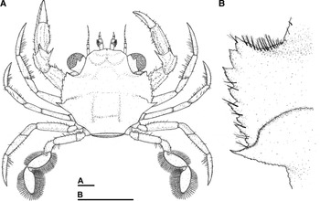
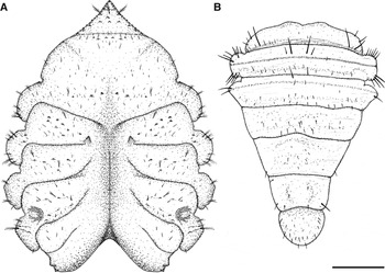
The general shape of the first stage is similar to that of the adult (Figure 1A). Carapace flattened dorso-ventrally and slightly convex, it has almost the same size in width and length, presents 8 lateral spines (finely serrated), besides the lateral pair commonly found in portunids (Figure 1B); small plumose setae on the orbital margin and small granules and sparse simple setae over the carapace surface, in addition to well-developed stalked eyes.
Achelous spinimanus (Latreille, 1819), first juvenile stage: (A) dorsal view; (B) lateral spines of carapace in detail. Scale bars: 0.5 mm.

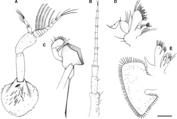
Antennule (Figure 2A) – developed basal segment bearing several plumose and simple setae, besides numerous small marginal granules; 2-segmented peduncle with sparse simple setae. Endopod 2-segmented (ventral flagellum) with 5 and 9 simple setae, respectively, on the proximal and distal segments. Exopod (dorsal flagellum) 8-segmented with 0, 12, 12, 10, 8, 6, 0 and 0 long aesthetascs, respectively; besides 0, 0, 1, 1, 0, 1, 4 and 2 simple setae, and also 3 terminal plumose setae on the last segment.
Achelous spinimanus (Latreille, 1819), first juvenile stage: (A) antennule; (B) antenna; (C) mandible; (D) maxillule; (E) maxilla. Scale bar: 0.2 mm.

Antenna (Figure 2B) – 3-segmented antennal peduncle, provided with sparse simple setae, besides small marginal and terminal granules on the first segment. Antennal flagellum 9-segmented with 0, 2, 5, 4, 1, 4, 2, 3 and 4 simple setae, all of them terminal.
Mandible (Figure 2C) – well-chitinized blade, 2-segmented palp with 2 plumose setae on proximal segment and 17 serrate setae, 5 plumose setae, 2 simple setae and 1 plumo-denticulate seta on distal segment.
Maxillule (Figure 2D) – coxal endite bearing 6 plumose setae, 4 simple setae of several sizes and 13 serrate setae; basial endite with 4 small simple setae and 1 long seta on the proximal margin, 12 serrate setae, 2 small simple, 10 cuspidate, 3 plumose and 2 plumo-denticulate setae on distal margin. The endopod is 2-segmented with 4 plumose setae and 1 simple seta on proximal segment, and 1 plumose seta and 3 simple setae on the distal segment. Two simple setae on protopod margin.
Maxilla (Figure 2E) – bilobed coxal endite with 9 plumose setae on the proximal segment, 3 simple setae and 2 plumose setae on the distal segment; basial endite, bilobed, with 3 slightly plumose setae and 7 simple setae on the proximal segment, 2 plumose setae, 2 slightly plumose setae and 10 simple setae on the distal segment. The endopod has 4 plumose setae on distal margin. The exopod (scaphognathite) shows 86 plumose marginal setae and 57 simple surface setae.
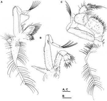
First maxilliped (Figure 3A) – coxal endite with 19 plumose, 12 serrate and 13 simple setae; basial endite with 47 plumose setae, 17 plumo-denticulate and 4 small simple setae. The endopod is unsegmented with 6 plumose setae and 34 simple setae of several sizes; 2-segmented exopod with 6 plumose, 5 simple and 5 cuspidate setae on the proximal segment, 7 plumose and 3 simple setae on the distal segment. The epipod is large, with 77 long simple setae and 14 small simple setae.
Achelous spinimanus (Latreille, 1819), first juvenile stage: (A) first maxilliped; (B) second maxilliped; (C) third maxilliped. Scale bar: 0.2 mm.

Second maxilliped (Figure 3B) – 5-segmented endopod with 10, 17, 2, 7 and 4 plumose setae, 2 and 7 serrate setae and 1 and 4 cuspidate setae on the fourth and fifth segments, respectively; 1 plumo-denticulate setae on the third and on the fourth segment, besides 0, 6, 3, 5 and 0 simple setae. The exopod is 2-segmented with 14 plumose setae, 17 simple setae and 9 cuspidate setae on the proximal segment; 5 simple setae and 7 plumose setae on the distal segment. The protopod shows 7 plumose setae and 4 simple setae; and the epipod is rudimentary and smooth.
Third maxilliped (Figure 3C) – 5-segmented endopod with several protuberances on the first and second segments, several setae on each segment: 29, 9, 12, 25, 13 serrate setae, 29, 11, 11, 6, 0 plumose setae, 46, 20, 17, 8, 4 simple setae from the proximal to distal segment. The exopod is 2-segmented with several marginal protuberances, 8 plumose setae and 41 simple setae on the proximal segment and 8 plumose and 5 simple setae on the distal. The protopod has 34 plumose setae of several sizes and 21 small simple setae. The epipod shows 7 plumose setae, 3 plumo-denticulate setae and around 40 small simple setae on the proximal portion, 28 long simple setae and 2 long serrate setae on the distal portion.
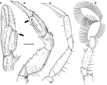
Chelipeds (Figure 4A, B) symmetrical, with small spines on external margin and 4 strong inner ones on merus, and 3 terminal spines on carpus with the dorsal one being the longest; its propodus presents a spine (Figure 4, arrows) in the inner margin at the level of the dactyl, characteristic of this species, as well as simple and plumose setae sparsely distributed. Second, third (Figure 4C) and fourth pereiopods are similar, with sparse simple and plumose setae present (the dactyls show several grouped setae on the inner margin). Fifth pair of pereiopods (Figure 4D) with a spine on the coxa; both propodus and dactyls flattened; dactyls are paddle-shaped with several marginal plumose setae.
Achelous spinimanus (Latreille, 1819), first juvenile stage: (A) cheliped (lateral view); (B) cheliped (dorsal view); (C) third pereiopod; (D) fifth pereiopod. Arrows indicate the propodus spine, characteristic of this species. Scale bar: 0.4 mm.

Thoracic sternites (Figure 5A) with simple and plumose setae sparsely distributed over surface, concentric and semi-circular wrinkles on the seventh somite (near the coxa).
Achelous spinimanus (Latreille, 1819), first juvenile stage: (A) sternum (ventral view); (B) abdomen (dorsal view). Scale bar: 0.4 mm.

Abdomen (Figure 5B) with 6 somites, each one wider than long, with several sparse simple setae; telson with small simple setae. Pleopods absent on the inner face.
Morphology of the second to ninth juvenile stage of A. spinimanus
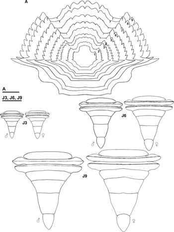
The proportion increases between carapace width and length of the juvenile individuals in each successive ecdysis, as the individuals grow up. From the ninth stage on, the carapace reaches similar proportions to those observed in the adult phase (Figure 6A).
Achelous spinimanus (Latreille, 1819): (A) carapace development throughout all juvenile stages obtained (numbers represent the stage of the juvenile phase); (J3–J6–J9) development of the abdomen shape of both males and females in third, sixth and ninth stages, respectively. Scale bars: A, 2.5 mm; J3–J9, 2 mm.

The abdomen increased in size during the growth time and begins to show slight changes in shape, associated with sex, from the ninth stage on. Nevertheless, such changes do not allow the visual differentiation of the sex of individuals, as can be observed in male and female adults (Figure 6 J3–J9).
The main morphological changes verified from the second juvenile stage onwards refer, mostly, to the secondary sexual characteristics. The pleopods, which were absent in the first juvenile stage, rise in different numbers and degree of development, according to each sex.
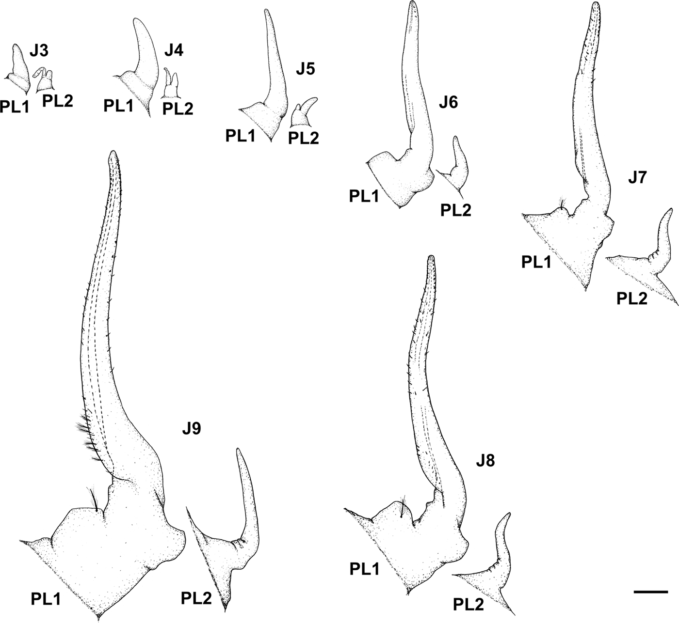
For males, in the third juvenile stage, a pair of pleopods appears on the first (PL1) and second (PL2) abdominal somite, respectively, being PL2 biramous (Figure 7 J3). In the fourth and fifth stages (Figure 7 J4–J5) there are no significant morphological changes, with exception of increase in sizes, especially of PL1. In the sixth stage (Figure 7 J6) the PL2 becomes completely uniramous and a few simple setae appear on the distal portion of PL1. From the seventh to ninth stages (Figure 7 J7–J9) the modifications that occur on both the PL1 and PL2 are limited only to the gradual increase in size, the appearance of simple and plumose setae and emergence of some protuberances on the distal portion of PL1. In the ninth juvenile stage, the general morphology of pleopods still differs from that of adults, mainly regarding the number of setae.
Achelous spinimanus (Latreille, 1819), PL1 and PL2: pleopods of the first and second male abdominal somites, respectively, from third (J3), fourth (J4), fifth (J5), sixth (J6), seventh (J7), eighth (J8) and ninth (J9) stages on. Scale bar: 0.2 mm.

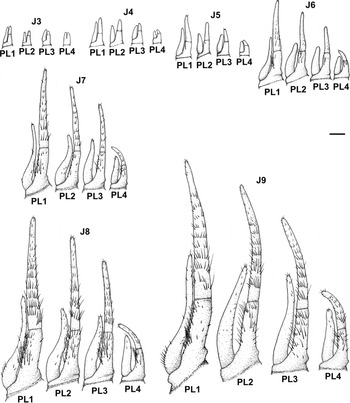
For females, in the third juvenile stage 4 pairs of biramous pleopods appear from the second to fifth abdominal somite (PL2, PL3, PL4 and PL5), all of them with endopods partially segmented and smooth (Figure 8 J3). In the fourth and fifth stages (Figure 8 J4–J5), the pleopods slightly vary in size and PL2, PL3 and PL4 acquire setae. In the sixth stage (Figure 8 J6), the endopods of the pleopods exhibit a true segmentation and increase in size and number of setae. From the seventh to ninth stages (Figure 8 J7–J9), the main modifications are restricted only to the rapid increase of number of setae and size of pleopods.
Achelous spinimanus (Latreille, 1819), PL2 to PL5: pleopods from the second to fifth female abdominal somites, respectively, from third (J3), fourth (J4), fifth (J5), sixth (J6), seventh (J7), eighth (J8) and ninth (J9) stages on. Scale bar: 0.2 mm.

We can point out some structures, which show other alterations. The most significant changes are: (1) on the endopod of the first maxilliped with a foliaceous shape and a groove in the apical region, particularly in the inner margin from the third stage on; and (2) on the first segment of the antennal peduncle, with the apical region showing a projection that resembles the antennal scale, in the advanced stages.
No significant morphological change occurs on the remaining body appendages. However, there is an increase in the number of setae on segments of each appendage. Denticule-serrulate setae on the epipod of the third maxilliped appear from the third juvenile stage on. Setae on the epipod of all maxillipeds from the fourth stage on are of the type denticule-serrulate or harpoon-shaped setae.
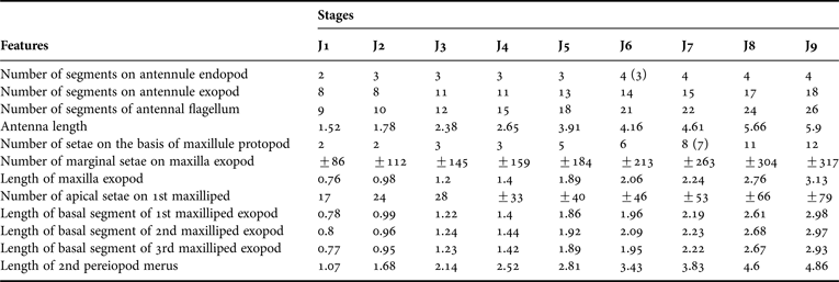
The most notable morphological characters that allow the identification of the first nine stages of the juvenile development of A. spinimanus are shown in Table 2.
Achelous spinimanus (Latreille, 1819).

Main morphological features that allow the identification of the first nine juvenile stages (J1 to J9). Length measurements represent average values, in mm; numbers in parentheses indicate alternative values of low frequency; ‘±’ indicates ‘approximately’.
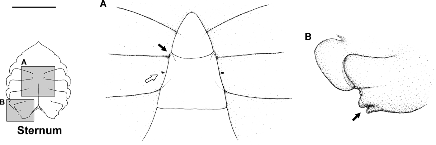
Regarding female gonopores, their vestigial aperture comes from the third juvenile stage (Figure 9A, white arrow). In males, it was not possible to verify the emergence of the gonopores.
Achelous spinimanus (Latreille, 1819), third juvenile stage: (A) white arrow, the rudimentary female gonopores; black arrow, anterior ‘closing mechanism’ position; (B) posterior ‘closing mechanism’ position, pointed by the black arrow (see schematic drawing of the position of the abdomen over the posterior ‘closing mechanism’ in Bolla Jr et al., Reference Bolla, Fransozo and Negreiros-Fransozo2014). Scale bar: 0.5 mm.

In both sexes, the abdomen remained sealed to sternum throughout the juvenile stages obtained. The presence of 2 pairs of ‘closing mechanisms’ or ‘sternal buttons’ on the sternum was verified. There is an anterior one (located on the fifth sternite, at the level of the distal region of the sixth abdominal somite) (Figure 9A, black arrow); and another, posterior one (located on the eighth sternite, at the level of the lateral region of the second abdominal somite) (Figure 9B, black arrow). Such structures, together with cementing substances present in the contour of the abdomen, do not allow the extension of the abdomen before the beginning of the reproductive period.
The size of the chelipeds did not differ between sexes throughout the obtained juvenile stages.
DISCUSSION
Although the species studied here have recently changed to the resurrected genus Achelous, its early juvenile stages share morphological features with other species belonging to the genus Portunus, at least, for those species previously studied regarding their juvenile development. To better distinguish possible unique characteristics of these two genera we need to complete further studies.
Descriptions of the carapace of portunids show that, just in the first post-larval stage, P. anceps exhibits 8 lateral spines, as well the common pair of spines characteristic of this family (Lebour, Reference Lebour1944a). For P. pelagicus, besides the common features shared with P. anceps, Yatsuzuka & Sakai (Reference Yatsuzuka and Sakai1980) also found a serrulate rostrum, as occurs for C. ornatus and C. danae studied by, respectively, Bolla Jr et al. (Reference Bolla, Negreiros-Fransozo and Fransozo2008) and Bolla Jr et al. (Reference Bolla, Fransozo and Negreiros-Fransozo2014), and for A. spinimanus (present study).
Definitions of specific morphological characteristics for each species, both portunids as well as representatives from other groups, are extremely complex due to the low number of previous studies on juvenile development among Decapoda. However, such definition is particularly crucial, as a tool for identification of juveniles of these decapods, for monitoring of areas of population growth, especially for threatened and/or invasive species.
For morphological descriptions of juvenile decapods, the authors take into account the number of segments and setae present on body appendages. Nevertheless, due to the difficulty in finding distinctive characters with light microscopy, the study of the juvenile phase has been limited to the count of setae on appendices and the mapping of its distribution throughout the body of the animal (Rieger & Beltrão, Reference Rieger and Beltrão2000).
In this study, some appendages and morphological characteristics proved to be of great importance due to some peculiarities, which are observed with minimal manipulation of specimens and, probably, could be used for future comparisons and species identification. We can point out the following characters in this procedure: (1) the spines of internal margin of merus of the cheliped (A. spinimanus and P. pelagicus show 4 spines, whereas C. danae, C. ornatus and C. sapidus show only 3); (2) the long superior spine of carpus of the cheliped (present in A. spinimanus and P. pelagicus and absent in C. danae, C. ornatus and C. sapidus); and (3) the segmentation of the mandible palp (which is 2-segmented in A. spinimanus and P. pelagicus and 3-segmented in C. danae, C. ornatus and C. sapidus). These morphological features can provide separations among the species of the genus Callinectes and the genera Portunus and Achelous, but not only between the last two genera.
Another two easy differentiations detected for each species are: (1) the number of segments on antennal flagellum (C. sapidus, A. spinimanus, C. danae and C. ornatus show 8, 9, 10 and 10(11) segments, respectively); and (2) the number of segments on the exopod of antennule – A. spinimanus is the only one among the species of these two genera that has 8 segments (other species have 7 segments), in addition to a set of aesthetascs. This feature probably could be used for quick identification, while new descriptions are not provided for the other species of this group.
Concerning the sternum, the presence of concentric and semi-circular wrinkles on the seventh somite of the first juvenile stage, observed in A. spinimanus, C. danae and C. ornatus, could be explained as a remnant of sternal spine found in the same place during the megalopa stage, and it is characteristic of this larval phase for the family Portunidae (Kurata, Reference Kurata1975). Additionally, such wrinkles was not found in the later stages, reinforcing this idea; however, further studies are necessary to clear up the origin of these structures. In C. sapidus and P. pelagicus, the authors did not report the presence of these wrinkles, probably because they did not note this feature.
The superior abdominal closing mechanism (located on the fifth thoracic sternite) of the juvenile phase of A. spinimanus, C. danae and C. ornatus is in accordance with the more typical system of sealing found in Eubrachyura (Guinot & Bouchard, Reference Guinot and Bouchard1998). Nevertheless, nothing is mentioned in the literature about the inferior closing mechanism (on the eighth sternite), except for C. danae, in which this feature is also present (see Bolla Jr et al., Reference Bolla, Fransozo and Negreiros-Fransozo2014 for details). Thus, this mechanism could be exclusive to the family Portunidae, but we could only confirm this hypothesis with further studies of other species within and outside this family.
The sexual dimorphism in the abdomen shape of males and females adults is not evident throughout the stages obtained for A. spinimanus. For C. sapidus, C. ornatus and P. pelagicus studied, respectively, up to the 11th, 12th and third juvenile stages, the sexual dimorphism in the abdomen shape was also not observed. Thus, probably this feature appears in later stages for the Portunoidea species, in contrast to representatives of other superfamilies. For instance, in Inachus dorsettensis Pennant, 1777 (Majoidea) and Pachygrapsus transversus Gibbes, 1850 (Grapsoidea), the morphological sexual differentiation occurs from the third and sixth juvenile stages on, respectively (Ingle, Reference Ingle1977; Flores et al., Reference Flores, Negreiros-Fransozo and Fransozo1998); in Cyrtograpsus angulatus Dana, 1851 (Grapsoidea), the sexual differentiation begins from the fourth stage on, becoming more apparent from the eighth juvenile stage on (Rieger & Beltrão, Reference Rieger and Beltrão2000); and in Calappa tortugae Rathbun, 1933 (Calappoidea), the secondary sexual characters appear from the second juvenile stage on (Negreiros-Fransozo et al., Reference Negreiros-Fransozo, Wenner, Knott and Fransozo2007).
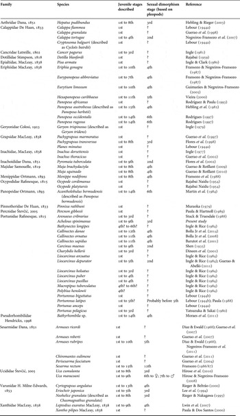
The presence of rudimentary pleopods in the first juvenile stage can be different among the Portunoidea species studied; this feature is present in C. sapidus, C. ornatus and P. pelagicus, but it is absent in C. sapidus and A. spinimanus. However, with the exception of P. pelagicus in which the rudimentary pleopods are present up to the last stage studied (third), in all other species the pleopods totally degenerate from the second juvenile stage. They reappear (two pairs for males and four pairs for females) in later juvenile stages that can vary according with species, as can be seen in Table 3. Among Portunoidea, this occurs from the third stage in A. spinimanus and from the fourth stage in C. danae, C. ornatus and C. sapidus; this, apparently, could be a particular character of these genera. Nevertheless, only with a high number of species studied of distinctive genera could one get a satisfactory definition.
List of species with some stages of the juvenile phase known to date.

‘?’ = the authors did not mention or did not see the stage in which the sexual differentiation occurs.
Another interesting differential characteristic is the biramous condition of the second pair of the pleopods of males (PL2), from its first appearance to the fifth stage in A. spinimanus, until the eighth stage in C. danae and until the sixth stage in C. ornatus. In C. sapidus, the PL2 remain biramous, at least, until the seventh stage (the authors did not report if they still are biramous in the later stages). With respect to the first pair of pleopods of males (PL1), this is uniramous as of its origin in all Portunoidea species studied, except for C. sapidus, in which the PL1 is also biramous until the seventh juvenile stage. These features of pleopods, both the rudimentary ones and the biramous conditions of male pleopods, are useful for species identification within the group, since they can be easily observed.
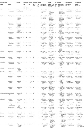
Table 4 presents the comparative studies of the main characteristics that can be used to differentiate the species with juvenile development previously studied, and which occur in the Brazilian coast.
Diagnostic features that allow the differentiation and identification of the first juvenile stage of species with occurrence in the Brazilian coast, with stages of the juvenile phase known to date.

Seg = number of segments; (E) = endopod; (Ex) = exopod; (P) = protopod; (CE) = coxal endite; (BE) = basal endite; (Ep) = epipod; ? = the authors did not mention; ± = approximately; Nu = numerous; ab = absent. Commas separate setae by segment.
The description of early stages of decapod crustacean species are useful in the identification of settlement areas of such species, which is necessary to the fisheries management in many areas. In addition, it could contribute towards systematic studies, since it aids in the elaboration and validation of identification keys for adult specimens (Martin et al., Reference Martin, Felder and Truesdale1984). Morphological studies also collaborate for effective separation of species of interest for carciniculture, besides their identification in planktonic or benthic samples (Guerao & Abelló, Reference Guerao and Abelló2011; Bolla Jr et al., Reference Bolla, Fransozo and Negreiros-Fransozo2014).
ACKNOWLEDGEMENTS
We thank the members of NEBECC for their assistance during this study. All sampling in this investigation was conducted in accordance with Brazilian applicable state and federal laws.
FINANCIAL SUPPORT
We are grateful to the Brazilian Council of Scientific and Technological Development (CNPq) for the first author's Master of Science scholarship (#553174/2008-8) and to the São Paulo Research Foundation (FAPESP) for providing the financial support during neuston collections (#2004/15194-6).